
Abstract
The incorporation of Quality-by-Design (QbD) principles in tissue-engineering bioprocess development toward clinical use will ensure that manufactured constructs possess prerequisite quality characteristics addressing emerging regulatory requirements and ensuring the functional in vivo behavior. In this work, the QbD principles were applied on a manufacturing process step for the in vitro production of osteogenic three-dimensional (3D) hybrid scaffolds that involves cell matrix deposition on a 3D titanium (Ti) alloy scaffold. An osteogenic cell source (human periosteum-derived cells) cultured in a bioinstructive medium was used to functionalize regular Ti scaffolds in a perfusion bioreactor, resulting in an osteogenic hybrid carrier. A two-level three-factor fractional factorial design of experiments was employed to explore a range of production-relevant process conditions by simultaneously changing value levels of the following parameters: flow rate (0.5–2 mL/min), cell culture duration (7–21 days), and cell-seeding density (1.5×10³–3×10³ cells/cm2). This approach allowed to evaluate the individual impact of the aforementioned process parameters upon key quality attributes of the produced hybrids, such as collagen production, mineralization level, and cell number. The use of a fractional factorial design approach helped create a design space in which hybrid scaffolds of predefined quality attributes may be robustly manufactured while minimizing the number of required experiments.
Introduction
The number of potential process variables and the sensitivity of cell-based products mean that their manufacturing would be particularly dependent on choosing and maintaining the optimum bioprocess conditions. 7 The use of bioprocess development tools such as statistical design of experiments (DoE) 8 and windows of operation 9 has recently been suggested for efficiently defining the regions of process operation that would yield desired in vitro quality characteristics of TE constructs, characteristics that could be ultimately linked with tissue formation and function in vivo. An important element of this strategy for TE applications would be to identify certain key process parameters and evaluate their impact on TE construct quality attributes developing, at the same time, corresponding predictive process models that would highlight a process space, termed as design space, wherein tailored TE constructs could be manufactured in a standardized and validated way. Statistical DoE provides efficient procedures for planning multifactor experiments in such a way that the data obtained can be analyzed to yield consistent and objective conclusions on the involvement of all investigated parameters. Furthermore, it requires fewer number of experimental runs, while, at the same time, covering a broader knowledge space than a one factor at a time approach. DoE has been used for optimizing medium compositions for stem cell culture,10–12 defining bioprocess related shear–stress thresholds for human cell cancer vaccines, 13 optimizing critical TE-related process parameters such as cell-seeding efficiency,8,14 and also as a tool for evaluating in vivo bone formation on cell-seeded calcium phosphate (CaP) carriers. 15
Recently developed osteogenic hybrid materials have shown potential to serve as clinically relevant bone reparative units, providing a biomimetic template to more effectively mediate in vivo bone regeneration.16,17 Enhanced in vivo bone-forming capacity and biological activity were recently observed when implanting such materials, produced in either static 16 or bioreactor setups. 17 Moreover, recently developed bioinstructive osteogenic media containing CaP have shown promise in further improving the performance of hybrid materials in an in vitro biomimetic model. 18
Perfusion bioreactors have been successfully used in bone TE to date, to develop controlled culture environments eliminating diffusion limitations observed in static culture19,20 by introducing convective nutrient and oxygen transport throughout the construct volume due to flow through the scaffold.21–25 Furthermore, due to fluid flow, necessary biomechanical stimuli may also be provided to the cultured cells, enhancing extracellular matrix production and its mineralization,21,26–28 as well as improving its spatial distribution within the scaffold.23,29 However, a more systematic study on the effect of individual bioreactor process variables on process variation as well as linking individual statistically significant effects of a specific process parameter on corresponding quality characteristics of the produced TE construct needs to be further examined. It should be stressed that bioreactor process design and further development should take into consideration the fact that this unit operation would be one among a series of operations with the final aim of in vivo bone formation recently discussed by Lenas and Luyten. 2 Hence, its performance would be affected by preceding unit operations such as a cell-seeding process and would affect subsequent unit operations such as in vivo bone formation where the mouse may be seen as a living bioreactor or a decellularization process step as recently suggested. 10
In this work, factorial DoE was employed as a statistical tool to explore a process design space constituted by important perfusion bioreactor process parameters (input variables) such as cell-seeding density, medium flow rate, and time of bioreactor operation. The evaluation of the level of impact of each process parameter on osteogenic hybrid material quality attributes (output variables) such as DNA content, collagen content, and mineral content followed. As raw materials, a titanium alloy scaffold (Ti6Al4V) with a regular pore structure and a clinically relevant cell type derived from human periost (hPDC) with proven in vivo bone-forming properties15,16,30 were used. A bioinstructive culture medium supplemented with Ca2+ and Pi ions was also employed to drive hPDC cultures on Ti6Al4V scaffolds through an enhanced proliferation stage, followed by an osteogenic differentiation stage as shown by Chai et al. 10 The in vitro production of such laboratory-scale-size osteogenic hybrids based on a robust perfusion bioreactor technology could become a part of a clinical-scale manufacturing strategy for regenerative units.
Materials and Methods
Ti6Al4V scaffolds
An inert titanium alloy biomaterial was produced by additive manufacturing of selective laser melting, resulting in highly porous Ti6Al4V scaffolds (Ø=6 mm; h=6 mm) and will be further referred to as Ti scaffolds. 31 The Ti scaffolds were produced and subsequently analyzed as described by Pyka et al. 32 and had a porosity of 73%±1%, surface area of 6.5±0.2 cm2, strut diameter of 245±2 μm, and pore size of 755±3 μm. Before use, scaffolds were cleaned ultrasonically, immersed for 10 min in acetone, 10 min in ethanol 70%, and 10 min in distilled water, subsequently oxidized for 12 h at 60°C in a 5 M sodium hydroxide (Sigma-Aldrich) solution, rinsed with distilled water, and finally sterilized in a steam autoclave. Before cell seeding, all scaffolds were prewetted by vacuum impregnation in a cell culture medium for 2 h in a humidified incubator at 37°C, and dried overnight in a nonhumidified incubator. 33
hPDC culture
hPDCs were isolated from periosteal biopsies of different donors as described previously. 34 This procedure was approved by the ethics committee for Human Medical Research (Katholieke Universiteit Leuven) and with patient-informed consent. hPDCs were expanded in the Dulbecco's modified Eagle's medium with high glucose (Invitrogen) containing 10% fetal bovine serum (BioWhittaker) and 1% antibiotic–antimycotic (100 units/mL penicillin, 100 mg/mL streptomycin, and 0.25 mg/mL amphotericin B; Invitrogen). The seeding density used for the two-dimensional (2D) hPDC expansion was 6000 cells/cm2. hPDCs were passaged at 80%–90% confluency. At the time of experiment, cells from passage 6 were trypsinised with Tryple Express (Invitrogen) and seeded in the scaffolds. A static drop-seeding protocol was used for seeding cells onto prewetted 3D Ti scaffolds. Three-dimensional hPDC culture on Ti scaffolds lasted for 7, 14, and 21 days under static or dynamic culture conditions. (1) For static culturing, bone TE constructs were positioned in 50-mL falcon tubes (disposable 50-mL Falcon tubes; BD Biosciences) containing 10 mL of the cell culture medium, which was the same amount used for dynamic culture, and incubated at 37°C in a humidified and CO2-controlled incubator. (2) For dynamic culturing, bone TE constructs were cultured in an in-house developed perfusion bioreactor (Scheme 1) equipped with seven parallel perfusion circuits. Each bone chamber, holding a single scaffold, was connected to an individual medium reservoir (disposable 50-mL Falcon tubes; BD Biosciences) containing 10 mL of cell culture medium via a Tygon® (Cole Parmer) tubing and a two-stop tubing (BPT; Cole Parmer) connected to a peristaltic pump (IPC-24; Ismatec SA). In both static and bioreactor cultures, the medium was refreshed every 2 days for the entire culture period. The bioinstructive medium used for cell culture this study was a supplemented form of the expansion medium containing Ca2+ (6 mM) and Pi (4 mM) and ascorbic acid with a concentration of 0.05 ng/mL. 10
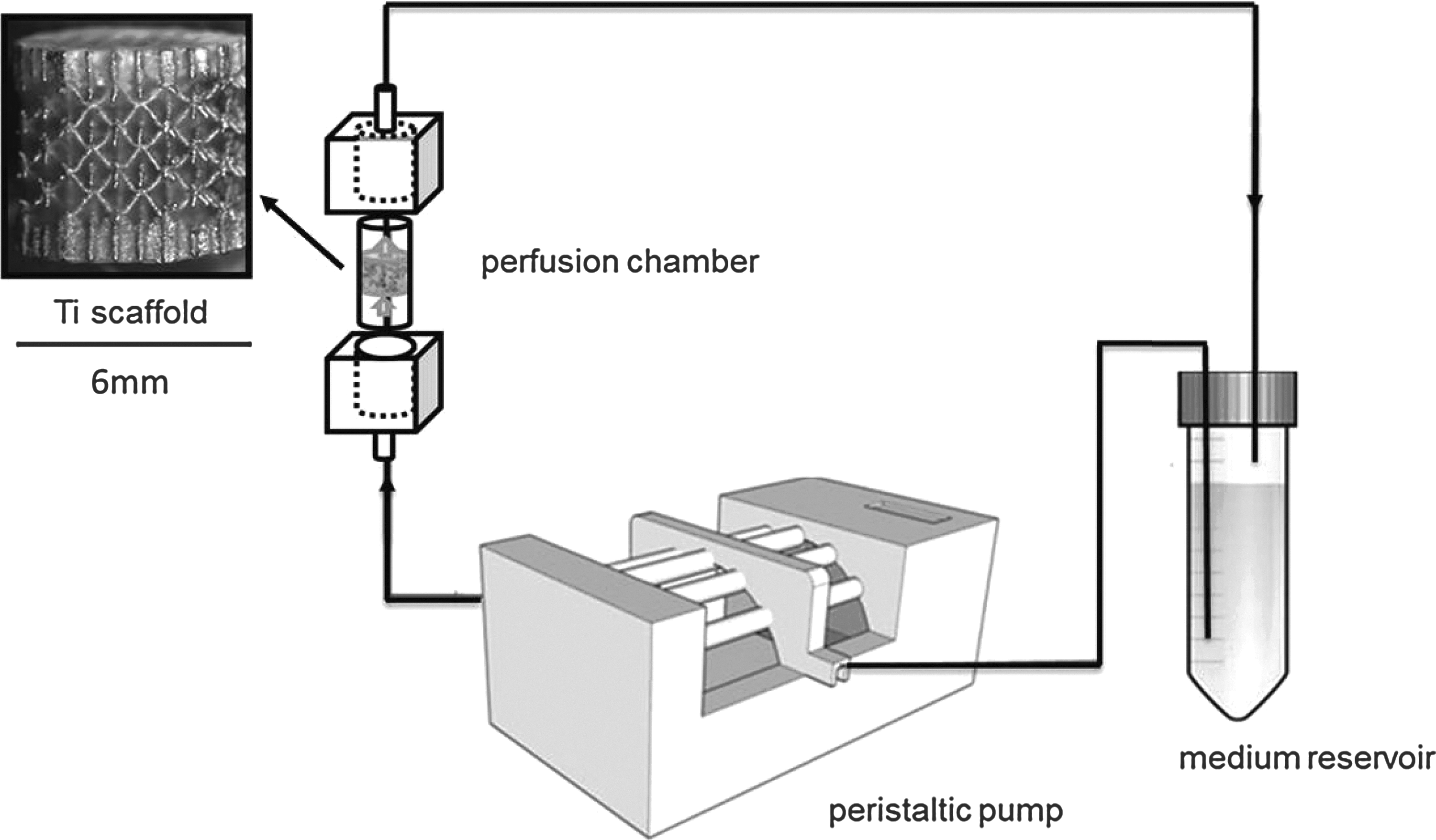
Representation of the bioreactor setup used for this work. The closed perfusion system simulations consisting of a reservoir, a peristaltic pump (IPC-N; Ismatec SA), and a bioreactor, all connected by the pump tubing. The 6×6-mm Ti6Al4V scaffold was positioned in the bioreactor perfusion chamber. The peristaltic pump circulated the medium through the construct at different flow rates (0.5–2 mL/min).
Monitoring of hPDC growth and viability on 3D porous Ti6Al4V scaffolds
hPDC growth on 3D porous Ti scaffolds was monitored by a nondestructive analysis of the metabolic activity of each sample at defined time points using alamarBlue™, 8 (Invitrogen) according to the manufacturer's instructions (n=4). Scaffolds containing cells were harvested every 2 days submerged and incubated in 1ml AlamarBlue™ assay while as a control wells containing only the assay were incubated at the same time. After 1-h incubation at 37°C, the metabolic activity of the cells was quantified by measuring absorbance values of the supernatant liquid at 570 nm. Higher absorbance values indicated higher metabolic activity and cell growth. All sample absorbance values were normalized to those of the control samples.
DNA content measurement
The DNA content analysis was obtained by using a highly quantitative and selective DNA assay (Quant-iT™dsDNA HS kit; Invitrogen). Cell-containing scaffolds were washed with phosphate-buffered saline (PBS) and lysed in 350 μL RLT lysis buffer (Qiagen) by 60 s of vortexing. The lysed samples were stored at −80°C before analysis, thawed at room temperature, and spun down for 1 min at 13000 rpm. DNA concentrations were quantified with a Qubit®Fluorometer (Invitrogen). 8 Unattached cells in the residual seeding medium were spun down 10 min at 180 g, rinsed with 350 μL PBS, spun down again, and resuspended in 350 μL cell lysis buffer. The cell-seeding efficiency was determined by the ratio of DNA content values derived from the cells attached on the Ti scaffolds over the sum of DNA content values of seeded scaffolds and DNA content values of the suspension, multiplied by 100%. Following this methodology, an average cell-seeding efficiency of 60% was achieved, as has been reported elsewhere. 33
Characterization of 3D osteogenic extracellular matrix production: mineralization assay
For each bioreactor condition, mineralization of the cell cultures was assessed by alizarin red staining (pH 4.2) after 7, 14, and 21 days of culture. Briefly, cell cultures were rinsed with PBS, followed by fixation in 4% formalin (in PBS) for 10 min, and washed with distilled water before staining with an alizarin red solution for 1 min. Nonspecific staining was removed by rinsing in distilled water. The deposited minerals were quantified by dissolving the dye in 10% cetylpyridinium chloride solution with constant agitation (n=3). Subsequently, 1 mL triplicates of the solution from each sample were transferred to a 96-well plate, and the absorbance readout at 570 nm with a microplate reader was obtained (TECAN). 35
Characterization of 3D osteogenic extracellular matrix production: collagen assay
The 3D collagen matrix production and matrix mineralization on porous Ti scaffolds were characterized by Picrosirius Red staining (1 mg/mL Sirius Red in saturated Picric acid, n=3). 36 The stained samples were thoroughly washed with distilled water to remove unbound dye and dried at 37°C before qualitative analysis by stereomicroscopy. For quantitative analysis, the Picrosirius red dye was dissolved in 0.2M NaOH/Methanol (1:1 ratio) with mild shaking overnight, and the optical density was measured at 492 nm using a microplate reader (TECAN) for 1 mL triplicate samples for each condition.
Scanning electron microscopy observation
The morphology and CaP crystal structure of the mineralized matrix were characterized by scanning electron microscopy (SEM) coupled with energy-dispersive X-ray (EDAX) analysis (FEI XL30 FEG) at 10 kV. Briefly, the cultures were rinsed twice with PBS, fixed with 2.5% glutaraldehyde (in PBS) for 1 h, and postfixed in 1% osmium tetroxide for 2 h before dehydrated in 50%, 75%, 95%, and 100% ethanol solutions. Finally, the samples were chemically dried with hexamethyldisilazane for 3 min and gold-sputtered before SEM analysis.
Factorial analysis
The three critical quality attribute (CQA) responses, DNA, collagen, and mineral content results, were analyzed with statistical software (JMP). Each response was an average value of triplicate measurements on the hybrid scaffolds produced in three parallel bioreactor runs per condition. The experiments were run in a random order to minimize the effects of unexpected variability in the observed responses. Care was also taken when choosing the factor levels that differences between high and low would be statistically significant. Because several factors are involved, multivariate analysis of variance was used to test the significance of each term in the equation and the goodness of fit of the regression model. The statistical significance level was set at p<0.1 in all cases. Regarding the first process parameter, flow rate, the low-level value was determined as 0.5 mL/min, while the high-level value as 2 mL/min. The purpose for selecting these levels was that within this flow regime range, flow enhances collagen production and mineralization.
37
Regarding the time of culture, 7 days were considered as the low value reflecting the early time period for hPDC growth, while 21 days were selected as high level, since hPDCs will be fully differentiated by this time point. The seeding density values were selected based on preliminary experimentation (data not shown) to establish minimum cell densities that would allow subsequent cell growth in the bioreactor. A low level of 105 cells (for each scaffold) was the minimum robust cell-seeding density for the type of scaffold used in this study, while 2×10⁵ cells were chosen as the maximum to maintain the same level of seeding efficiency, since higher seeding densities result in lower seeding efficiencies.
8
The design matrix used for this experimental work is shown in Table 1. For the fractional factorial design used in this study, it should be pointed out that main factor effects were aliased with the interactive effect of the remaining factors, hence assuming linear behavior between process parameters and quality attributes for the process space under investigation. Due to the limitations of this design type, partial least-square fitting was used, missing out in this way any possible curvature of the response surface. All values are reported as mean±standard deviation. General linear-model parameter estimation was performed by least-squares estimation. The main effects are included in the statistical model following,
For each bioreactor experimental run, a corresponding static culture was performed on parallel noted as S1, S2, S3, S4, and S5.
where Y is the predicted response; b is the parameter estimate; X is the coded value of the factor levels; and e is the residual error. Statistical models were accepted when there was no lack of fit and no correlation in the residual plots, and the residuals were normally distributed.
Statistical analysis
In Figure 2, data are expressed as mean±standard deviation. The statistical difference of means of multiple independent groups was evaluated by one-way analysis of variance, and the two independent groups were compared by the unpaired Student's t-test (two-tailed), both by establishing the statistical significance at p<0.05.
Results
Perfusion bioreactor versus static culture: monitoring cell growth via metabolic activity
The Alamar blue assay was used to monitor the metabolic activity of hPDCs during cell culture periods that spanned from 7 to 21 days. Figure 1 shows the metabolic activity of cells that were cultured dynamically under perfusion in a bioreactor setup and those cultured in a static setup. Statically cultured hPDCs showed limited metabolic activity (no significant increase in respect to day 1) during the first 5 days of culture (for all experimental conditions), followed by a slow increase in metabolic activity that was enhanced toward the final days of culture, resulting in a 2-fold increase in respect to day 1 for B4 and 1.7-fold increase for the B5 experimental condition. On the other hand, a high metabolic activity was observed in the bioreactor for the whole culture period for all experimental culture conditions that continued until day 15 for the B3 (3.2-fold in respect to day 1), B4 (3.4-fold increase in respect to day 1), and B5 (2.6-fold increase in respect to day 1) experimental conditions, with metabolic activity values constantly increasing for all time points. After day 15, a flattening-off of this trend and a decrease of metabolic activity in respect to day 14 were seen for experimental conditions B4 and B5. When the two culture strategies are compared, it can be seen that cells cultured in dynamic conditions demonstrate a significantly higher (p<0.05) metabolic activity during the whole culture period for the B1 and B2 experimental conditions (7-day total culture duration), which is maintained until day 16 of culture for the B4 and B5 experimental conditions (21-day total culture duration). However, this difference seems to be less distinct and is not significant by day 20.
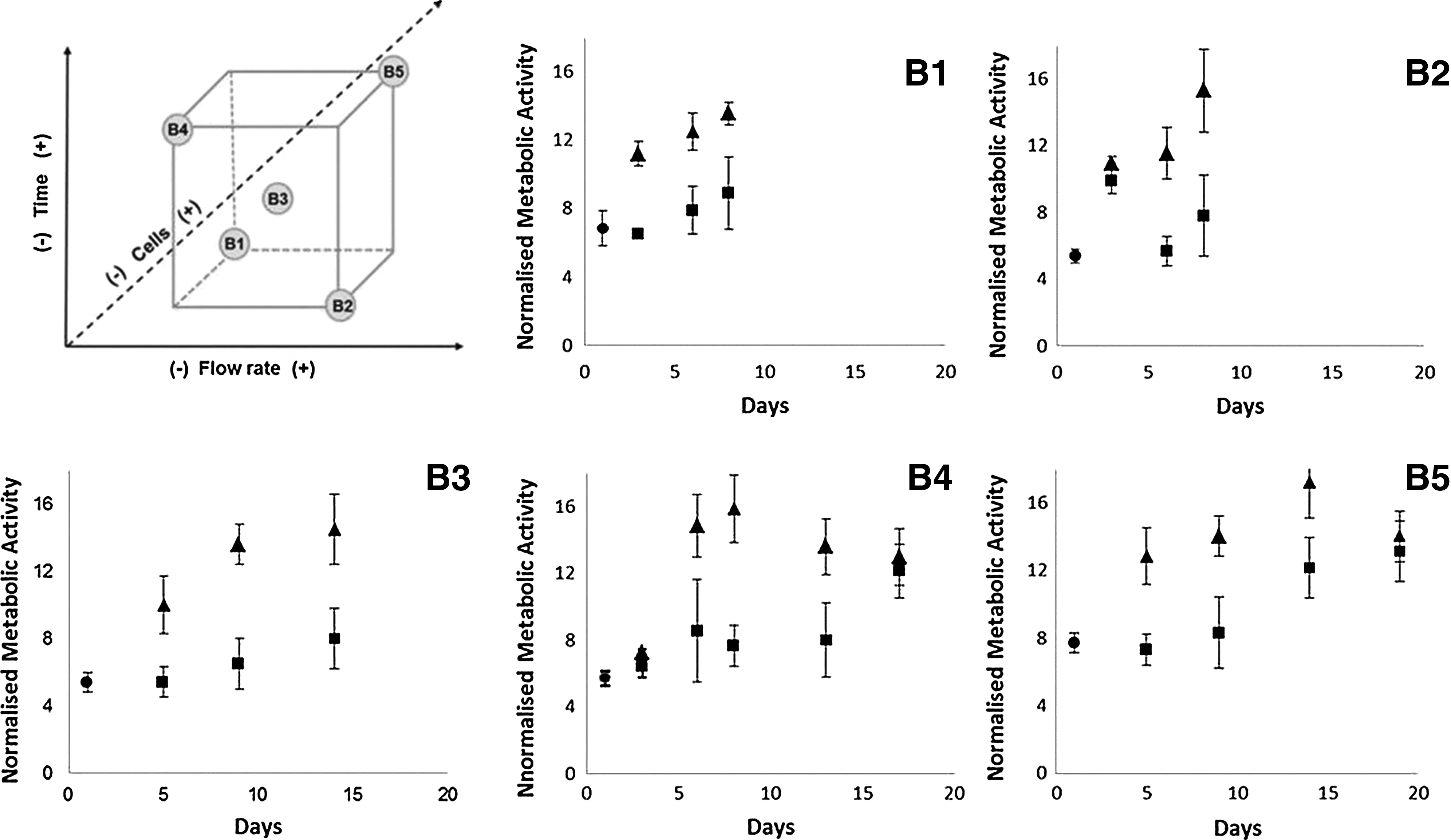
Human periosteum-derived cell (hPDC) growth as indicated by metabolic activity measurements, on Ti6Al4V scaffold for all experimental conditions of the fractional factorial design shown in the schematic representation of the upper left corner, comparing in all cases static (▪), with perfusion bioreactor (▴), culture conditions (n=4). Statically seeded scaffolds were used as the initial culture condition for both static and bioreactor setups and are denoted as (•).
Perfusion bioreactor versus static culture: quality attributes of as-produced hybrid scaffolds
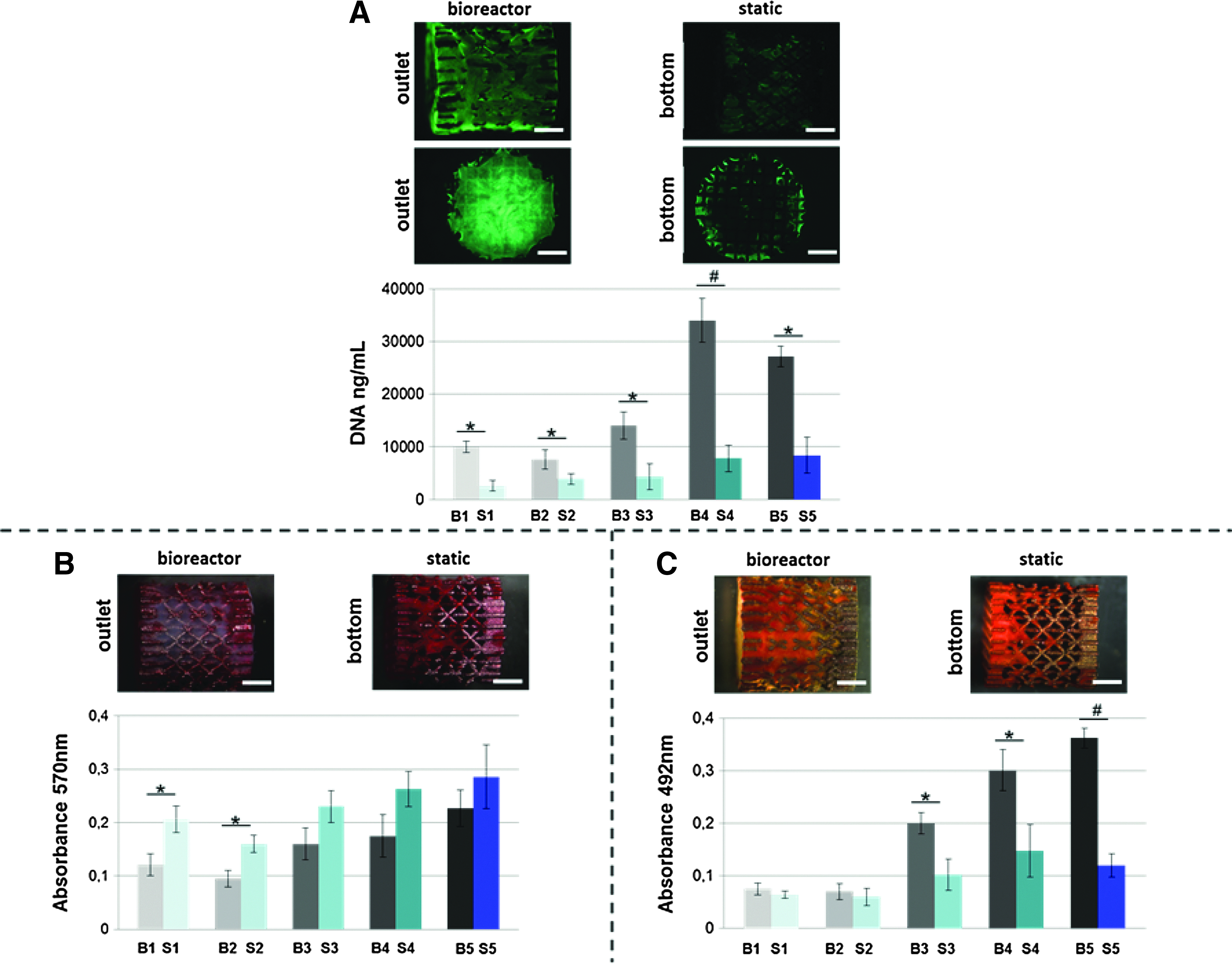
Manufactured hybrid scaffolds were characterized in terms of total DNA content while the 3D collagen matrix production and matrix mineralization that was produced during culture on porous Ti scaffolds were also assessed (Table 2). Figure 2 illustrates a gradual closure of the scaffold macropores from day 7 (B2) to day 21 (B4). Furthermore, an increased presence of dead cells in a 21-day static culture-produced construct (S4) in respect with the corresponding bioreactor condition (B4) was also observed. Representative SEM images further highlight the gradual change of the morphology of the cell–collagen–CaP structure from day 7 (B2) to day 21 (B4) with increased presence of deposited CaP nodules (B4) and gradual decrease in pore size, maintaining however a porous morphology (mineral nodules were confirmed to be CaP as shown by EDAX analysis in the image insets). Static culture (S4) results in a more bulky deposition of CaP nodules, often agglomerated, within which cells are difficult to be distinguished. Based on DNA content measurements, perfusion cell culture induced significantly higher proliferation (p<0.05) of hPDCs than static culture as seen by final DNA content measurements (for all experimental conditions) in Figure 3A. It is worth noting that 21-day bioreactor runs resulted in a 4-fold (B4) and 3-fold (B5) expansion of the cell number in comparison with the initially seeded cell number. Furthermore live/dead images show a distinct difference in the overall morphology of the resulting hybrid scaffolds as shown by representative images of B4 and S4 experimental conditions. A homogeneous or controlled viable cell distribution throughout a scaffold is important to guarantee a successful tissue construct. Images may provide a first qualitative understanding regarding homogeneity. Side-view images as well as bottom or outlet (bioreactor) images indicate that under perfusion, a much more homogeneously distributed and dense cell population may be observed (Fig. 3A), while static culture conditions yielded lower cell numbers and a more fragmented presence of cells on the scaffold, located more toward the periphery of the scaffold. Cell growth was initiated on the surface of the scaffold and gradually bridged out into the internal pore space leading to total closure of the scaffold macropores. Dynamically cultured hybrid scaffolds showed lower mineral content than static culture-derived hybrid scaffolds for all experimental conditions as shown in Figure 3B and significantly lower for B1, B2, and B3 (p<0.05). In the corresponding images, it is clear that in the static case, the stain is more intense, but again, inhomogeneity is evident for the resulting construct. A collagenous matrix was found to be deposited throughout the porous Ti scaffolds in a 3D manner as seen in the representative images (Fig. 3C). A significantly increased collagen content in perfusion-derived hybrid scaffolds was measured: 2-fold for B3 in respect to S3 and B4 to S4 (p<0.05 for B3 and B4) and 3-fold for B5 in respect to S5 (p<0.01 for B5) experimental runs.

Close-up live/dead images (scale bar: 1 mm), SEM micrographs, and EDAX spectra showing hybrid construct micromorphology of representative bioreactor and static conditions. (SEM, scanning electron microscopy; EDAX, energy-dispersive X-ray; C, cell; CaP, mineral grains; 3500× (scale bar 40 μm) and 14000× (scale bar: 10 μm) magnification, respectively. Color images available online at
In vitro engineered three-dimensional (3D) osteogenic hybrids using porous Ti6Al4V scaffolds, comparison between perfusion bioreactor culture and static controls.
Values shown in this table are average values of three independent experimental runs.
Multilevel factorial analysis for the manufacturing of hybrid scaffolds in a perfusion bioreactor: main effects
To study the effects of the three selected bioreactor process parameters termed as factors (culture time, number of seeded cells, and flow rate), on the process output variables, that is, hybrid construct quality attributes (DNA content, collagen content, and mineral content), we used a two-level three-factor fractional factorial analysis (indicated in Table 1) with a JMP Software package to detect the main effect of each factor on every output variable and to highlight those factors that would be most influential for the design of a production process. Positive and negative effects of every factor on each of the output variables and their significance are shown in Figure 4. For all output variables, time had a statistically significant positive effect (p-values are shown in Fig. 4), which is expected, since cell growth, extracellular matrix deposition, and mineralization are all time-dependent phenomena. For the range of conditions studied in this work, flow rate and seeding density seemed to have a negative effect on DNA content, although not significant effect (p>0.1). Flow rate was seen to have a significant positive effect (p<0.019) on the collagen content of the hybrid constructs, while the seeding density had a nonsignificant positive effect. Regarding mineral content, the seeding density had a significantly positive effect (p<0.088), while flow rate had a positive, but not significant, effect.
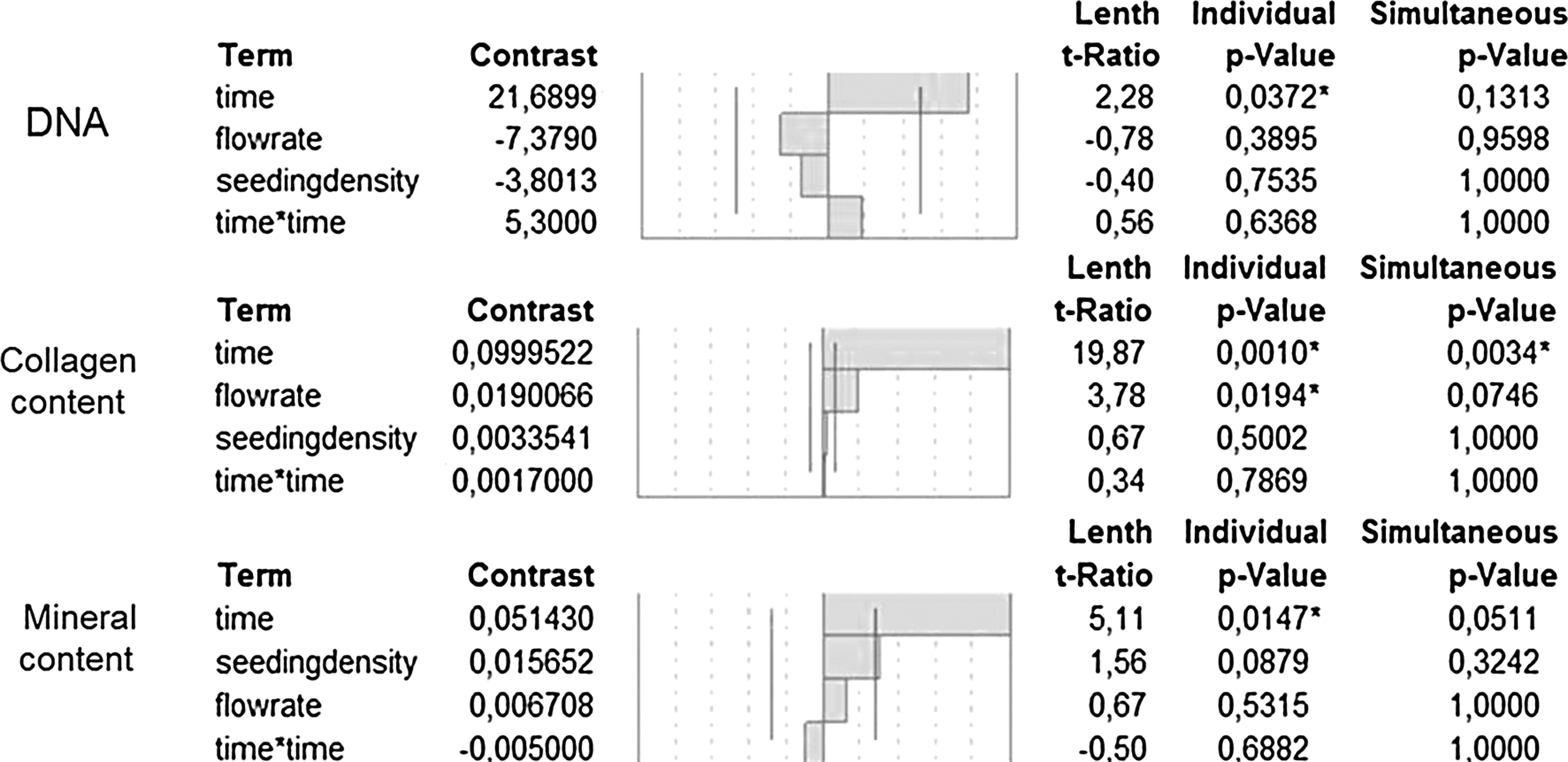
Multilevel factorial analysis screening of the main effects of experimental factors on output variables: DNA content, collagen content, and mineral content. Positive and negative effects of each factor on each of the output variables are also shown. p-values indicate statistically significant levels of the overall main effects of each factor on the respective output variable. The significance threshold is indicated by the vertical lines and was set at p<0.1.
Response surfaces: DNA content
For a quantitative result analysis, a combined response surface methodology (RSM) was employed. Figure 5A shows the distribution of living cells for all experimental conditions that were used for the factorial design. From top to bottom, images show an increase in cell growth with the progression of time (shown in Fig. 5A), but also a gradual closure of the initially open Ti scaffold pores. Experimental data were analyzed, and a response surface model (statistical model) was subsequently created based on a least-square fit. The response surface quantitatively correlates the effect of the experimental factors (process parameters) of the design on final DNA content. This correlation is valid for the process space that is covered by the high and low values chosen in the design for each factor. Figure 5B shows the relationship between the flow rate and seeding density on the final DNA content for a given time point (day 21). This time point was chosen as an indicative time point, since it is routinely used as an endpoint for bioreactor operation time for osteogenic cell-type cultures. Such predictions could be made for any day of the culture period, as it may be observed in the second plot of Figure 5B. Figure 5C shows an interaction plot between the three factors used for the design where the slope of each line indicates the significance of each factor for the levels used. The goodness-of-fit of the model through the experimental data is finally demonstrated in Figure 5D.
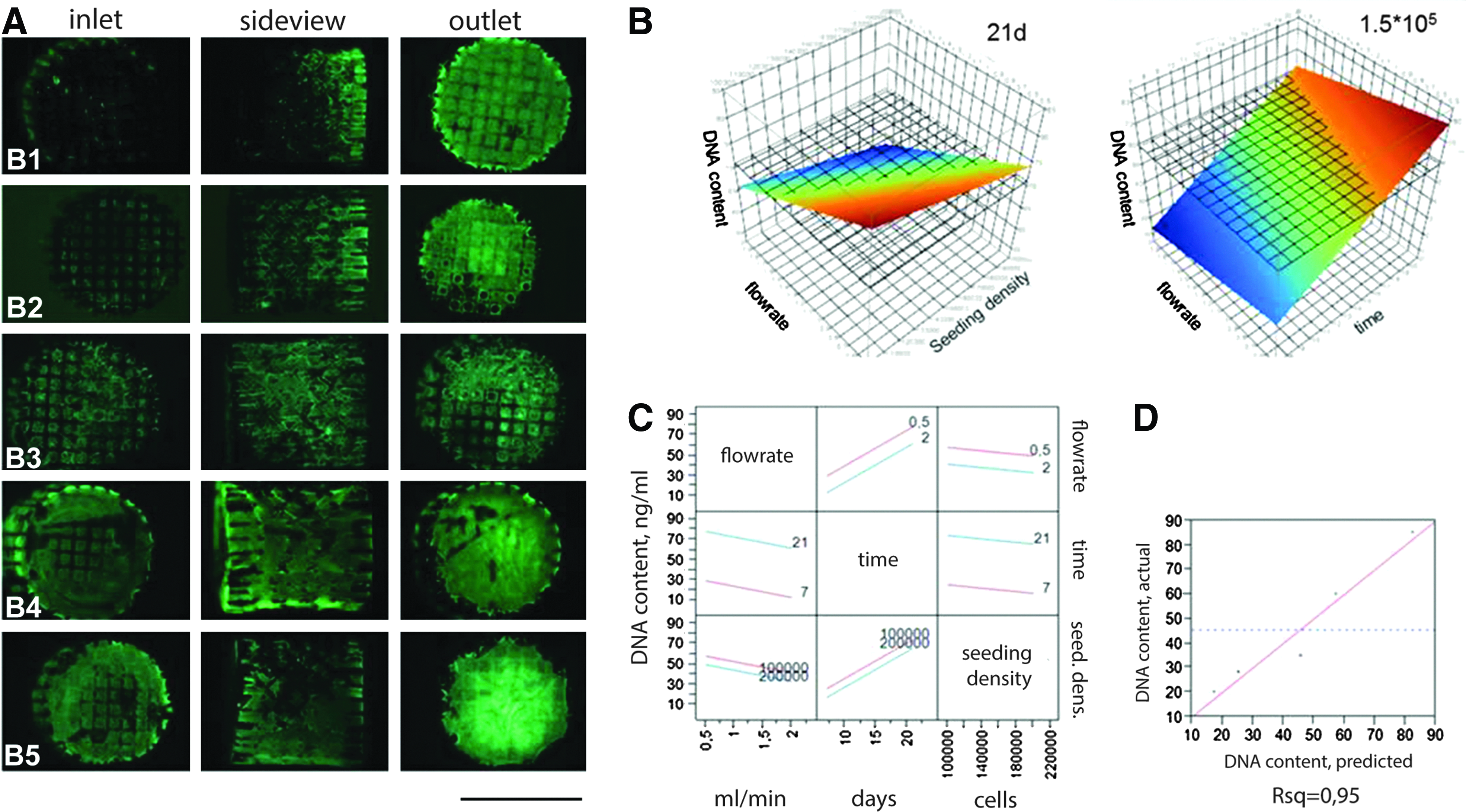
DNA content of perfused hybrid scaffolds.
Response surfaces: collagen content
Collagen content is one of the most important variables, and its control is crucial for the quality of the hybrid scaffold. Images in Figure 6A show the distribution of collagen throughout the Ti scaffold over time (from top to bottom). A statistical model was created by best fitting through the experimental data points of the design, and response surfaces were created for the process space outlined by the high and low levels of the design. The response surface plot shown in Figure 6B shows the dominant-positive effect of the flow rate in collagen content in respect to the weaker positive effect of seeding efficiency for a given timepoint (day 21 shown). In the second case, the significant effect of time (p<0.001) and flow rate (p<0.019) is also highlighted for a fixed-starting cell-seeding density (seeding density 1.5×10³ cells). The interaction plot in Figure 6C further clarifies the ranking of the investigated process parameters, whereas Figure 6D shows the goodness of fit.

Collagen content of perfused hybrid scaffolds.
Response surfaces: mineral content
Mineral content is also an important quality characteristic linked to the bone-forming capacity of hybrid scaffolds. Due to the use of a CaP-containing culture medium, mineral content can be attributed to the nonphysiological deposition of suspended CaP on the cell monolayer and produced a collagen matrix, but also to the physiological mineralization of the extracellular matrix. Figure 7A shows the distribution of mineral content after staining with alizarin red. An increase in homogeneity may be seen with the progression of culture time. Statistical models were obtained, and response surface models were created as shown in Figure 7B, correlating the selected experimental factors to hybrid scaffold mineral content. The effect of seeding density and flow rate on the mineral content for day 21 is shown, while the effect of time and flow rate are also demonstrated for a fixed cell-seeding density. The interaction plot Figure 7C gives a further overview of the relations between the factors, while 7D shows the goodness-of-fit of the model through the experimental datapoints.
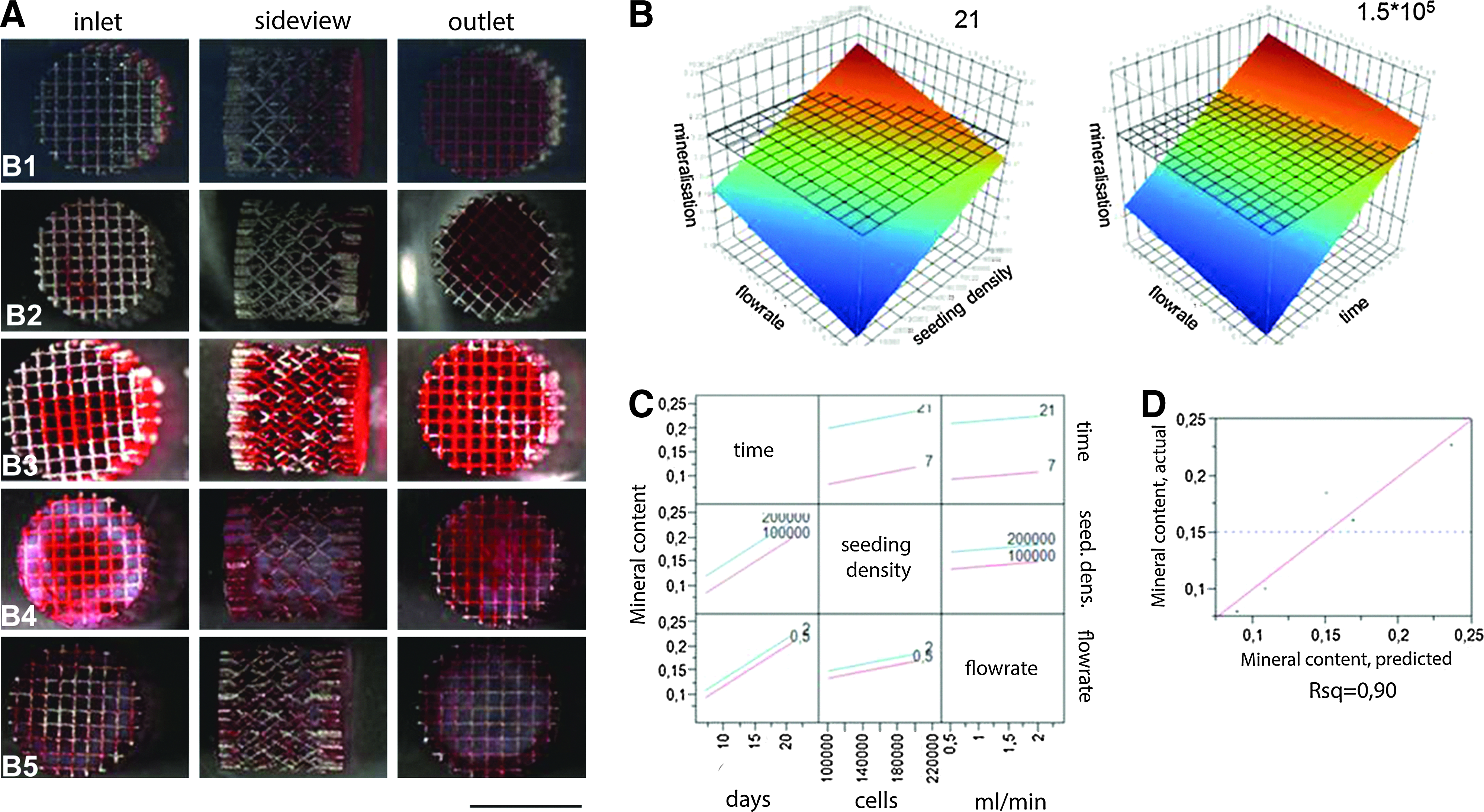
Mineral content of perfused hybrid scaffolds.
Highlighting a design space: case study
As already observed each process response (CQA) is influenced to a different extent by every experimental factor. By combining the statistical models developed and the respective response surfaces, we can define an overall optimized process space, a design space, wherein the produced hybrid scaffolds will meet prerequisite criteria. In Figure 8, such a case study is demonstrated. It shows for a specific time point (21 days of culture) a combination of process parameters (flow rate and seeding density) that would allow the manufacturing of a hybrid construct the following cell–mineral–collagen specifications: (1) a minimum of 28200 ng/μL DNA (i.e., 106 cells), (2) 0.22 absorbance at 570 nm indicative of a certain level of mineral content, and (3) 0.2 absorbance at 492 nm indicative of a certain level of collagen content. As an integrated representation, Figure 8 illustrates the possible combinations (white area) of operating process parameters that would allow the production of a hybrid scaffold with acceptable CQA. By changing further the time of culture in parallel with the flow rate and seeding density, a design volume could be finally obtained.

Case study—defining an integrated design space bordered by limitations set by specifying a threshold value to each of the selected critical quality attributes (CQAs). The CQA set specification was (1) collagen content that would result in absorbance value of 0.2 at 490 nm, (2) DNA content: 28000 ng/mL, and (3) mineral content that would result in absorbance value of 0.22 at 570 nm. The white area highlights that allowed combinations of flow rates and seeding densities that would result in a hybrid scaffold that would minimally meet the previous requirements after t=21 days of culture. Color images available online at
Discussion
In this study, we evaluated the use of a perfusion bioreactor setup as a means to improve the quality characteristics of produced 3D tissue-engineered osteogenic hybrid scaffolds in respect to those produced in a static culture setup when cultured in a CaP-containing medium. Furthermore, we systematically investigated the effect of three important perfusion bioreactor process parameters, namely, DNA, collagen, and mineral content on the final quality attributes of osteogenic hybrid scaffolds containing a primary hPDC population. 30 These attributes were selected as three crucial quality attributes for the hybrid scaffold in vivo bone-forming capacity. This work is a first step toward a systematic quality-engineering approach to establish and control sources of process variation and prioritize process factors for optimization. A design space wherein hybrids of predefined quality attributes may be manufactured in vitro was also outlined.
Metabolic activity measurements were used to monitor cell growth over time. Cell growth in static culture was limited, and in some cases resulted in scaffolds containing no cells (∼20% batch failure). This was attributed to the continuous precipitation of CaP nodules over the cell surface during the culture period, which included regular medium refreshment every 2 days. This led to the eventual formation of CaP layers covering the cell population and thus decreasing oxygen and nutrient diffusion conditions that would be detrimental for hPDCs. Similar observations have been made for 2D culture when using the same Ca-P medium composition, where cell growth was subsided due to the adverse effect of the CaP precipitate formed on the cell monolayer. 18 However, for cells cultured in the bioreactor, fluid flow enhanced oxygen and nutrient transfer,26,38 resulting in a burst of hPDC growth for the first 14 days of culture while no culture (batch) failures were observed in any bioreactor experimental run. Nevertheless, this initial burst of growth was followed by a period where the cells showed a declining metabolic activity. This may be explained by the fact that during the last week of culture, cell activity is typically associated with matrix maturation and differentiation of osteoblastic cells, while DNA synthesis decreases.38,39 Another explanation might be that the Alamar blue assay and its performance may be reaching a threshold when more mature 3D cellular structures are evaluated due to diffusion limitations within the more dense and complex hybrids. 40 Hence, customized dynamic protocols suited for bioreactor operation that can measure online metabolic activity of more complex 3D cell systems should be developed in parallel to overcome existing analysis limitations. However, the present technique can already be useful as a first screening bioprocess tool for assessing noninvasively the state of bioreactor cultures, identifying underperforming batches and outliers.
For this study a titanium alloy (Ti6Al4V) was selected as a case study scaffold. The regular structure of these scaffolds ensures a narrow hydrodynamic shear–stress distribution throughout the scaffold geometry, minimizing thus spatial variability in respect to irregular scaffold geometries. 8 Hence, it seemed a suitable option for the quality engineering approach followed in this study to use regular and reproducible scaffold architecture, contributing further to the control of sources of variation entailed in a bioreactor process culture step. A recent study further improves this aspect by proposing Ti scaffold surface treatment for further eliminating remaining surface anomalies. 32 To further eliminate any possible sources of variation, scaffold positioning within the perfusion bioreactor chamber was kept strictly the same across batches, ensuring identical fluid flow patterns.
Cell content has been linked with osteogenicity and final in vivo bone formation15,16; thus, DNA content as a measure of cellularity is an important quality characteristic. Perfusion through the scaffold was seen to have a significant positive effect on final DNA content resulting in a 3-fold (p<0.05) B5 and 4-fold (p<0.05) B4 increase after 21 days of culture in respect to corresponding static control cultures for all experimental conditions. A 2.5-fold increase of DNA content after 7 days of culture between perfused and statically cultured cells has been reported for MC3T3-E1 osteoblast-like cells 22 and for stromal cells 38 when cultured in a serum-containing medium. However, for increasing values of flow rates for the range studied (0.5 to 2 mL/min), the DNA content was seen to be affected negatively, although not statistically significant. Similar observations showing a weak negative effect of flow rate on the DNA content have been recently reported for a 15-fold increase in the magnitude of flow rates used, 29 although significant negative effects have also been observed for increasing flow rate values. 22 McCoy et al. 41 have recently investigated the detachment of cells from scaffolds during perfusion culture, which was attributed to shear–stress levels and to a bridged cell–scaffold attachment morphology. The average shear–stress values where cell detachment was observed were 0.0176 Pa, which was comparable to the average shear–stress values developed in this work (τlow=0.0052 Pa for Qlow=0.5 mL/min and τhigh=0.028 Pa, for Qhigh=2 mL/min). We should point out that in our study, due to the regular geometry of our scaffold and the larger pore size (755 μm compared to 375 μm), the percentage of bridged cells, prone to detachment, should be minimum in respect to the fully spread attachment morphology. However, the weak negative trend observed in our data between the increase in flow rate and DNA content could be attributed to this factor. Microscope observations of stained hybrid scaffolds (live/dead staining) showed increased presence of dead cells in the statically cultured scaffold; while a much lower presence of dead cells was seen in perfusion-produced hybrids, enhanced cell viabilities in dynamically cultured scaffolds have also been observed by Du et al. 42
Collagen content is a measure of the extracellular matrix produced by proliferating cells, and it is also an important CQA for a hybrid scaffold, since it provides a biomimetic template that enhances in vitro 3D osteoblastic differentiation when reused as a scaffold, 43 while the level of its maturation before implantation has been linked to vascularization of an implant. 44 Hybrids produced in a perfusion bioreactor contained a higher amount of collagen that was statistically significant for B3, B4, and B5 in respect to S3, S4, and S5 experimental conditions, respectively. Furthermore, for the flow rate range studied here (0.5–2 mL/min, scaffold diameter=6 mm, pore size=800 μm), increased values had also a statistically significant positive effect (p<0.0194) on collagen production. Fluid flow has been seen in many instances to enhance mineralized matrix synthesis within tissue-engineered constructs during perfusion culture in a dose-dependent manner,21,29,45 while an increase of shear stresses that had been decoupled from volumetric flow was also seen to increase mineralized extracellular matrix deposition on titanium fiber meshes.27,28
The final CQA used for the characterization of the produced hybrids was mineral content. Static scaffolds contained higher amounts of minerals in respect to the perfusion-produced ones. This may be attributed to the fact that in static culture, CaP accumulates over time resulting in a bulk deposition, while under perfusion due to increased mass transport, there is a deposition of smaller CaP nodules avoiding a bulk accumulation. However, over time, a statistically significant increase in mineral content was seen for perfusion-produced hybrids. With time, cell growth and extracellular matrix production created a network upon which CaP nodules from the medium were trapped. In addition, physiological deposition of minerals on the collagen matrix due to osteoblastic cell differentiation may have also contributed to this increase. Flow had a positive effect, although not statistically significant, whereas the seeding density influenced positively and significantly mineral content. A possible explanation could be that a higher cell-seeding number resulted in a quicker formation of extracellular matrix (ECM), and hence with time, also more CaP deposits from the medium. Furthermore, flow has been seen to mineralize ECM during perfusion culture as already mentioned, and this could further add to the quantity of minerals.
Besides the aforementioned quality attributes, homogeneity in cell, collagen, and mineralized matrix distribution throughout the scaffold should also be an important quality characteristic for perfusion bioreactor produced hybrids that should be taken in consideration when designing a process. However, we are currently lacking the tools to quantitatively assess such an important characteristic. Established cell assays developed for use in 2D systems cannot be transferred to 3D setups. The development of customized and validated 3D assays for a noninvasive monitoring of such characteristics is necessary. Images may give a qualitative representation of levels of homogeneity demonstrating, in our case, total closure of the initial Ti scaffold pores after 21 days of culture and increased homogeneity for the experimental conditions B4 and B5. An improved homogeneity from earlier on in the culture could be possibly achieved by using oscillatory rather than unidirectional flow. 42 The application of imaging techniques such as microcomputed tomography (micro-CT) could be further incorporated in DoE studies. Recently, micro-CT-compatible bioreactors have been designed for monitoring mineralization and cell differentiation in vitro.46,47
The DoE approach is an efficient method for evaluating the effects of process parameters (factors) on response variables. To date, this type of multifactorial analysis has been rarely applied as a process development tool for process steps involved in TE applications. Recently Chen et al. 8 employed a DoE methodology for a dynamic seeding process step and succeeded in screening important factors such as scaffold geometry comparing structured and unstructured pore distributions, or cell type comparing hPDCs to SAOS cells, while, at the same time, optimizing seeding process responses such as cell-seeding volume and cell-seeding efficiency. A similar study systematically investigated a cell-seeding step, using C2C12 mouse myoblast cells, by a randomized factorial experimental design with three factors: seeding time, seeding number, and seeding setup with cell seeding volume, metabolic activity, and cell-seeding efficiency as the response parameters. 14 In this work, we attempted to further use this methodology for the subsequent bioreactor culture process step to systematically characterize a suitable process space for manufacturing designed tissue-engineered constructs, such as the hybrid osteogenic scaffolds that we produced. We do realize though that the use of a fractional factorial rather than a full-factorial design aliases (i.e., does not decouple) interactive effects of remaining factors to the main effect of a factor not making use to the full extent of the possibilities of such a strategy. Furthermore, due to the limitations of this design type, partial least-square fitting was used missing out in this way any possible curvature of the response surface. The reason for using this type of design was its efficiency, since it decreased by 37.5% (five experiments instead of eight required for a full-factorial design), the time required for gathering necessary data something that are valuable for bioprocess development when dealing with time-consuming experiments involving bioreactor setups and cell culture. Furthermore, although linear approximations were obtained by the RSM models, information regarding possible optima that may lie outside the process space studied was obtained.
TE is undergoing a major conceptual and methodological transformation in an effort to implement in vitro processes that mimic in vivo tissue development. We believe that multifactorial DoE will be an excellent tool for optimizing such sequential processes and will help to achieve the goal of consistent tissue-engineered products. The DoE approach used in this study was designed to screen three important factors that influence the bioreactor culture process, all exerting various effects on the different response variables (DNA, collagen, and mineral content); hence, trade-offs between individual optima were made. However, to decide thoroughly on a gold standard for CQA of a produced hybrid, feedback from successful in vivo experiments is necessary. The strategy of first screening for important factors and interactions, followed by their optimization using RSM, is of interest when optimizing global and individual TE process steps. The eventual implementation of real-time monitoring tools at full-scale following the recent Process Analytical Technology guidelines 48 will allow manufacturing decisions to be made based on online data during processing, rather than on a discrete time basis or endpoint analysis, as it is being practiced at present. This will help to further monitor only critical process variables that should be carefully controlled, as seen in this study, during future good manufacturing practice to successfully meet regulatory requirements.
Footnotes
Disclosure Statement
No competing financial interests exist.