




Abstract
Mechanical stimulation during cartilage tissue-engineering enhances extracellular matrix (ECM) synthesis and thereby improves the mechanical properties of tissue engineered (TE) cartilage. Generally, these mechanical stimuli are of a fixed magnitude. However, as a result of ECM synthesis and spatial variations thereof at both the macroscopic and microscopic scales, the internal mechanical conditions in the constructs change with time. Consequently, the physical signals in the environment of the cells will vary spatially and temporally, even though macroscopically the same loading is applied to the construct. The purpose of the present study was to numerically quantify such effects and thereby reveal the importance of adjusting loading regimes during cartilage tissue-engineering. A validated nonlinear fiber-reinforced poroviscoelastic swelling cartilage model that can accommodate for effects of collagen reinforcement and swelling by proteoglycans was used. At the microscopic scale, ECM was gradually varied from localized in the pericellular area, toward equally distributed throughout the surrounding interterritorial matrix. At the macroscopic tissue scale, ECM was gradually varied from predominantly localized in the periphery of the TE construct toward homogeneously distributed. Both concentration of ECM in the pericellular area and concentration of ECM in the periphery of a construct alter the physical signals up to an order of magnitude compared to those at the onset of the culture. Of particular interest, is the effect of elevated osmotic swelling pressure in the pericellular area, which shields not only the cells from receiving external mechanical compression, but also directly induces tension on the cells. Based on the present computational simulations, it is therefore, proposed that cartilage TE studies should consider ECM distribution as an important factor when developing loading protocols for cartilage culturing process. For instance, the level of mechanical compression should gradually increase to sufficiently deform chondrocytes over time, in case there is matrix accumulation in the pericellular area.
Introduction
R
The mechanical properties of native articular cartilage are mainly determined by two constituents in its ECM: negatively charged glycosaminoglycans (GAGs) immobilized in the form of large proteoglycan (PG) aggregates, and collagen fibers.13–18 Collagen resists tension, including the tension that is induced by the swelling potential of the PGs, which attract water through Donnan osmotic pressure. Together, PGs and collagen allow cartilage to withstand high compressive forces.
Chondrocytes, the only cell type in cartilage, are responsible for ECM synthesis. Chondrocytes respond to changes in their micromechanical environment as signals for regulating their metabolic activity and gene expression. ECM synthesis of chondrocytes can be enhanced by subjecting these cells to appropriate mechanical triggers.19–26 Dynamic compressive loading at moderate levels (2%–10% strain or 0.5–1.0 MPa pressure) and physiological frequencies (0.01 to 1.0 Hz) stimulates the biosynthesis of collagen and PG.23,27–29 There are experimental observations showing that the beneficial efficacy of such mechanical stimuli varies during the ongoing time of culture.30,31
This phenomenon may occur because the physical signals in the environment of the cells change as a consequence of the development of ECM in the constructs and around the cells. Initially after seeding chondrocytes in commonly used scaffold materials, such as agarose or alginate, there is no ECM and the mechanical properties of the extracellular environment are rather uniform. 32 During the first days of culture, islands of new matrix develop around chondrocytes, which later grow out to larger aggregates.33–35 In addition to the consequent local, microscopic differences in ECM density, macroscopic nonhomogeneites in ECM distribution develop. Matrix content in the periphery of TE cartilage constructs are generally higher than in the center, at least partly as a consequence of spatial variations in nutrition.36–38
As a consequence of these temporal and spatial variations in matrix content, the cells are likely to receive different physical signals depending on their location and stage of culture. Thus, theoretically, if the same external mechanical loading is applied on the construct throughout the duration of culture, the cells will be exposed to different mechanical conditions over time. Consequently, if the chondrocytes are to be stimulated similarly throughout the culture time, the loading regime will have to be adjusted over time. However, even though in principle that would be possible, this is not current practice.30,31,38–41 The main reason for keeping mechanical stimulation constant over time is that it is unknown to what extent they need to be changed. To determine this, we require quantitative insight into the consequence of ECM development for mechanotransduction of externally applied strains down to the chondrocyte level. The significance of these questions is clear from studies showing that even modest variations in mechanical loading may have significant effects on ECM synthesis by chondrocytes and other cells.21,31,42,43
The aim of the present study is therefore, to quantify the influence of macroscopic and microscopic ECM distribution on the mechanical conditions and physical signals in the environment of the cells in TE constructs during unconfined compression. This ultimately reveals the importance of dynamically adjusting loading regimes during cartilage tissue-engineering.
Methods
Macroscopic scale model
Cylindrical chondrocyte-agarose constructs (radius=2 mm and height=4 mm) consisting of an inner core and an outer layer were modeled using an axisymmetric finite element mesh (Fig. 1a). Constructs with small (r=1.26 mm, h=2.52 mm) or large (r=1.6 mm, h=3.2 mm) inner core were considered. In the construct with the small inner core, the volume of the core was one-third that of the outer layer. In the construct with the large inner core, the volume of the inner resembled that of the outer layer. Due to symmetry, the top half of the construct was modeled. First, a construct with 2% agarose and no ECM was considered representing the initial stage of culture. This case was then compared to three different cases in which the distribution of ECM was either localized in the outer layer (highly inhomogeneous), 60% ECM in the outer layer and 40% ECM in the inner core (slightly inhomogeneous), or homogeneously distributed throughout the construct, while keeping total ECM amount constant. Compression was applied by an impermeable plate, which was connected to the top surface by frictionless contact. The displacements of the nodes at the symmetry axis were confined in radial direction. Bottom nodes were confined in vertical direction. The nodes on the sample lateral edge were prescribed to zero pore pressure to simulate free fluid flow. There was no boundary condition prescribed between the inner core and the outer layer. The finite element mesh for the entire construct was considered as one part and only ECM concentration was allocated for each node depending on whether this node was located in the outer layer or in the inner core. Simulations consisted of a free swelling equilibrium step followed by a 10% ramp unconfined compression, applied in 0.5 s to simulate maximum loading during a 1 Hz dynamic compression regime.

Axisymmetric macroscopic scale finite element model of a cylindrical tissue engineered construct (radius=2 mm and height=4 mm) consisting of an inner core and an outer layer, where the top half of the construct was modeled due to symmetry
Microscopic scale model
Representative three-dimensional cubic volumes (200×200×200 μm3) of 2% agarose-chondrocytes constructs with five randomly located inclusions embedded in a surrounding interterritorial matrix were simulated. In the model at the microscopic scale, similar to the model at the macroscopic scale, four cases were distinguished. Case I represented the condition immediately after cell-seeding, when matrix was not yet synthesized. In cases II, III, and IV, an equal amount of matrix was present in the construct, but the distribution of matrix in the construct varied. Given that most matrix in the early tissue development is localized in the close environment of the cells, 44 a pericellular area with thickness of 25 μm was defined in which it was assumed that all of the matrix was located (case II) or 60% of the matrix was located, while the other 40% was equally distributed in the surrounding interterritorial matrix (case III). Finally, in case IV it was assumed that all the matrix was homogenously distributed throughout the tissue, that is, that there was no difference between the pericellular area and the surrounding interterritorial matrix. Cell radius was set to 5 μm.45–47 Idealizing the tissue as a periodic arrangement of the representative cubes,45,48–50 the lateral edges of the cubes were forced to remain straight during the deformation process. Bottom nodes were confined in the vertical direction. Loading protocol was considered to be the same as that in macroscopic scale model (i.e., a free swelling equilibrium step followed by unconfined compression). In addition to a stress-strain analysis, the cell deformation index was calculated by dividing the dimensions of cell parallel to the axis of compression by that perpendicular to this axis. 51
Material model
To model cartilage TE construct material, a composition-based fibril-reinforced poro-elastic swelling model was adopted, which consisted of a fluid phase and a porous solid matrix with swelling properties.13,14,52 The porous matrix of the biphasic tissue consisted of a swelling nonfibrillar ground substance, which contains mainly PGs and agarose ground substance, and a fibrillar part representing the collagen network. The material model was implemented in ABAQUS (v6.9; Pawtucket, RI). The mechanical behavior of the material model was the direct consequence of the composition (fluid fraction, solid matrix fraction and fixed charge density) of the tissue.
The governing stress equation of Wilson et al.
14
was adjusted such that the agarose could be included as a separate constituent. The resulting governing stress equation was:
where μf was the water chemical potential,
For agarose gel, the same compressible Neo-Hookean model of the nonfibrillar matrix was used of which the compressibility was dependent on the solid fraction:
14
where Gagr was the shear modulus of the solid matrix in the biphasic agarose gel and J was the determinant of the deformation gradient tensor
For collagen fibers, a strain-dependent viscoelastic model was used. 13 At each integration point 13 fibers were included; 3 running in the directions of the orthogonal axis, 6 running in directions between those axes with 45 degrees from each of the two orthogonal axis in each plane and 4 running in the spatial direction between the orthogonal axes with 60 degrees from each axis. 52 This configuration of the collagen fibers was used to implement a quasi-isotropic description of the collagen fibers, which represented a random orientation of the collagen fibers in cartilage TE constructs.
The osmotic pressure gradient Δπ was calculated from the effective fixed charge density (cF,exf) as53,54:
with φα osmotic coefficients, γα activity coefficients, cext the external salt concentration, T the temperature, and R the gas constant.
Effective fixed charge density was expressed as a function of the tissue deformation, as:
with nf the total fluid fraction, nexf the extra-fibrillar fluid fraction, nf,0 the initial fluid fraction, and cF,0 the initial fixed charge density in mEq per mL total fluid.
For more details on the constitutive equations of the nonfibrillar matrix, collagen and osmotic swelling behavior the reader is referred to Wilson et al.13,14,52
Input parameters
For the shear modulus of the solid matrix in agarose biphasic ground substance, Gagr=0.35 MPa was used, which was based on a simulation where Gagr was manually fitted in an unconfined compression test so that the agarose biphasic gel had overall Young's modulus of 15 kPa.
30
Overall permeability of the constructs was assumed to be 3.5×10⁻¹³ m
4
N−1s−1.
45
In the microscopic scale model, cells were considered to be biphasic with aggregate modulus of 1 kPa, Poisson's ratio of 0.4 and permeability of 10−15 m
4
N−1s−1.45,46,55 Input parameters ncol, ns,0, nag, and cF,0 were calculated as follows. In a construct with initial total volume Vtot,0 and total mass mtot, collagen volume Vcol was calculated as:
with β col collagen wet weight (ww) fraction, ρ tot construct mass density, mcol collagen mass, and ρ col collagen mass density. Mass density of collagen and GAG was assumed to be 1.4 mg/mm3.56,57 Mass density of the construct was assumed to be 1.02 mg/mm3. 38
The exact values of GAG and collagen content may depend on culture conditions, such as cell seeding density, culture medium or mechanical loading protocol. Representative values were selected in the range of those suggested in the literature. Collagen and GAG weight fraction, β col and β GAG , respectively, were chosen to be each 1% of the ww, which were in the range of experimental data at early stages of cartilage TE culture.31,38,58
Using Eq. (5), and considering β GAG =β col =β=0.01, the parameters mcol, mGAG, VGAG and Vcol were calculated.
Total solid volume Vs was then calculated as:
where Vagr was volume of the solid agarose gel in the construct, which was assumed to be 2% of the total initial volume of the construct.
Assuming biphasic saturation, initial fluid volume and fluid fraction were given as:
and initial solid fraction ns,0 with respect to total initial volume was then calculated as:
Collagen, GAG and agarose fractions with respect to total solid matrix volume Vs were calculated as:
To calculate initial fixed charge density cF,0, it was assumed that GAGs contained 77% chondroitin sulfate and 23% keratan sulfate. Initial fixed charge density was then calculated as:
with valencies zCS=2 (mEq/mmol) and zKS=1 (mEq/mmol) and molecular weights MWCS=0.513 (g/mmol) and MWKS=0.464 (g/mmol). 59
Input parameters calculated for the macroscopic scale model with small and large inner core are summarized in Tables 1 and 2, respectively. Input parameters calculated for the microscopic scale model are summarized in Table 3.
Compared to the case (Homog.) in which ECM was homogeneous distributed in the construct, two different cases (I–II) tow different cases were considered in which the distribution of ECM was either localized in the outer layer or 60% ECM in the outer layer and 40% ECM in the inner core, respectively, while keeping total ECM amount constant.
ECM, extracellular matrix.
Compared to the case (Homog.), two different cases were considered in which the distribution of ECM was either localized in the pericellular area or 60% ECM in the pericellular area and 40% ECM in the surrounding interterritorial matrix, while keeping total ECM amount constant.
Results
Free swelling condition: macroscopic scale
In the construct with the small inner core, in free swelling condition (Fig. 2a–d), localization of ECM in the outer layer of the construct resulted in elevated osmotic swelling pressure (Fig. 2a) in the outer layer. This pressure was 33% higher compared to that in the construct with homogeneous ECM distribution where osmotic swelling pressure was homogeneous with magnitude of 7 kPa. Dispersion of 40% of the ECM from the outer layer toward the inner core decreased the osmotic swelling pressure in the outer layer to be comparable to that in the homogenous case but it significantly increased the osmotic pressure in the inner core up to twofold than in the homogenous case. As a result of such osmotic swelling pressure distributions, highly inhomogeneous stress and strain fields were developed in the constructs. Compared to that in the homogeneous case, peak volumetric strain was increased by 25% (Fig. 2b) and peak deviatoric strain (Fig. 2c) as well as von Mises stress (Fig. 2d) was increased by one order of magnitude.
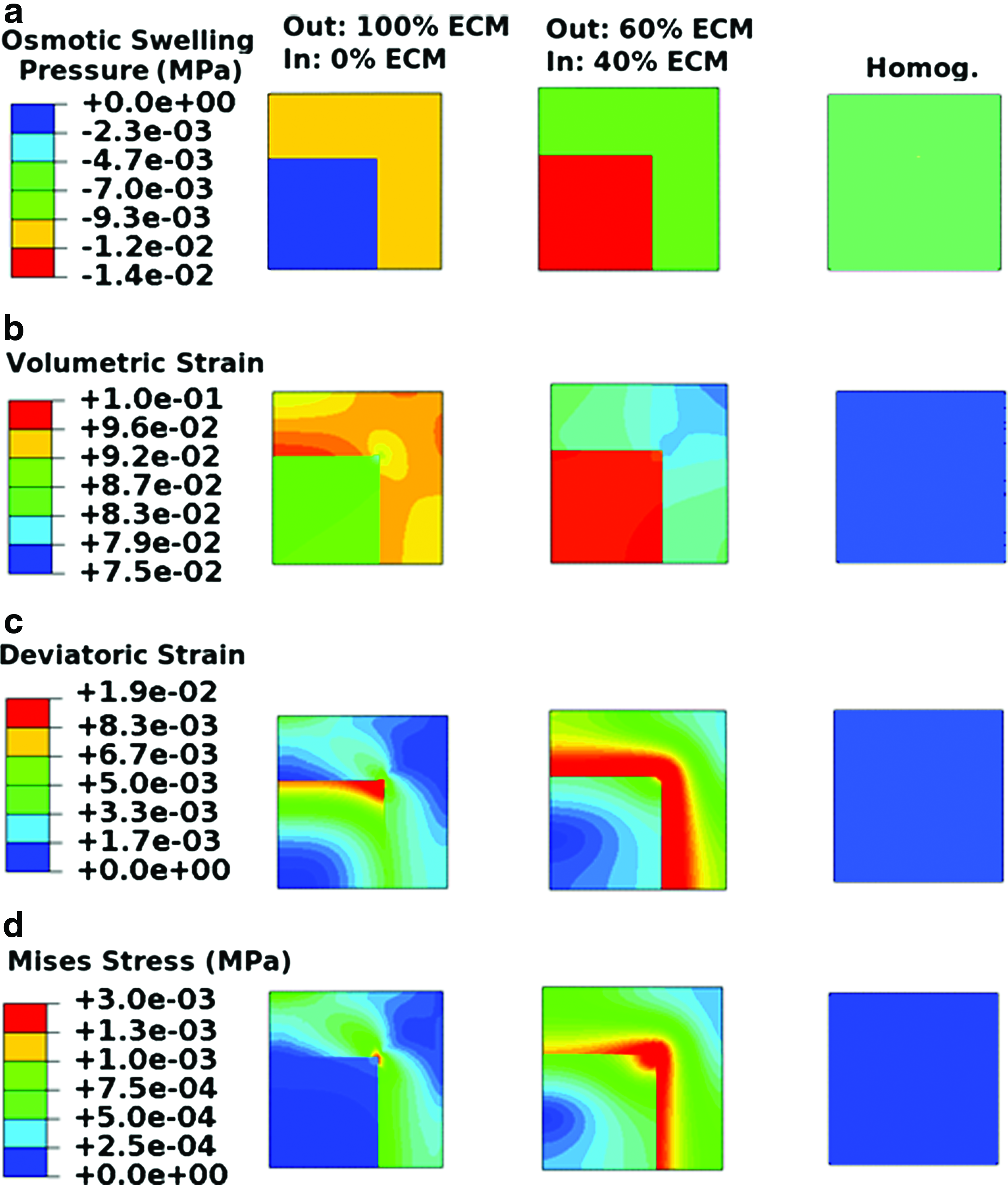
Macroscopic osmotic swelling pressure
With a large inner core, localization of the ECM at the construct periphery resulted in an osmotic pressure of 20 kPa in the outer layer (Fig. 3a), which was 30% higher than that in the construct with small inner core. This elevation in the osmotic pressure increased local volumetric strains up to 12% (Fig. 3b). Compared to the construct with the small inner core, the peak deviatoric strain increased from 1.9% to 2.7%, while the peak von Mises stress remained the same (Fig. 3c, d). With the large inner core containing 40% of the ECM, strain and stress fields (Fig. 3b–d) were close to those in the homogenous case (Fig. 2).

Macroscopic osmotic swelling pressure
Loading condition: macroscopic scale
Under 10% unconfined compression (Fig. 4a–d), in the absence of ECM, agarose experienced deviatoric strain of 5%, von Mises stress of 1.4 kPa, and negligible volumetric strain and pore pressure. In the construct with small inner core, localization of ECM in the outer layer of the construct increased peak volumetric strain and deviatoric strain by 120% and 70%, respectively (Fig. 4a, b). Further, the peak von Mises stress was shifted from the center of the construct to the outer layer, with 20% increase in the magnitudes (Fig. 4c). Moreover, the peak pore pressure at the center of the construct was decreased from 110 to 37 kPa (Fig. 4d). Dispersion of 40% of the ECM from the outer layer toward the inner core decreased the peak magnitudes of deviatoric strain and von Mises stress and resulted in a more similar distribution of strains and stresses to that in the construct with homogeneous ECM distribution (Fig. 4b, c). However, volumetric strain (Fig. 4a) was elevated in the inner core with peak magnitudes 30% higher than that in the homogeneous case. With homogeneous ECM distribution, volumetric, and deviatoric strains were ∼5% (Fig. 4a, b). Stress and pore pressure fields showed peak values of 100 and 110 kPa, respectively, with higher magnitudes toward the center of the construct (Fig. 4c, d).
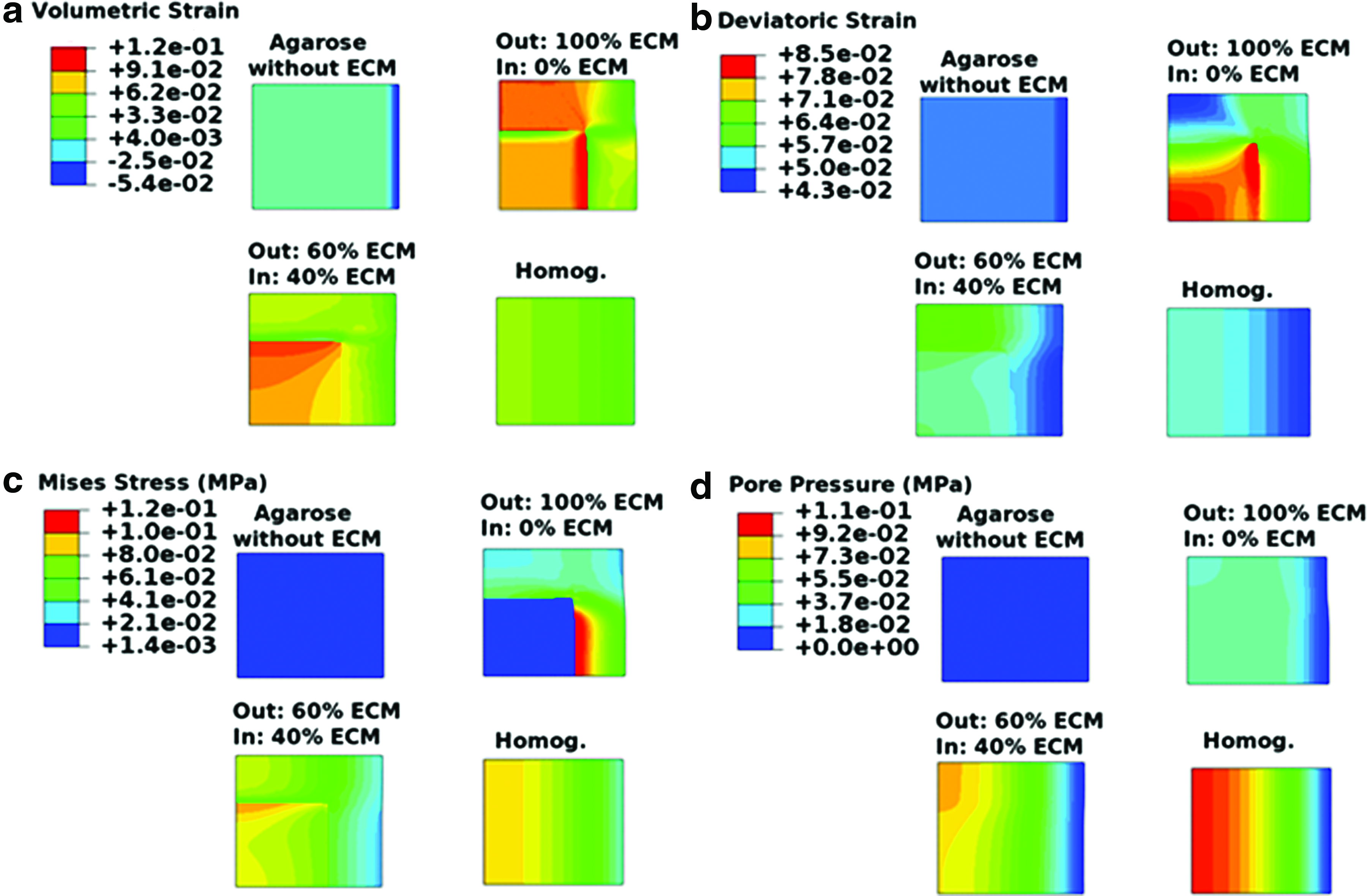
Macroscopic volumetric and deviatoric strain
In the construct with large inner core, when ECM was localized in the outer layer, the peak values of the volumetric and the deviatoric strains remained close to that in the construct with small inner core (Fig. 5a, b). The increase in the size of the inner core did not change the distribution of the stress and the pore pressure but it influenced their peak values. The von Mises stress and pore pressure were an order of magnitudes lower in this case (Fig. 5c, d). Dispersion of the 40% ECM toward the inner core resulted in more homogenous strain and stress fields (Fig. 5a–c).
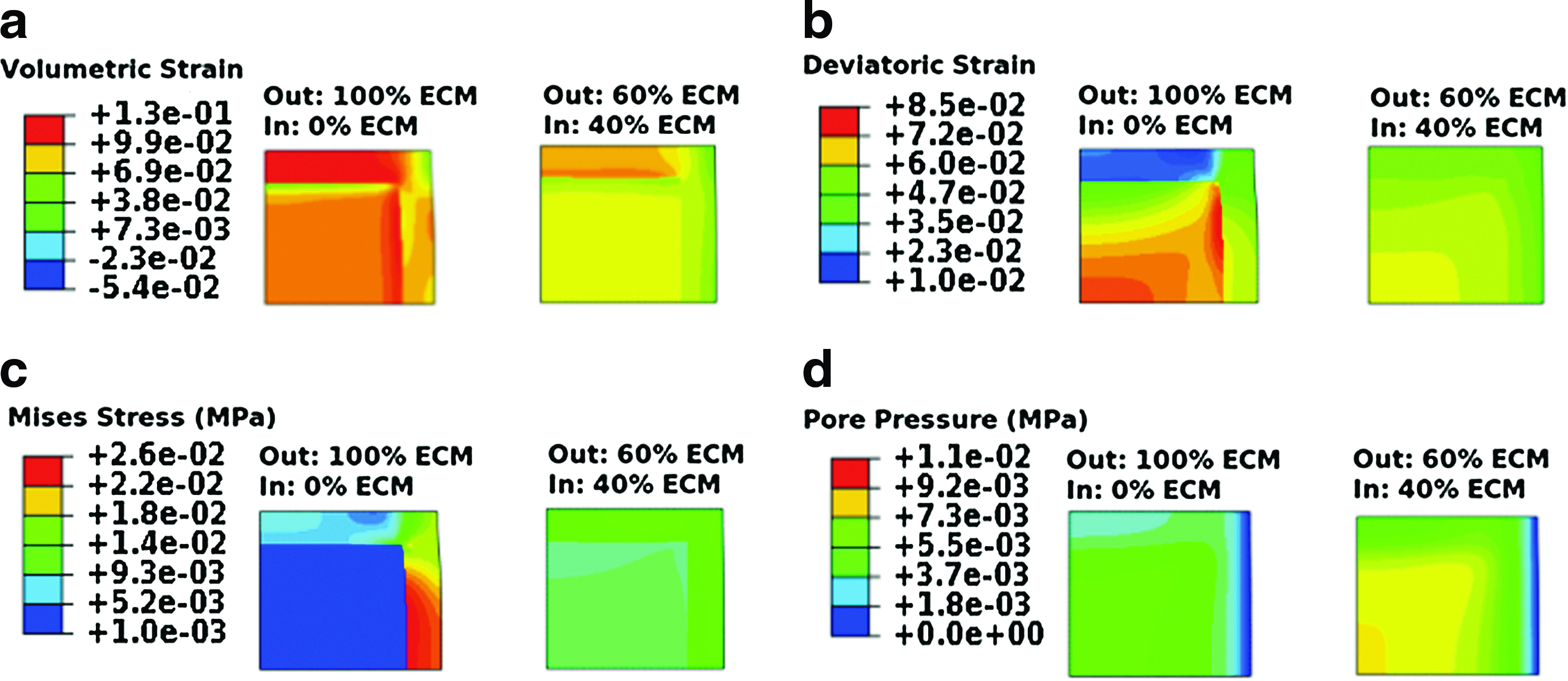
Macroscopic volumetric and deviatoric strain
Free swelling condition: microscopic scale
At microscopic scale, in free swelling condition (Fig. 6a–c), localization of ECM in the pericellular area resulted in considerable elevation of osmotic swelling pressure (Fig. 6a) with magnitude of 490 kPa around the cells, which was two orders of magnitudes higher compared to that in construct with homogeneous ECM distribution where osmotic swelling pressure was homogeneously distributed with magnitude of 7 kPa. Dispersion of the 40% of the ECM from the pericellular area toward the surrounding interterritorial matrix decreased the osmotic swelling pressure to 200 kPa. Highly concentrated osmotic swelling pressure around the cells induced volumetric strain (Fig. 6b) up to 29% on cells, which was significantly higher than the 12% strain, which cells experienced in the case of homogeneous ECM distribution. ECM distribution did not have a considerable effect on deviatoric strain in free swelling condition (Fig. 6b).
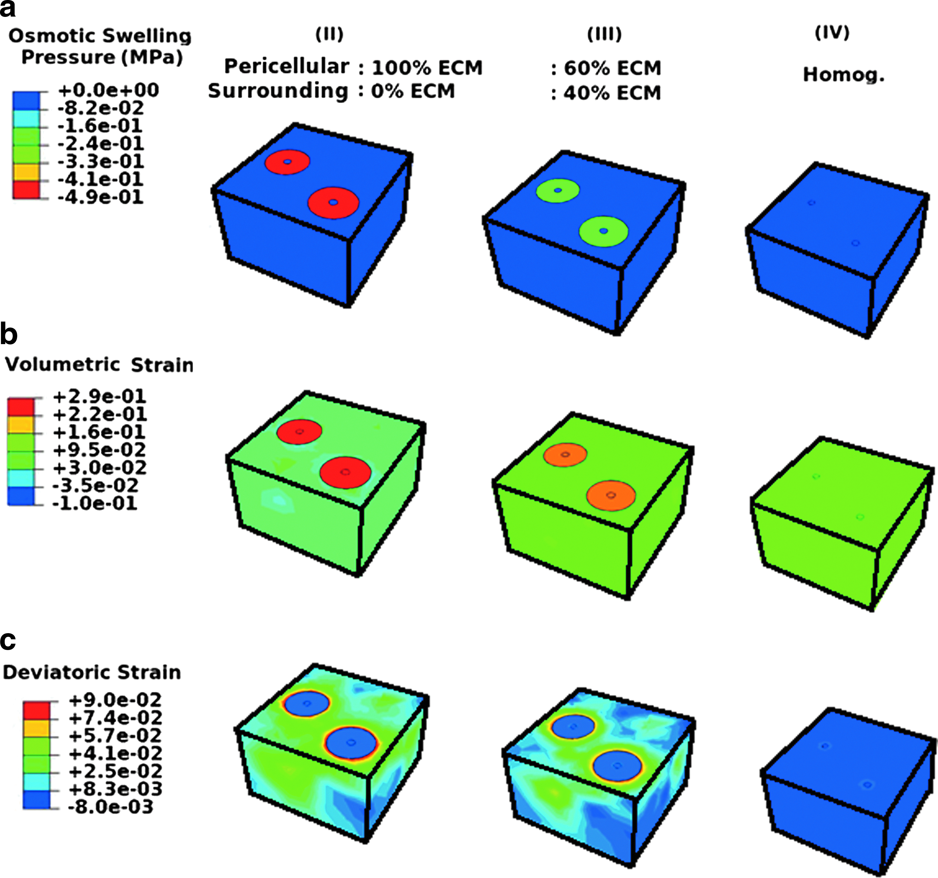
Microscopic osmotic swelling pressure
Loading condition: microscopic scale
Under loading (Fig. 7a, b), cells embedded in agarose experienced volumetric strain of −3.7%, deviatoric strain of 8.9% and deformation index of 0.72. When ECM was localized in the pericellular area, considerably different strain was induced on cells with volumetric strain of 30% and negligible deviatoric strain. Furthermore, this localization of ECM around the cells shielded the cells from being stimulated as indicated by a cell deformation index of 1.0. Dispersion of the 40% of the ECM from the pericellular area to the surrounding interterritorial matrix decreased the volumetric strain to 23% and increased the deviatoric strain to 4.3%. Furthermore, the shielding effect was reduced leading to the cell deformation index of 0.94. With homogeneous ECM distribution, volumetric strain was 23%, deviatoric strain was 19% and cell deformation index was 0.71 (Fig. 7c).

Microscopic volumetric
Discussion
The results indicate that inhomogeneities in ECM distribution in cartilage TE constructs associated with the concentration of PGs and therefore, with the density of fixed charges, give rise to gradients in osmotic swelling pressure in the construct at both macroscopic and microscopic scales. The preload free swelling condition of the construct may lead to significant variations in mechanical loads/strains within the constructs (Figs. 2 and 3). Combination of such preload swelling condition and externally applied loading results in complex strain and stress fields. The macroscopic tissue scale localization of the ECM in the outer layer of TE cartilage constructs results in the local elevation of the physical signals within the constructs up to an order of magnitude (Figs. 4 and 5). Therefore, cells in different locations experience considerably different mechanical conditions, when the same global mechanical deformation is applied during culture. With the dispersion of the ECM toward the inner core of the constructs, the local elevation of the mechanical stimulation reduces and stimulation of the cells becomes more uniform.
Microscopic scale evaluations showed that localization of ECM in the pericellular area results in a highly nonlinear elevation in osmotic swelling pressure around the cells. This shields not only the cells from receiving external mechanical deformation (i.e., deformation index close to 1.0, under 10% external compression), but also imposes a continuous tension on the cells in free swelling. When ECM is homogenously distributed at the microscopic scale, cells experience the deformation level similar to that at the onset of the culture (i.e., deformation index of 0.71, under 10% external compression). However, the transferred mechanical strains are essentially different from those at the onset of the culture (Figs. 6 and 7).
It has been shown that chondrocytes sense physical signals through mechanosensors of stretch-activated ion channels, the cytoskeleton, and nuclear deformation. 19 Chondrocyte and nuclear deformations are integrally linked to the deformation of the ECM.60,61 The present study shows significant effects of ECM production and distribution on the physical signals applied on cells during culture.
We speculate that some unexplained experimental observations with regard to the effect of mechanical stimulation of TE cartilage could be directly or indirectly attributed to the differences in mechanical conditions perceived by the cells, depending on the distribution of ECM at either the macroscopic or microscopic scale. One observation is that mechanical loading appears to be a more effective trigger for ECM synthesis during first 2–3 weeks of culture than at later time points. This phenomenon may be related to the elevation in osmotic swelling pressure in the pericellular area, after the initial synthesis of ECM in this zone. As shown in the present study, this swelling pressure substantially changes the micromechanical environment of the cells, which is essentially different from the one cells experience at the onset of the culture, when no or limited ECM is synthesized. Interestingly, treatment with chondroitinase ABC revived ECM synthesis.62,63 This may be partly related to a change in the pericellular mechanical environment. Another experimental observation is that supplementing TGFß in chondrocyte-agarose constructs grown in FBS-containing medium increased the efficacy of mechanical stimulation. 64 Part of this effect on mechano-responsiveness may be attributed to the more homogeneous matrix that develops in TGFß-supplemented medium, 35 possibly as a result of reduced collagen cross-linking by TGFß. 65 Finally, a common experimental observation is that ECM content is highest in the construct periphery. This has been attributed to the availability of critical nutrients 38 because diffusivity reduces with matrix development, 66 or to nutrient utilization by peripheral cells. 37 The present study shows that such inhomogeneities cause significant variation in mechanical conditions between cells. In addition to nutritional effects, this variation in mechanical condition may induce more inhomogeneities in the constructs.
Optimal physical signals to be applied on cells during culture to enhance their synthetic activities are yet to be elucidated. In general, physiological loading involves a combination of compression, shear, tension, fluid pressure, and swelling pressure, and each might have a role in the synthesis of ECM. As an example, it has been shown that sulfate incorporation under indentation loading of articular cartilage is increased both under a compressing indenter and also in the noncontact area at the vicinity of the indenter. 67 This could suggest that both compressive and tensile forces/strains could stimulate PG synthesis. In current practice, unconfined compression with constant level of loading during the culture is the most common loading regime in cartilage TE studies. With this loading, it is generally observed that mechanical stimulation is more effective during the first weeks of culture. Therefore, the calculated cell deformations at the onset of culture may be closer to the optimal deformation than those computed for deformation in the presence of ECM. Thus, an optimal loading protocol for cartilage tissue engineering may require updating the loading protocol over time, such that the externally applied stimuli perturb the cells in a similar way as they are during the first week of culture. Whether this requires an increase or a decrease of the mechanical stimuli depends on the prevailing ECM distribution in the experiment. When ECM accumulation in the pericellular area is apparent, the present results predict that the level of external loading should be increased over culture time. Also, the results show that macroscopic dispersion of the ECM in cartilage TE constructs should be enhanced to avoid inhomogeneous stimulation of the cells within the constructs under external loading. This may possibly be achieved by using suitable scaffold materials with improved mass transport properties in combination with appropriated growth factor likes TGF-ß, which causes the dispersion of ECM through constructs. 35
To quantify the effect of local and global variations in ECM, content on the stresses and strains received by chondrocytes is experimentally challenging. Theoretical models of cells and tissues are valuable because they can provide information on biophysical parameters at the cell level 46 and help to correlate specific mechanical parameters to the resulting effect on cell behavior. 19 In addition, a numerical approach enables explicit control of different parameters and therefore, allows studying the influence associated with variations in a single parameter. Thus, it may be used to provide insights on isolated effects of for instance matrix density or distribution. This is challenging to obtain in experiments where such effects may be masked by simultaneously occurring effects. To perform such numerical evaluation requires mimicking the mechanical state of the ECM in the engineered cartilage, including effects of osmotic swelling by PGs and the consequent straining of collagen. In fact, TE constructs are commonly exposed to dynamic mechanical loading for only a number of hours per day, and are otherwise kept under free swelling conditions.31,38,39 During the latter period, constructs may expand as a consequence of tissue synthesis and osmotic swelling. When investigating the effect of ECM inhomogeneities on mechanical conditions at the microscopic scale, the effects of this swelling period may be essential to account for because they change the prestrain in the tissue. Therefore, the present study accommodated for the role of osmotic swelling pressure in relationship with the ECM distributions. An important result of the present study is that osmotic swelling pressure should not be disregarded in such models.
The present constitutive material model has previously been validated for mature cartilage,13,14 and the same material properties for GAG and collagen were used. In this material model, the influence of cross-linking was inherent in the fitted material properties of the collagen network. Therefore, the lack of collagen network cross-linking in TE constructs was inherently represented in the lower content of collagen in the material model. It has been shown that this material model can predict the bulk modulus of cartilage TE constructs in a range close to those measured in the experiments. 68 Yet, it cannot be excluded that for instance crosslink density per amount of collagen is less in TE constructs than in mature cartilage. Such effects may be incorporated when quantitative data on the effect of crosslink density on the mechanical properties of the collagen network in both mature and tissue engineered cartilage becomes available. Furthermore, for full quantitative validation of the proposed tissue development model, the ECM distribution at the macroscopic and microscopic scale of the constructs should be corroborated to experimental data under various conditions. Also, internal tissue deformations and distributions of osmotic swelling pressure would be valuable. Unfortunately, availability of such quantitative data is insufficient to perform quantitative validation. Here to validate the numerical predictions on cell deformations, the predicted cell deformation index (at equilibrium) is compared with experimental measurements of Lee and Bader. 51 In their study, agarose-chondrocyte constructs were subjected to 5%–20% strain and cell shape was assessed at the onset of culture and after 6 days. Good agreement is obtained between the numerical predictions and the measured cell deformation index at the onset of the culture when no ECM is present (Fig. 8). With ECM synthesis after six culture days, at 10% compression, the experimentally measured deformation index is reduced from 0.82 to 0.95, which is well in the range of the numerical predictions (Fig. 7cI–II). Because of the profound difference in stiffness between cells and gel or matrix, the cell deformation index is rather insensitive to changes in other parameters, such as the Poisson's ratio. For example, changing the Poisson's ratio from 0.4 to 0.1 in the absence of ECM increases the cell deformation index less than 4% at 10% compression. Additionally, it was evaluated to what extend choices for matrix content, cell numbers and sizes of the pericellular area affected the conclusions. Combination of different amounts of GAG and collagen changes the results quantitatively, but not qualitatively. Therefore, the same contents and distributions for GAG and collagen were chosen to enable easier interpretation of the results. Also the number of cells with different distributions and sizes of the pericellular area did not change the general trend of the results and the conclusion. Therefore, these were arbitrarily chosen in the presented simulations. Finally, it is assumed that PG and collagen synthesis occur at the same time and don't interact. Different synthesis rates of PGs compared to that of collagen may lead to changes in the mechanical behavior of TE constructs.11,62,63 Future studies may explore temporal effects of matrix synthesis experimentally by modulating biochemical stimuli, 65 or computationally by using growth models in which the gradual deposition of ECM components is accounted for. 69
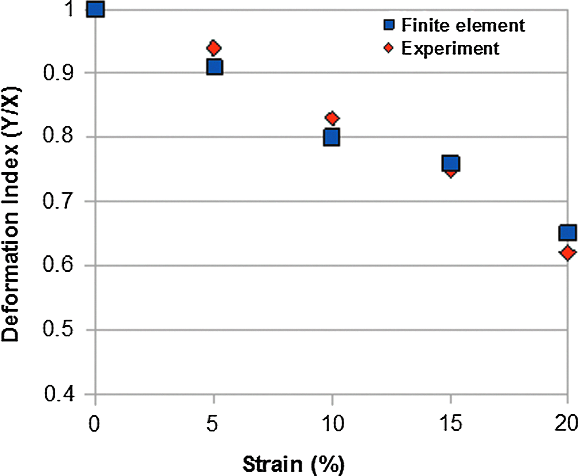
Comparison between model predictions for cell deformation index in 5%–20% strain against experimental measurements of Lee and Bader (1995). Color images available online at
The conclusions regarding effects of matrix inhomogeneites on the mechanical conditions inside TE constructs at the macroscopic and microscopic scale also hold in more complex cases; for instance when both effects occur simultaneously which one dominates will depend on the actual distribution of the matrix, as may be visualized histologically. To couple cell scale and tissue scale inhomogeneites in one modeling framework, a more complex and computationally expensive multiscale modeling approach. Fortunately, such extensive approach is not required for making practical use of the present insights. For instance, this study illustrates which kind of matrix distribution likely enhances or reduces physical signals in the environment of the cells. This information may be valuable when interpreting the histological matrix distribution at the macroscopic and the microscopic scale in a study where the response to mechanical perturbation reduced over time. If for instance ECM is observed to accumulate in the pericellular area, then it may help to increase the magnitude of the externally applied mechanical perturbation, or to enhance matrix dispersion by adding TGFß 35 or by using scaffolds with higher diffusion coefficients.44,45 However, if in addition matrix accumulates in the periphery of the construct, then adjusting the external loading may have distinct effects on the cells, depending on whether they are located in the center or in the periphery of the construct.
Conclusion
The present study shows significant effects of ECM development and distribution on the physical signals that cells may experience during culture. Both macroscopic and microscopic inhomogeneities and their changes over time during culture are essential, while variations in osmotic swelling pressure are particularly important. Optimal physical signals to be applied on cells during culture to enhance their synthetic activities are yet to be elucidated. However, because it is generally observed that mechanical stimulation is more effective during the first weeks of culture, it may be postulated that the mechanical condition at the onset of culture may be closer to the optimal condition than those in the presence of ECM. Thus, an optimal loading protocol for cartilage tissue engineering may require updating the loading protocol over time, such that the externally applied stimuli perturb the cells in a similar way as they are during the first week of culture. Whether this requires an increase or a decrease of the mechanical stimuli depends on the prevailing ECM distribution in the experiment. When ECM accumulation in the pericellular area is apparent, the results suggest increasing the level of external tissue deformation over culture time.
Footnotes
Acknowledgment
This study was supported with funding from the Dutch Technology Foundation STW (VIDI-07970).
Disclosure Statement
No competing financial interests exist.