
Abstract
Hyperpermeable tumor vessels are responsible for elevated interstitial fluid pressure and altered flow patterns within the tumor microenvironment. These aberrant hydrodynamic stresses may enhance tumor development by stimulating the angiogenic activity of endothelial cells lining the tumor vasculature. However, it is currently not known to what extent shear forces affect endothelial organization or paracrine signaling during tumor angiogenesis. The objective of this study was to develop a three-dimensional (3D), in vitro microfluidic tumor vascular model for coculture of tumor and endothelial cells under varying flow shear stress conditions. A central microchannel embedded within a collagen hydrogel functions as a single neovessel through which tumor-relevant hydrodynamic stresses are introduced and quantified using microparticle image velocimetry (μ-PIV). This is the first use of μ-PIV in a tumor representative, 3D collagen matrix comprised of cylindrical microchannels, rather than planar geometries, to experimentally measure flow velocity and shear stress. Results demonstrate that endothelial cells develop a confluent endothelium on the microchannel lumen that maintains integrity under physiological flow shear stresses. Furthermore, this system provides downstream molecular analysis capability, as demonstrated by quantitative RT-PCR, in which, tumor cells significantly increase expression of proangiogenic genes in response to coculture with endothelial cells under low flow conditions. This work demonstrates that the microfluidic in vitro cell culture model can withstand a range of physiological flow rates and permit quantitative measurement of wall shear stress at the fluid–collagen interface using μ-PIV optical flow diagnostics, ultimately serving as a versatile platform for elucidating the role of fluid forces on tumor–endothelial cross talk.
Introduction
T
Endothelial cells lining tumor vessels are directly exposed to wall shear stress (WSS), resulting from viscous forces of blood flow on the vessel wall. WSS directly regulates endothelial cell morphology, barrier function, and expression of angiogenic growth factors.11–13 However, within the context of the tumor microenvironment, the direct relationship between the endothelial response to shear stress and tumor angiogenesis is not well understood. Reports have provided evidence that interstitial flow regulates tumor cell growth and differentiation,14,15 although the effect of shear stress on tumor cell behavior has not been largely investigated. Other studies have shown that the interstitial flow regulates tumor cell migration and metastasis,16–23 in which, progressive changes in the tumor flow environment may enhance angiogenesis and promote tumorigenic activity of stromal cells such as fibroblasts, immune cells, and endothelial cells. While previous experiments have demonstrated the importance of tumor-endothelial signaling on angiogenesis,24–28 currently, it is not known to what extent shear forces affect endothelial cells during this pathological process. A better understanding of the role of fluid forces on tumor angiogenesis may offer insight into the development of improved therapeutic strategies to control vascularized tumors.
The development of physiologically relevant cell culture models, which facilitate the investigation of tumor–endothelial cross talk under dynamic conditions, is instrumental in this process. Conventional models of tumor angiogenesis are limited by the use of static, 2D culture systems that poorly reflect the pathological tumor microenvironment in vitro. Although in vivo models present a more physiologically representative environment, they can be complicated with many uncontrollable variables such as host cell invasion, mass transport, and external growth factors, 29 which may prevent isolation of the effect of a single stimulus, such as fluid shear, on the tumor response. More recently, promising three-dimensional (3D) culture systems have been developed for studying tumor progression in vitro, which have been shown to restore cellular morphologies and phenotypes characteristic of in vivo tumor development.30–33 Further advancements have led to the fabrication of microfluidic culture models, which replicate native 3D microenvironments in combination with gradient and flow control, and thus enable systematic investigation of both physiological and pathological phenomena in vitro.34–36
Measuring tumor blood flow in vivo and in vitro is important for assessing drug delivery, monitoring therapeutic response, and understanding tumor physiology. To date, there have been few successful methods for measuring shear stress gradients in vivo; therefore, little is known about how fluid shear stress regulates endothelial morphology, vessel permeability, or expression of angiogenic growth factors within the tumor microenvironment. As an intermediate solution, we designed a 3D microfluidic in vitro culture model to investigate shear stress-mediated endothelial organization and paracrine signaling with tumor cells. To accurately correlate the tumor response with the hydrodynamics of the microfluidic system, a method to quantitatively measure fluid velocity profiles and WSS is necessary. Microparticle image velocimetry (μ-PIV), a noninvasive flow measurement technique with a high spatial and temporal resolution, is a well-established optical flow diagnostic tool capable of accurately measuring instantaneous velocity fields that have a high dynamic range. 37 This method of optical flow measurement also has the potential to be translated to in vivo diagnostics, 38 but has not been previously utilized to measure in vivo tumor hydrodynamics or integrated into in vitro tumor culture models to determine flow velocity profiles or shear stresses. In a typical μ-PIV measurement, fluorescent tracer particles seeded in the flow are illuminated and imaged through a microscope objective by a high-speed camera. 39 Cross correlation of image pairs is used to statistically calculate particle displacements, which, combined with the known sampling frequency, is used to compute the velocity field.40,41 Next, instantaneous WSS is calculated to enable the relationship between tumor progression and fluid shear stress to be quantitatively investigated. Characterization of velocity profiles and WSS within the microfluidic culture model is important due to the fact that collagen hydrogels are compliant, porous, or may contain surface variations due to cell linings. These features can be expected to cause deviations from Poiseuille flow, and must be taken into consideration when correlating cellular responses to specific hydrodynamic stresses.
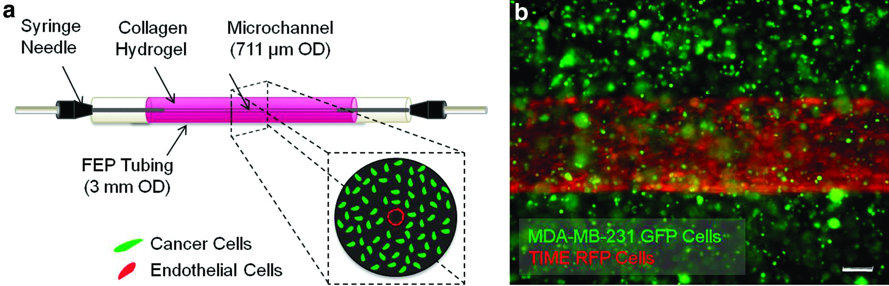
By integrating tissue-engineering strategies with cancer biology, microscale fluid mechanics, and optical flow diagnostics, we developed a 3D microfluidic for dynamic coculture of tumor and endothelial cells under a range of experimentally quantified WSS (Fig. 1a). Methods describing the microfabrication process of collagen hydrogels, which provide an optimal in vitro environment for the development of an endothelialized microchannel, inclusion of WSS, and coculture with tumor cells, are presented (Fig. 1b). This is the first application of μ-PIV in a tumor representative, 3D collagen matrix to measure flow velocity and shear stress in microchannels with cylindrical geometries. Shear-induced changes in endothelial morphology were assessed to demonstrate the integrity of the microfluidic tumor vascular model in 3D, dynamic culture. Gene expression analysis not only validates the ability of this system to yield sample volumes necessary for downstream molecular analysis, but also highlights the importance of the hydrodynamic tumor microenvironment in regulating angiogenic activity.
Tumor-endothelial coculture in microfluidic collagen hydrogels.
Materials and Methods
Scaffold design and fabrication
Type I collagen stock solutions were prepared by the following method: briefly, tendons were excised from rat tails and dissolved in a pH 2.0 HCl solution for 12 h at 23°C. The solution was centrifuged for 45 min at 30,000 g and the supernatant was collected. The concentration was determined by measuring the dry weight of the collagen stock solution.
The collagen stock solution was sterilized before scaffold fabrication by layering the solution over 10% (v/v) chloroform for 24 h at 4°C, after which, the top collagen portion was aseptically removed. 42 A working collagen solution of 8 mg/mL was prepared by neutralizing the stock solution with 10% (v/v) 10×DMEM mixed with 1N NaOH and deionized water (dH2O). This concentration was chosen because it has been shown to have an elastic modulus that closely matches neoplastic tissue stiffness 43 as well as supports both microfabrication and cellular remodeling.31,44 3D microfluidic collagen hydrogels were fabricated by pouring the neutralized collagen solution into fluorinated ethylene propylene (FEP) tubing fit concentrically with a 22G (711 μm) stainless steel needle and capped with polydimethylsiloxane (PDMS) sleeves (Fig. 2a). The collagen hydrogels were stabilized in standard 35-mm Petri dishes by boring opposing holes on either side of the dish through which the FEP tubing was inserted. A third hole was cut in the bottom of the dish and replaced with a 1.5-mm glass slide to improve optical access. Adapted from methods by Chrobak et al., 45 the collagen was allowed to polymerize for 20 min at 37°C before removal of the needle, creating a cylindrical microchannel embedded within the hydrogel (Fig. 2b, c). Before fabrication, the FEP tubing and PDMS sleeves were sterilized under UV with 70% ethanol for 1 h followed by exposure to air plasma (Harrick Plasma) for 2 min to activate hydroxyl groups on the FEP surface. To improve adhesion at the collagen/FEP interface, the FEP and PDMS sleeves were further treated with 1% (v/v) polyethyleneimine in dH2O for 10 min followed by 0.1% (v/v) glutaraldehyde in dH2O for 20 min.

Fabrication of microfluidic collagen hydrogels and perfusion setup.
The use of FEP tubing as an architectural mold for the microfluidic collagen hydrogel overcomes challenges associated with optical access and refractive index mismatching in typical tissue-engineering scaffolds or bioreactors. FEP has a refractive index that closely matches that of water, collagen, and cell culture media (Table 1), which minimizes optical distortion during image acquisition and flow measurements. Therefore, when the Petri dish is filled with water and the FEP housing the microfluidic collagen gel becomes submerged, refractive index matching across all curved surfaces affords optical clarity for high-resolution microscopy. Refractive indices for type I collagen and EGM-2 endothelial growth media (n=3) were measured using a digital refractometer (PA-202X; Misco) at 37°C and λ=589 nm.46–48
For measured values, a digital refractometer was used to determine the refractive index. Refractive index matching across all media minimizes optical distortion during image acquisition.
FEP, fluorinated ethylene propylene.
To introduce flow into the microfluidic culture model, 22G stainless steel needles were inserted through the PDMS sleeves and partially into the collagen microchannel. The inlet needle was connected to autoclaved Tygon® silicone tubing (1/16′′ ID), which connected to a syringe pump that controls the flow rate. The outlet needle was similarly connected to silicone tubing leading to a collection reservoir (Fig. 2d, e). In addition, a low protein binding air-eliminating filter set (Pall Life Sciences) was utilized at the tubing/microchannel interface to prevent bubbles from entering the microchannel during perfusion.
Average shear stress in normal microvasculature is 4 dyn/cm2.
49
It has also been published that cancer cell growth arrest and inhibited differentiation is observed after exposure to relatively high shear stresses of 12 dyn/cm2.
15
Therefore, a range of flow rates generating a target WSS (τW) of 1, 4, or 10 dyn/cm2 were introduced in the collagen microchannel. These shear stresses were first estimated based on an assumption of Poiseuille flow,
Cell culture
A human breast carcinoma cell line (MDA-MB-231) (American Type Culture Collection [ATCC]) and a telomerase-immortalized human microvascular endothelial cell line were used in this study. Telomerase immortalized microvascular endothelial (TIME) cells were provided as a generous gift from Dr. Shay Soker at the Wake Forest Institute for Regenerative Medicine (Winston-Salem, NC). A lentiviral vector system was used to genetically modify the cells to stably express either a green fluorescent protein (GFP) or red fluorescent protein (RFP) for real-time visualization during culture. Imaging was performed using an inverted fluorescence microscope (Leica AF6000) to observe cell localization, proliferation, and viability during culture.
MDA-MB-231 cells were cultured in the Dulbecco's Modified Eagle's medium, nutrient mixture F-12 (DMEM/F12) (Invitrogen) supplemented with 10% fetal bovine serum (Sigma-Aldrich), and 1% penicillin–streptomycin (Invitrogen). TIME cells were cultured in EBM-2 (Lonza) media supplemented with a growth factor BulletKit (Lonza CC-4176). All cell cultures were maintained in a 5% CO2/95% air atmosphere at 37°C within an incubator.
To fabricate cellularized microfluidic collagen gels, 1×10⁶ MDA-MB-231 cells/mL were resuspended in the neutralized collagen and carefully mixed before pouring the solution into the FEP tubing. During polymerization, the cells become suspended within the bulk of the collagen hydrogel. After gelation and removal of the needle, 10×106 TIME cells/mL were injected into the microchannel 2×at 10-min intervals and slowly rotated to encourage complete coverage of the lumenal surface. The microfluidic collagen scaffold was then connected to the syringe pump and flow was introduced into the microchannel. A 3-day graded increase in the flow rate, which corresponded with 36 h at τ=0.01 dyn/cm2 followed by 36 h at τ=0.1 dyn/cm2, was implemented to establish a confluent endothelialized microchannel (Fig. 3a, b), as this type of hydrodynamic loading has been shown to maintain endothelial integrity. 50 After the 72-h preconditioning, a rate of increase in shear of ∼5.5 (dyn/cm2)h−1 was used to reach a target WSS of τW=1, 4, or 10 dyn/cm2, after which, a constant flow rate was then used to maintain the target WSS for a total of 6 h.
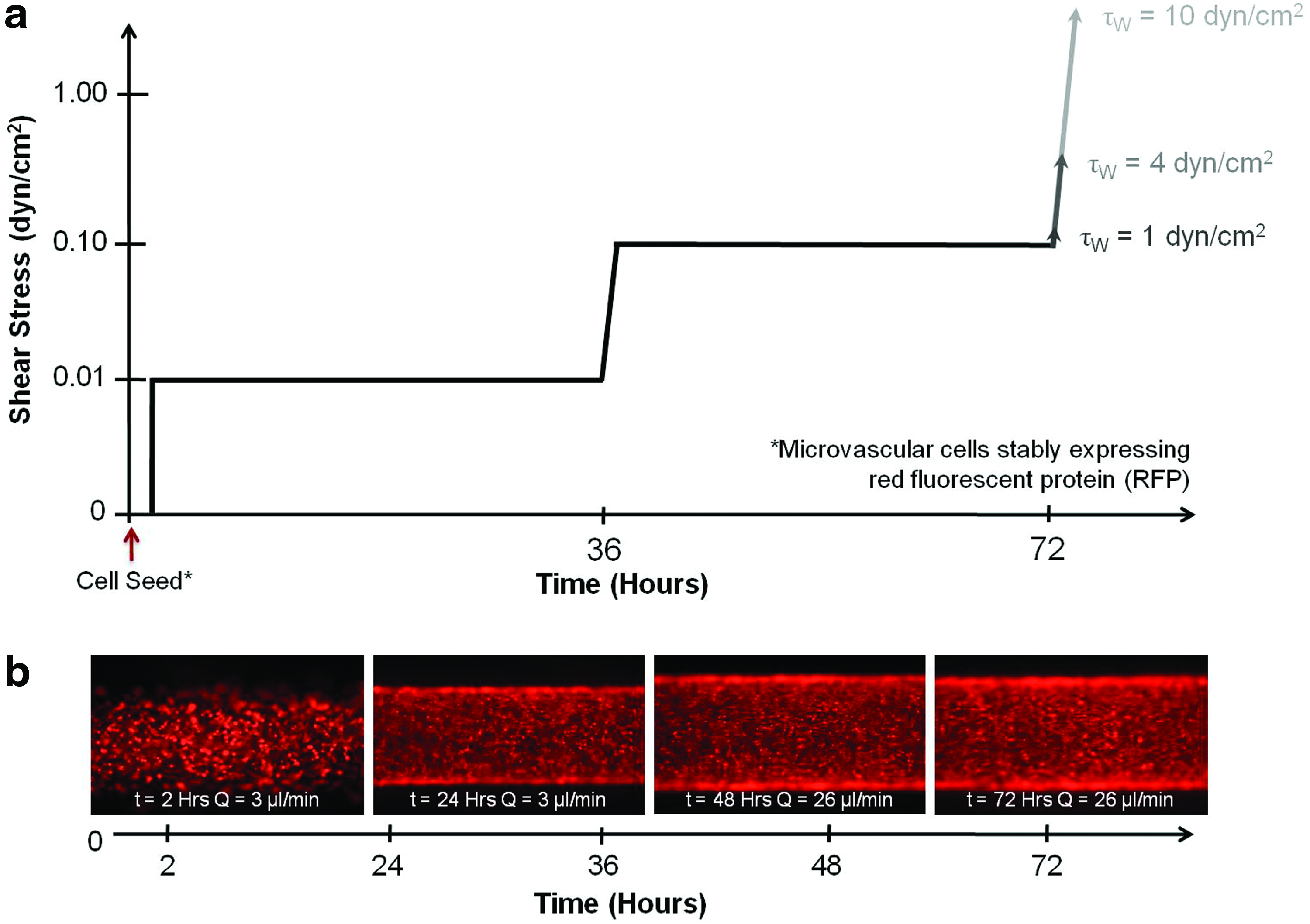
Shear stress preconditioning protocol to develop a confluent endothelium.
In addition, total oxygen consumption (3×10⁻⁹ mol/min) was estimated based on the tumor cell O2 consumption rate (5.4×10⁻¹⁷ [mol/s]cell−1), 51 total number of cells in the collagen scaffold (1×10⁶, volume of media in the microchannel (20 μL), and Henry's constant at 37°C (1.04×10⁻² mol/L), to ensure that at even the lowest flow rate (Q=3 μL/min), a sufficient amount of oxygenated media is provided to the cells. MDA-MB-231 viability in the collagen matrix was further verified using a calcein AM/propidium iodide LIVE/DEAD® assay (Invitrogen), at 0, 24, 48, and 72 h postflow (n=3) to monitor cell death at regions furthest from the microchannel. Images were taken in three random view fields per sample and analyzed using a custom-designed LabVIEW algorithm (National Instruments). 52 Nonfluorescently labeled MDA-MB-231 cells were used in the viability assay.
Morphological characterization
To visualize endothelial integrity after exposure to each target WSS, fluorescent labeling of F-actin and nuclei was utilized. Briefly, the endothelium was fixed by perfusing 3.7% paraformaldehyde and permeabilized by perfusing 0.5% Triton X-100 (Sigma-Aldrich) through the microchannel for 20 min, followed by incubation in a 1% bovine serum albumin blocking buffer (Santa Cruz Biotechnology, Inc.) for 30 min at 37°C. Cells were then stained with Oregon Green phalloidin (Invitrogen), a high-affinity probe for F-actin, for 20 min at room temperature, and DAPI (Vector Laboratories), to visualize nuclei. Imaging was performed using an inverted fluorescence microscope (Leica AF6000). Images were then analyzed with ImageJ software (National Institutes of Health, Bethesda, MD) to determine cell alignment by measuring the angle of orientation relative to the vertical access of the image.
PIV flow measurement
The design of the microfluidic collagen hydrogels enabled experimental measurement of fluid velocity in the microchannels using μ-PIV (Fig. 4a). μ-PIV optical flow diagnostics were performed in both acellular and endothelialized (TIME-GFP) microchannels. Measurements were conducted independently after the 72-h preconditioning period plus 6-h exposure to either τW=1, 4, or 10 dyn/cm2. Briefly, 3.0-μm red fluorescent particles (Thermo Scientific) were suspended in EMB-2 media and perfused through the microchannel at various flow rates to generate a target WSS (τW) of 1, 4, and 10 dyn/cm2 (Table 2). The flow rates were initially estimated based on Poiseuille assumptions and then validated using μ-PIV. The channel was illuminated through an epifluorescent inverted microscope (Zeiss) with a 532 nm double-pulsed laser (New Wave Research). The fluorescing particles were imaged using a 1MP camera (Photron APX-RS) using a 20×microscope objective. Five hundred image pairs were acquired at a frequency of 7.5 Hz (Fig. 4b). Mean subtraction and intensity normalization were performed on the images to enhance correlation accuracy. Image pair correlation was done using a two-pass multigrid sum-of-correlation robust phase correlation algorithm with discrete window offset and zero-mean windows and validated using a median universal outlier detection scheme.53–58 A set of 125 generated vector fields were ensemble averaged to obtain a mean velocity field (Fig. 4c). Shear stress at the channel wall was calculated based on the linear portion of the velocity gradient, determined by thin-plate spline radial basis functions,59,60 and the measured viscosity of culture media (μ=0.78±0.0057 cP).
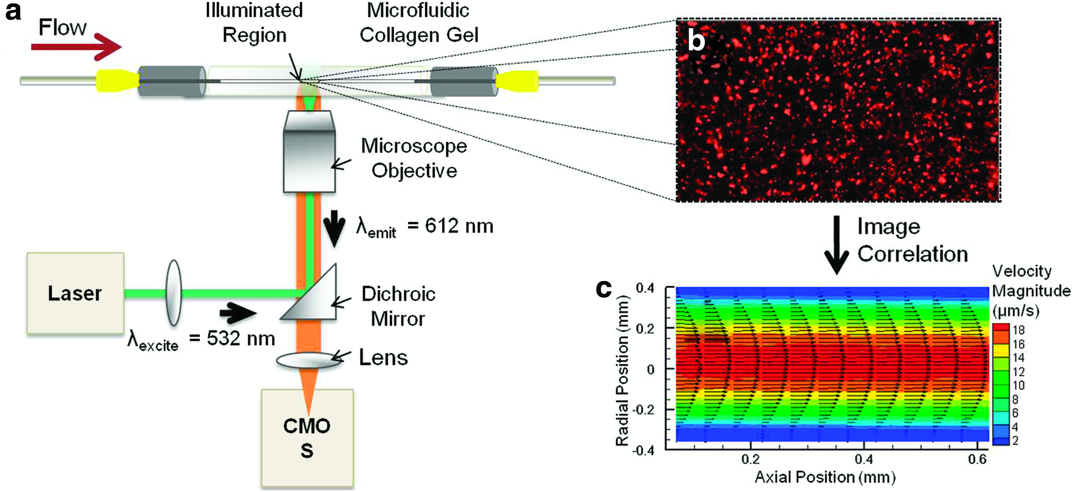
Schematic of microparticle image velocimetry (μ-PIV) flow diagnostics μ-PIV, a noninvasive flow measurement technique with a high spatial and temporal resolution,
37
was used to measure velocities in the collagen microchannel.
Quantitative RT-PCR
To determine the effect of 3D, dynamic coculture on tumor cell expression of angiogenic factors, expression levels of target genes in MDA-MB-231 cells were determined quantitatively by real-time RT-PCR as described previously. 26 A preliminary 24-h study was conducted in which, MDA-MB-231 tumor cells were cultured under either static or dynamic flow conditions in the microfluidic collagen hydrogels, in the presence or absence of microvascular endothelial cells, and compared to MDA-MB-231 monocultures on tissue culture-treated polystyrene (TCPS) as controls. To isolate mRNA from the tumor cell population, endothelial cells were trypsinized and removed from the microchannel lumen. Total tumor mRNA was then isolated by phenol–chloroform extraction. Briefly, cells were lysed using the TRI Reagent® Solution (Applied Biosystems/Ambion). A biphasic mixture was then formed upon the addition of chloroform, and isopropanol precipitation of RNA was utilized for RT-PCR. Reverse transcription and PCR amplification was conducted using gene-specific TaqMan PCR primers (Applied Biosystems) for matrix metalloproteinase 9 (MMP9), vascular endothelial growth factor (VEGFA), angiopoietin 2 (ANGPT2), and platelet-derived growth factor B (PDGFB), which are factors implicated in breast cancer angiogenesis and metastasis. 61 Evaluation of the 2−ΔΔCT indicates the fold change in gene expression, normalized to the GAPDH housekeeping gene and relative to the control group. 62
Statistical analysis
For the theoretical estimation of WSS based on Poiseuille flow, a propagation of error analysis was conducted to determine the effect of the variable's uncertainties (viscosity, μ; flow rate, Q; and radius, r) on the uncertainty of WSS calculation. Statistical analysis on the μ-PIV calculation of WSS was conducted by averaging all vectors (160) along the wall and computing the standard deviation, as described previously. 60
Gene expression analysis experiments were conducted with a total of four samples per group (n=4). Experimental groups were tested and analyzed independently and the data are expressed as mean value±standard deviation. Significance of results was verified using the Student's t-test, in which, a 95% confidence criterion was used to determine statistically significant differences between groups.
Results
Cell culture in microfluidic tumor model
Type I collagen hydrogels were selected as the scaffolding material to best replicate the structural architecture and mass transport properties of tumor tissue in vivo. Collagen can also be remodeled by the cells to accommodate long-term morphological changes and migration in three dimensions. A central microchannel embedded within the hydrogel functions as a single neovessel through which tumor-relevant hydrodynamic stresses are introduced. Endothelial cells seeded along the lumenal surface of the microchannel develop a confluent endothelium that provides a biological interface for exchange of growth factors with tumor cells seeded in the bulk of the hydrogel. This design enables tumor and endothelial cell coculture within physiologically relevant geometries, as well as the introduction of flow through the microchannel to expose the endothelial cells to a range of shear stresses. Last, the use of FEP in this model overcomes challenges associated with optical access in typical tissue-engineered scaffolds or bioreactor systems, such that live-cell imaging can be conducted without having the need to fix and dissect the scaffolding for analysis.
A successful 72-h preconditioning scheme was developed to encourage the development of a confluent endothelium. Following a 20-min static seed, the endothelialized microchannel was first exposed to a low flow rate Q=3 μL/min for 36 h before ramping up the flow to Q=26 μL/min for an additional 36 h. This scheme was based on an in vitro study investigating the integrity of endothelial cell-seeded vascular grafts, in which, a gradually graded shear stress was shown to reduce cell detachment, as well as stimulate endothelial elongation and orientation in the direction of flow. 50 A rate of increase in shear of ∼5.5 (dyn/cm2)h−1 did not perturb endothelial integrity or coverage of the microchannel lumen during preconditioning or exposure to the target WSS (τw=1, 4 or 10 dyn/cm2). After 72 h of preconditioning, the endothelial cells form a confluent monolayer on the microchannel lumen as characterized by F-actin staining. Following preconditioning, the endothelium withstood each τw for up to 6 h, demonstrating a qualitative increase in cell spreading and alignment in the direction of flow as a function of increased WSS (Fig. 5). Cell angle of orientation significantly increased (p<0.0001) for all τw relative to the precondition flow rate (data not shown).
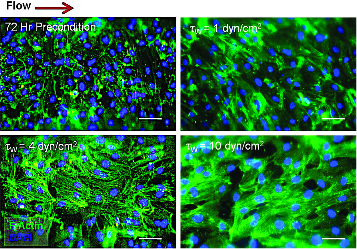
TIME cells develop a confluent endothelium and maintain integrity after exposure to each target WSS (τW). The endothelium with fluorescently labeled F-Actin and DAPI after the 72-h preconditioning scheme and plus an additional 6-h exposure of τW=1, 4, or 10 dyn/cm2 demonstrate increased alignment in the direction of flow as a function of increasing WSS. Scale bar 50 μm. Color images available online at
Tumor cell viability and proliferation was also confirmed in the microfluidic collagen hydrogels to ensure that a sufficient amount of oxygen and nutrients is available to sustain the culture for the duration of the 3-day experiment. Results demonstrate that tumor cells remain greater than 90% viable in the collagen matrix (Fig. 6a), with an observed increase in cell number from 0 to 72 h (Fig. 6b).

Tumor cell viability and proliferation in the collagen hydrogel.
Velocity and WSS measurements
μ-PIV experiments were successfully conducted in endothelialized and acellular microchannels to characterize the velocity profile and quantify WSS (τW=1, 4, and 10 dyn/cm2). Velocity data acquired from the μ-PIV experiments and postprocessed in MATLAB match closely to the estimated parabolic Poiseuille solution (Fig. 7a) confirming that fluid velocity through the endothelialized microchannels maintains a laminar flow profile. The tails observed at the wall for the experimental velocity data are likely due to measurement bias in the PIV processing. 59 Further experimentation and improved μ-PIV processing is necessary to interpret the low-velocity gradient measured at the microchannel wall.
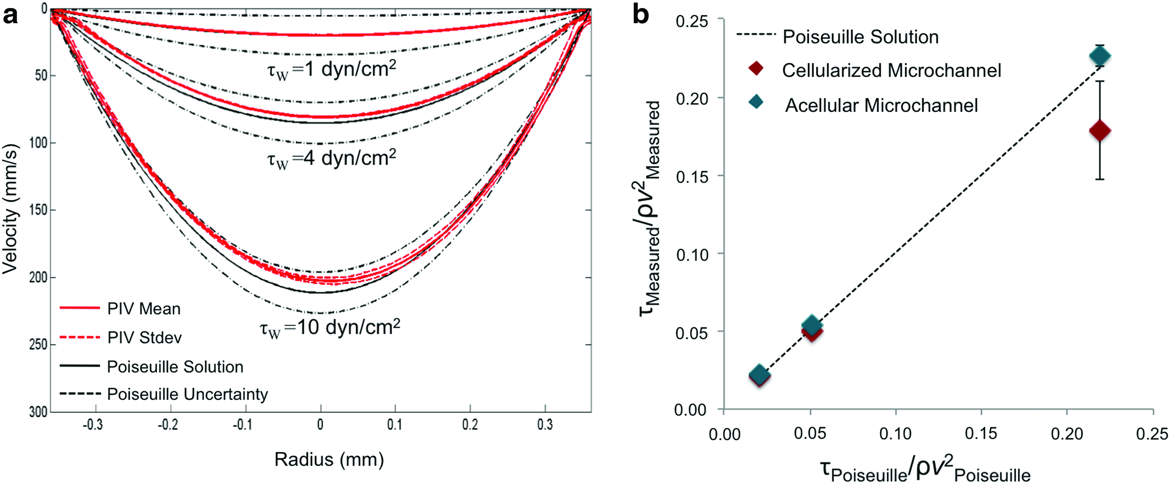
Experimentally measured velocity profiles and WSS in the microchannel.
WSS measurements were nondimensionalized by the characteristic fluid stress (ρv2) based on the centerline velocity, which allows for comparison between microchannels with varying diameters. This is important because during the 72-h preconditioning period, the diameter of the acellular microchannels widened (∼850 μm), decreasing the velocity gradient at the wall. In contrast, contractile forces of the endothelial cells better maintained the geometry of the microchannel under flow conditions. Experimental WSS measurements agree well with the theoretical values for all cases of τW, while the cellularized microchannel for τW=10 dyn/cm2 was slightly lower (Fig. 7b). This finding suggests that the endothelial cells may be influencing WSS; however, further experimentation is needed to investigate this effect.
Gene expression analysis
A preliminary 24-h gene expression analysis experiment was conducted to demonstrate the ability of the microfluidic culture system to yield a sufficient amount of cellular mRNA for quantitative RT-PCR. In contrast to conventional PDMS/hydrogel microfluidic devices, which typically contain less than 1000 cells, the design of this microfluidic tumor model contains a larger collagen volume that has the capacity for high cell density culture (>300,000 total cells), thereby enabling downstream molecular analysis that cannot be otherwise conducted using conventional methods in existing devices. RT-PCR results indicate that MDA-MB-231 monocultures grown in 3D microfluidic collagen hydrogels under static conditions, significantly increased gene expression of MMP9, a proteinase that degrades collagen, as well as PDGFB and ANGPT2, both of which are implicated in breast cancer angiogenesis, 61 relative to MDA-MB-231 monocultures grown on 2D TCPS controls (Fig. 8). The increased angiogenic capacity of tumor cells grown in 3D culture compared to 2D substrates has been attributed to centrally diminished oxygen availability in thick scaffolds and resultant induction of a hypoxic response.30,33 The 3D scaffolding also promotes integrin engagement important for angiogenic signaling.63,64 When a low flow rate of 50 μL/min (τ=0.2 dyn/cm2) is introduced through the microchannel, PDGFB gene expression was further upregulated, as well as a significant fold increase in VEGFA, relative to MDA-MB-231 monocultures grown in 3D static conditions. However, most interestingly, the addition of endothelial cells (cocultured on the lumenal surface of the microchannel) plus flow, significantly increased MDA-MB-231 expression of all proangiogenic factors investigated, relative to monocultures grown in 3D dynamic conditions. Comparatively, in the absence of flow, MDA-MB-231 cells significantly increased expression of VEGFA only during coculture with endothelial cells. These results are supported by evidence of a contact-independent mechanism of tumor–endothelial cross talk, in which VEGF mediates expression of the antiapoptotic protein, Bcl-2, which elicits reciprocal proangiogenic signaling. 27 Fluid shear forces and VEGF gradients have also been shown to cooperatively control endothelial sprouting and morphogenesis, 65 and likely play a significant role in tumor angiogenesis. Similarly, when all groups are normalized to TCPS control, results indicate that in the absence of flow, coculture decreases MDA-MB-231 expressed angiogenic genes; however, in the presence of flow, coculture increases gene expression (data not shown). These results highlight the importance of the hydrodynamic tumor vascular niche in regulating tumorigenesis, and warrant further investigation under physiologically relevant shear stresses.

Three-dimensional, dynamic coculture with endothelial cells significantly increases tumor-expressed angiogenic genes. After 24 h, MDA-MB-231 cells significantly upregulate matrix metalloproteinase 9 (MMP9), platelet-derived growth factor B (PDGFB), and angiopoietin 2 (ANGPT2) under 3D static conditions relative to 2D tissue culture-treated polystyrene controls. The introduction of a low flow rate (50 μL/min) further increased PDGFB as well as significantly increased vascular endothelial growth factor A (VEGFA) gene expression relative to 3D static conditions. The presence of endothelial cells without flow had a significant effect on tumor expressed VEGFA; however, in the presence of flow, all tumor-expressed proangiogenic genes were significantly upregulated during coculture, relative to MDA-MB-231 monocultures under 3D dynamic conditions. Relative mRNA to GAPDH mRNA expressed as a fold induction±standard deviation (n=4). *p<0.05.
Discussion
While previous studies have demonstrated the importance of tumor–endothelial cross talk under two-dimensional (2D), static culture conditions, a better understanding of the relationship between endothelial cells and the physical tumor microenvironment may be better accommodated in 3D, dynamic coculture models. This work demonstrates that microfluidic collagen scaffolds can be fabricated for coculture of tumor and endothelial cells under a range of physiologically relevant shear stress conditions. We have presented the design and fabrication of a 3D microfluidic tumor vascular model, characterized flow properties in the microchannel, and assessed endothelialization and tumor cell response to various flow conditions. While the average diameter of tumor microvessels is generally less than 100 μm, 5 this system is a viable tool for applying physiologically relevant flow shear stresses and measuring cellular responses. The microchannel diameter used in this study (711 μm) is still within the range of physiologically relevant mammary tumor capillaries, especially in the venous network (mean diameter of 650 μm). 66 Furthermore, a larger diameter microchannel is more stable during extended culture durations, less likely to collapse during perfusion, and reduces flow resistance, thereby avoiding pressure drops along the length of the channel that may be detrimental to cell health. Results demonstrate that tumor cells maintain viability in the bulk of the collagen hydrogel, while endothelial cells form a confluent monolayer along the microchannel lumen after 72 h of preconditioning at low flow rates. For longer durations of coculture (>3 days), a bilayered collagen hydrogel modification may be necessary to separate the tumor cells and endothelialized microchannels with a thin acellular collagen region, as direct contact of MDA-MB-231 cells and microvascular endothelial cells has proven to decrease endothelial viability in vitro.67,68
The microfluidic tumor vascular model can withstand a range of normal, high and low flow shear stresses that are relevant to the hydrodynamic tumor microvasculature, as evident by F-actin staining of the endothelium. Furthermore, RT-PCR results confirm that these cultures yield sufficient mRNA for downstream molecular analysis, and our preliminary findings underscore the importance of tumor–endothelial cross talk under dynamic culture conditions. Lastly, the 3D culture model is designed such that refractive index matching across all media affords optical clarity and the ability to obtain high-resolution images (both cell morphology and particle tracking for PIV) at various focal planes.
The velocity profile and WSS for three different flow conditions were successfully characterized in both endothelialized and acellular microchannels using μ-PIV. Experimental WSS measurements are in good agreement with the theoretical values, although the calculated WSS values were lower in acellular channels relative to endothelialized microchannels. This is mainly due to an increase in the diameter under flow without an endothelialized lumen, and emphasizes the importance of monitoring changes in microchannel geometry under different culture conditions, fluid forces, or duration of experiments. However, deviations from the theoretical estimation of WSS are not entirely unexpected because collagen is a compliant and porous biomaterial, and may contain surface variations along the microchannel lumen due to the endothelial cell lining or fabrication process. Resolution limitations in PIV estimation of WSS will also contribute to differences in experimental versus theoretical solutions.59,69,70 Moreover, although WSS in perfused culture models or bioreactors is conventionally estimated from the flow rate assuming Poiseuille flow, we have previously shown this assumption may not always hold true due to possible unsteadiness in flow and short vessel length typically used for in vitro models. 60 Challenges associated with optical access, refractive index mis-matching across nonplanar surfaces, and the need to use nonbiocompatible water/glycerin solutions for imaging has previously restricted the use of PIV for biological applications. Also, while previous studies have utilized particle tracking velocimetry in 2D microfluidic devices71,72 or 3D collagen-coated substrates, 65 this is the first experiment to integrate optical flow diagnostics into a 3D collagen matrix based on cylindrical microchannels rather than planar geometries. More specifically, this unique application of flow measurements within the context of a tumor-endothelial coculture model validates μ-PIV as a quantitative tool to correlate shear stress with tumor angiogenic responses in vitro. Future experiments that accommodate endothelial migration and branching off the lumenal surface of the microchannel to form 3D tubules in the collagen hydrogel will be achieved by increasing culture duration, adjusting the ratio of tumor and endothelial cells, and supplementation of angiogenic growth factors. μ-PIV can then be used to measure localized shear stress in endothelial branches to provide insight on how shear gradients may influence endothelial tubule formation within the tumor microenvironment.
The next generation of 3D culture systems utilizes a combination of microfluidic technologies and tissue-engineering approaches to enable the development of in vitro models fabricated with biomaterials that permit exchange of solutes with cells, provide specific barrier properties of the endothelium, and accommodate remodeling during angiogenic progression.31,36,73 Diffusion rates, cytokine gradients, shear stresses, and microscale cellular niches can be artificially recreated in microfluidic scaffolds to study physiological processes, such as wound healing, neovascularization, or tumor development, under controlled conditions.74,75 Moreover, the flow properties in microfluidic devices are dominated by viscous rather than inertial forces. 76 Therefore, mass transport is governed by local diffusion rates, whereby convective contributions are negligible and the supply of nutrients, gases, and therapeutic drugs to cells can be controlled and analyzed. In particular, these characteristics enable microfluidic scaffolds to be used as a tool to model the physical tumor microenvironment, in which the effects of matrix permeability, hypoxia, and shear stress on tumor responses can be systematically investigated. This research area is a burgeoning field that has led to the development of novel biomimetic microdevices designed to study the dynamics of cancer progression, namely, interstitial flow on tumor cell migration patterns17,22,35,77–80 and vascular responses. 34 Many of these models have provided insight into the field of cancer research by identifying novel mechanisms of flow-guided endothelial alignment, sprouting, and vessel formation.65,81,82 However, to the best of our knowledge, there are no existing in vitro culture models that are specifically designed to investigate the effect of flow shear stress on tumor-endothelial signaling or vascular organization within the context of the tumor microenvironment.
State-of-the-art microfluidic devices for tissue engineering and cancer research have been primarily based on microchannels formed by soft lithography in PDMS and sealed with glass coverslips for image acquisition.17,35,36,65,78,83,84 While these systems provide dynamic 3D cell culture environments with complex planar geometries, the low cell density and scaffold thickness limitations inherent to silicon microfabricated devices severely restrict dynamic cellular processes and prevent downstream molecular analysis of tumor-expressed genes and proteins. Furthermore, existing devices are prohibitively expensive and difficult to modify for specific biological applications. 85 We have overcome these limitations by eliminating the need for costly photolithography, modified the microfabrication design to increase cellular capacity and sample yield for gene and protein expression analysis, and replaced planar geometries with 3D cylindrical microchannels to improve physiological fidelity of the tumor model. The development of physiologically relevant in vitro tumor models can potentially impact cancer research applications of cytotoxicity testing and therapeutic targeting, as well as provide a platform for elucidating fundamental mechanisms of tumor physiology and progression under controlled culture conditions.
Footnotes
Acknowledgments
We would like to thank Dr. Scott Verbridge for many insightful discussions on this work. We would also like to thank Andrea Martin for generously donating the Sprague-Dawley rat tails. Funding for this study was provided by the National Science Foundation Early CAREER Award CBET 0955072 and a National Institute of Health Grant IR21CA158454-01A1.
Disclosure Statement
No competing financial interests exist.