
Abstract
Tissue-engineering approaches to cultivate corneal endothelial cells (CECs) or induce CECs from stem cells are under investigation for the treatment of endothelial dysfunction. Before clinical application, a validation method to determine the quality of these cells is required. In this study, we quantified the endothelial pump function required for maintaining the corneal thickness using rabbit CECs (RCECs) and a human CEC line (B4G12). The potential difference of RCECs cultured on a permeable polyester membrane (Snapwell), B4G12 cells on Snapwell, or B4G12 cells on a collagen membrane (CM6) was measured by an Ussing chamber system, and the effect of different concentrations of ouabain (Na,K-ATPase specific inhibitor) was obtained. A mathematical equation derived from the concentration curve revealed that 2 mM ouabain decreases pump function of RCECs to 1.0 mV, and 0.6 mM ouabain decreases pump function of B4G12 on CM6 to 1.0 mV. Ouabain injection into the anterior chamber of rabbit eyes at a concentration of <2 mM maintained the corneal thickness, while those over 3 mM significantly increased the corneal thickness. B4G12 cell sheets transplanted into rabbit eyes treated with 0.6 mM ouabain maintained the corneal thickness, while 3.5 mM ouabain significantly increased the corneal thickness. Taken together, pump function >1.0 mV is required to maintain the corneal thickness. These results can be used for standardization of CEC pump function and validation of tissue-engineered CEC sheets for clinical use.
Introduction
The corneal endothelium consists of a single layer of hexagonal cells with a thick basement membrane (Descemet's membrane) covering the posterior surface of the cornea in a well-arranged mosaic pattern. 8 Corneal hydration is determined primarily by the balance between the movement of the aqueous humor across the corneal endothelium into the stroma and the subsequent pumping of the fluid out from the stroma. 8 The accumulation of fluid in the stroma due to disturbance of this balance may lead to bullous keratopathy. The active transport of fluid out from the stroma depends on cation transport by both corneal endothelial and epithelial cells, and the Na+- and K+-dependent ATPase (Na,K-ATPase) expressed in the basolateral membrane of corneal endothelial and epithelial cells is primarily responsible for this pump function.8,9 Therefore, tissue-engineered CECs will need sufficient pump function to maintain corneal transparency. However, there are no objective parameters to validate the quality of CEC pump function. The purpose of this study is to establish such a standard of CEC pump function that can be used for validating various types of tissue-engineered CECs intended for clinical use.
Herein we report how to quantify pump function of human and rabbit CECs electrically using the Ussing chamber system, and determine how much pump function would be required to maintain the corneal thickness in using a rabbit cornea model.
Materials and Methods
Cell culture
All animals were handled in full accordance with the ARVO (Association for Research in Vision and Ophthalmology) Statement for the Use of Animals in Ophthalmic and Vision Research. Descemet's membrane together with rabbit CECs (RCECs) was stripped off en bloc from the rabbit cornea with forceps. After washing with phosphate-buffered saline (PBS), Descemet's membranes with RCECs were unfolded and attached on 0.1% gelatin- and 10 μg/mL laminin (Sigma-Aldrich)-coated polycarbonate 35-mm dishes, and cultured in Human-Endothelial-Serum-Free-Medium (Invitrogen), supplemented with 1.0% fetal bovine serum (Sigma-Aldrich) and 10 ng/mL FGF2 (Sigma-Aldrich) under a humidified atmosphere of 5% CO2 at 37°C. RCECs reached semiconfluence in 2 weeks, were replated on 35-mm dishes or Snapwell inserts (Corning), and were cultured for subsequent experiments.
The human CEC line B4G12 cells, 10 which were immortalized by SV40 large T- and small T-antigens, were purchased from DSMZ, and cultured in Human-Endothelial-SFM, supplemented with 1.0% fetal bovine serum and 10 ng/mL FGF2 under a humidified atmosphere of 5% CO2 at 37°C.
The medium was changed every 2 to 3 days. B4G12 cells reached semiconfluence in a week, were replated on 35-mm dishes, Snapwell inserts, or type 1 aterocollagen sheets CM6 (Koken), and were cultured for subsequent experiments. All dishes and flasks used for culture were polystyrene, noncoated vessels obtained from Asahi Techno Glass. For the Ussing Chamber study, cells were dissociated into single cells, suspended at a cell density of 2×10⁵ cells/cm2, plated, and cultured for an additional 1 week on 0.1% gelatin- and 10 μg/mL laminin-coated polycarbonate Snapwell inserts with a membrane pore size of 0.4 μm, or on similarly coated type 1 aterocollagen sheets CM6.
Preparation of human CEC sheets on collagen sheet
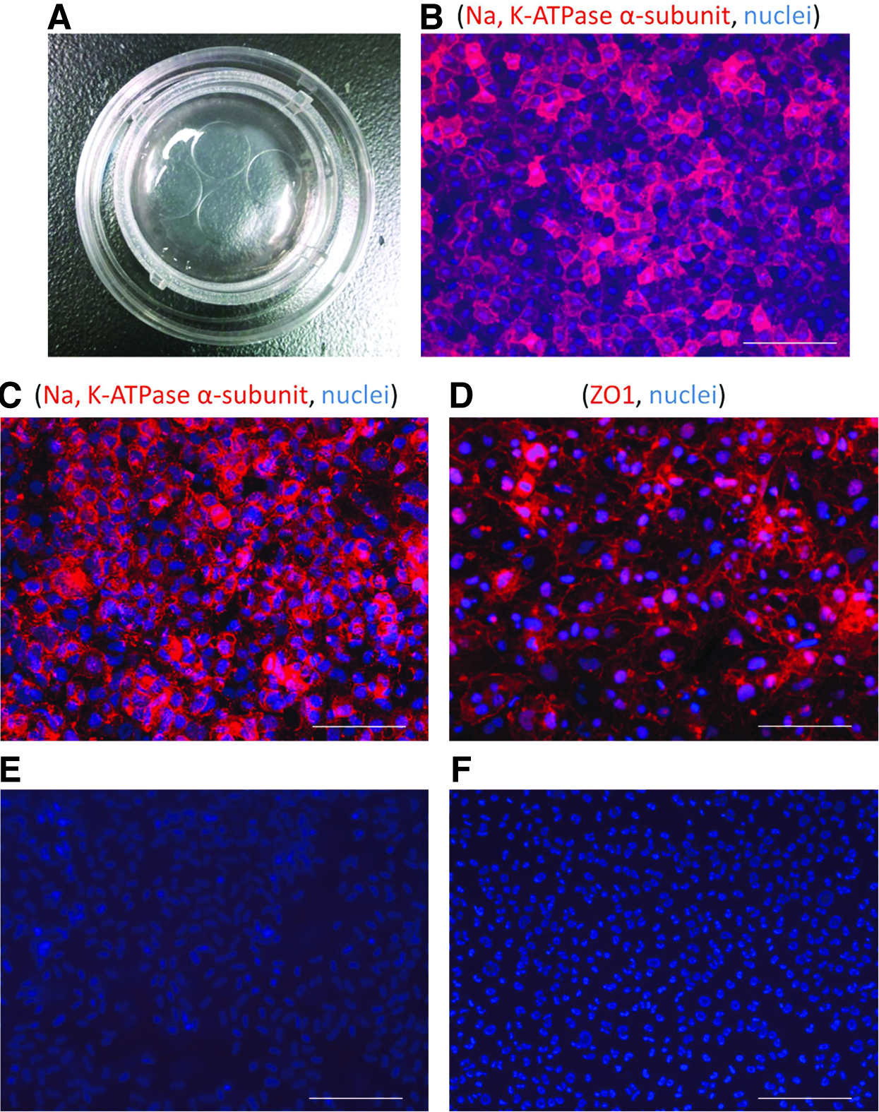
Four samples of B4G12 cell sheets of 8-mm diameter could be punched out from one B4G12 cell sheet on CM6 (Fig. 1A); so, the following in vitro pump function measurement of the B4G12 cell sheet by the Ussing chamber system and the in vivo pump function evaluation by the B4G12 cell sheet transplantation model were performed using the same B4G12 cell sheet.
Immunocytochemistry
B4G12 cells and RCECs cultured on the 3.5-mm culture dish were fixed at room temperature for 10 min in 4% formaldehyde in PBS. After three washes with PBS, samples were incubated for 30 min in Morphosave (Ventana Medical Systems). After another two washes with PBS, the samples were incubated for 30 min in 10% normal donkey serum to block nonspecific binding. This was followed by overnight incubation at 4°C with the 1:100-diluted mouse anti-Na,K-ATPase α1 subunit antibody (Novus Biologicals) or with the 1:100-diluted rabbit anti-ZO-1 antibody (Invitrogen), and then washed three times in PBS. The cells were then incubated for 1 h in a 1:200 dilution of the Cy3-conjugated donkey anti-mouse IgG antibody (Jackson ImmunoResearch, final concentration 30 μg/mL) and again washed three times in the dark. The samples were mounted on dishes with an antifading mounting medium containing 4′,6-diamino-2-phenylindole (DAPI), and the slides were inspected with the microscope (Axio Imager; Carl Zeiss, Inc.). Immunostainings for negative control were performed without primary antibodies.
B4G12 cells harvested from the corneas in the CEC sheet transplantation model were also stained by the same method using the mouse anti SV40 large T-antigen antibody (BD Pharmingen) as the primary antibody and 1:200 dilution of the Cy3-conjugated donkey anti-mouse IgG antibody as the secondary antibody, and mounted with the antifading mounting medium containing DAPI. Localization of transplanted B4G12 cells was observed by the Axio Imager. Immunostainings for negative control were performed without a primary antibody.
In vitro measurement of pump function by the Ussing chamber system
The pump function of confluent monolayers of B4G12 cells or RCECs was measured with the use of an Ussing chamber as described previously with minor modifications.11–13 Before measurements, cell densities were manually counted by a microscope. The cells cultured on Snapwell inserts were held by a specific holder (P2302), whose measurement area was 1.12 cm2. The CM6 collagen sheets with cells were cut in 8-mm-diameter circles using a biopsy punch (Kai Industries) and clasped by a specific holder (P2303A), in which the measurement area was 0.12 cm2. The entire setup was then placed in an Ussing chamber EM-CSYS-2 (Physiologic Instruments). The cell surface side was in contact with one chamber, and the Snapwell membrane side or collagen sheet side was in contact with another chamber. The chambers were carefully filled with 4 mL of Krebs-Ringer bicarbonate (120.7 mM NaCl, 24 mM NaHCO3, 4.6 mM KCl, 0.5 mM MgCl2, 0.7 mM Na2HPO4, 1.5 mM NaH2PO4, and 10 mM glucose bubbled with a mixture of 5% CO2, 7% O2, and 88% N2 to pH 7.4). The chambers were maintained at 37°C by an attached heater. The short-circuit current (SCC) was measured by narrow polyethylene tubes positioned close to either side of the Snapwell, filled with Krebs-Ringer bicarbonate and 4% agar gel, and connected to silver electrodes. These electrodes were connected to a computer through the Ussing system EVC4000 (World Precision Instruments) and iWorx IX/408 data acquisition system (iWorx Systems). SCC was recorded by Labscribe 2 Software for Research (iWorx Systems). After the Snapwell inserts with cells were placed in the chamber, SCC was continually measured for 15 min until SCC reached a steady state. Next, the cells on the Snapwell membrane were loaded with 1 mV currents through the electrodes three times, and the SCC change was recorded. Transendothelial resistance (TER) was calculated from the average SCC change and loaded voltage according to the Ohm's law.
Next, 100 μL of the Krebs-Ringer solution in the cell surface-side chamber was drained, and same amount of the specific Na,K-ATPase inhibitor ouabain solution (25 mM, diluted in the Krebs-Ringer solution with 37.5% ethanol, Sigma-Aldrich) or a solvent (the Krebs-Ringer solution with 37.5% ethanol) was added to the chamber. This process was repeated 21 times, and the final ouabain and ethanol concentration in the chamber was 10.3 mM and 15.5%, respectively. SCC was continually recorded during this procedure.
Finally, recorded SCC during ouabain addition was translated to potential difference (PD) by multiplying TER.
In vivo pump function measurement by ouabain treatment to rabbit eye model
All animals were treated in accordance with experimental procedures approved by the Committee for Animal Research of Keio University School of Medicine and Tokyo Dental College Ichikawa General Hospital. Japanese white rabbits (female, 2.5 kg body weight, Shiraishi Experimental Animal Breeding Farm) were anesthetized intravenously with a mixture of diazepam (0.5 mg/kg, Takeda Pharmaceutical) and pentobarbital sodium (30 mg/kg Kyoritsu Seiyaku). The anterior chamber of rabbits was washed with 1 mL of the Krebs-Ringer solution containing various concentrations of ouabain. The solution (total amount 1.0 mL) was repeatedly injected into the anterior chamber and drained five to six times through the paracentesis. The ouabain solution replaced the aqueous humor and remained in the anterior chamber after the procedure. The anterior chamber of rabbit eyes were shallow so that this process was enough to replace the aqueous humor with the ouabain solution. The central corneal thickness was measured with an ultrasound pachymeter (Tomey), and intraocular pressure (IOP) was measured by Accupen (White Medical) before and after the wash on 1, 6, 24, 48, and 120 h.
Transplantation of human CEC sheets into a rabbit model
All animals were treated in accordance with experimental procedures approved by the Committee for Animal Research of Keio University School of Medicine and Tokyo Dental College Ichikawa General Hospital. Japanese white rabbits (female, 2.5 kg body weight, Shiraishi Experimental Animal Breeding Farm) were anesthetized intravenously with a mixture of diazepam (0.5 mg/kg, Takeda Pharmaceutical) and pentobarbital sodium (30 mg/kg Kyoritsu Seiyaku). Transplantation was performed in the left eye of each animal only.
Corneal buttons were prepared by an 8.0-mm Barron donor cornea punch (Barron Precision Instruments) from the center of donor rabbit corneas purchased from Funakoshi Co, Ltd., and the Descemet's membrane along with the entire endothelium was stripped from the corneal buttons. B4G12 cell sheets with carrier collagen sheets CM6 were harvested by the same size punch, and soaked in the Krebs-Ringer solution with various concentrations of ouabain for 10 min. Subsequently, they were placed on the stromal bed of corneal buttons. Within 5 min, the sheets stably attached to the stroma. The recipient central cornea was excised by Hassburg-Barron Vacuum Trephine (Barron Precision Instruments) and the corneal buttons with B4G12 cell sheets were then placed on the graft bed of the originally surgically operated eye and sutured with sixteen interrupted sutures (10–0 nylon). Antibiotics (0.3% ofloxacin) and steroids (0.1% betamethasone) were applied topically three times a day. After transplantation, eyes were carefully observed by slit-lamp microscopy and serial photographs were obtained. The central corneal thickness was measured with an ultrasound pachymeter (Tomey), and IOP was measured by Accupen (White Medical) on 1, 2, and 5 days after transplantation. The central corneal thickness measurement and IOP measurement were performed by two different investigators who were masked to the details of transplantation in each group. Three eyes from three individual rabbits were used for each experimental group. Finally, rabbits were sacrificed, the operated eyes enucleated, and the corneas of each operated eye were cut and embedded into paraffin and mounted on dishes or glass slides. Immunocytochemistry was performed as described above, and localization of transplanted B4G12 cells with SV40 Large T-antigen staining was observed by the Axio Imager.
Statistical analysis
Data are presented as mean±SD and were compared by the Student's t-test or multiple t-test with the Bonferroni correction following ANOVA with the use of Excel 2007 software (Microsoft). The Gauss–Newton method, coefficient of determination, and other statistical analysis were calculated or drawn by Excel 2007 and JMP 8 software (SAS). A p value of <0.05 was considered statistically significant.
Results
Na,K-ATPase and ZO1 expression in CECs
Figure 1B and C show immunocytochemistry of the Na,K ATPase α1 subunit in RCECs (Fig. 1B) and in the human CEC line B4G12 cells (Fig. 1C), confirming that the Na,K-ATPase α1 subunit was expressed on the lateral side of both cells. ZO1 expression of B4G12 cells on CM6 was also confirmed by immunocytochemistry (Fig. 1D).
In vitro pump function measurement by Ussing chamber system
Figure 2A–C show SCC, TER, and PD of RCECs and B4G12 cells cultured on Snapwell, which were measured before ouabain treatment by the Ussing chamber system. The cell densities of RCECs or B4G12 cells on Snapwell were 3211.1±50.9 and 3177.8±96.2 cells/mm2, respectively. The TER of B4G12 cells was significantly higher compared with RCECs; however, there was no significant difference between SCC and PD. These results suggested that TER may differ between these cells, however, the difference in TER was too small to affect the difference of SCC or PD. Net pump function as PD values of RCECs and B4G12 cells were 1.43±0.15 and 1.25±0.02 mV, respectively.

Figure 2D–F show SCC, TER×(measurement area), and PD of B4G12 cells cultured on Snapwell or CM6. Net pump function as PD values of B4G12 cells on CM6 was 1.34±0.27 mV. Since measurement areas of Snapwell and CM6 were different and resistance is inversely proportional to the measurement area, TER×(measurement area) (ohm×cm2) should be compared for standardized comparisons. Under these conditions, there were significant differences in SCC and TER between both culture conditions, while PD did not differ significantly. These results suggested that electrical resistance of the collagen membrane on CM6 was significantly lower than the polyester membrane of Snapwell (Fig. 2E), and might have affected SCC. However, PD values were not affected by difference in resistance between Snapwell and CM6.
Figure 3A shows representative tracing of SCC (μA/well) obtained with RCECs in an Ussing chamber. Figure 3B–G show the relationship between ouabain concentrations and SCC and PD of RCECs on Snapwell, B4G12 cells on Snapwell, and B4G12 cells on CM6. Both SCC and PD of these cells decreased as ouabain concentrations increased. (SCC and PD slightly decreased by administration of the solvent of the Krebs-Ringer solution with 37.5% ethanol as well [Supplementary Fig. S1; Supplementary Data are available online at
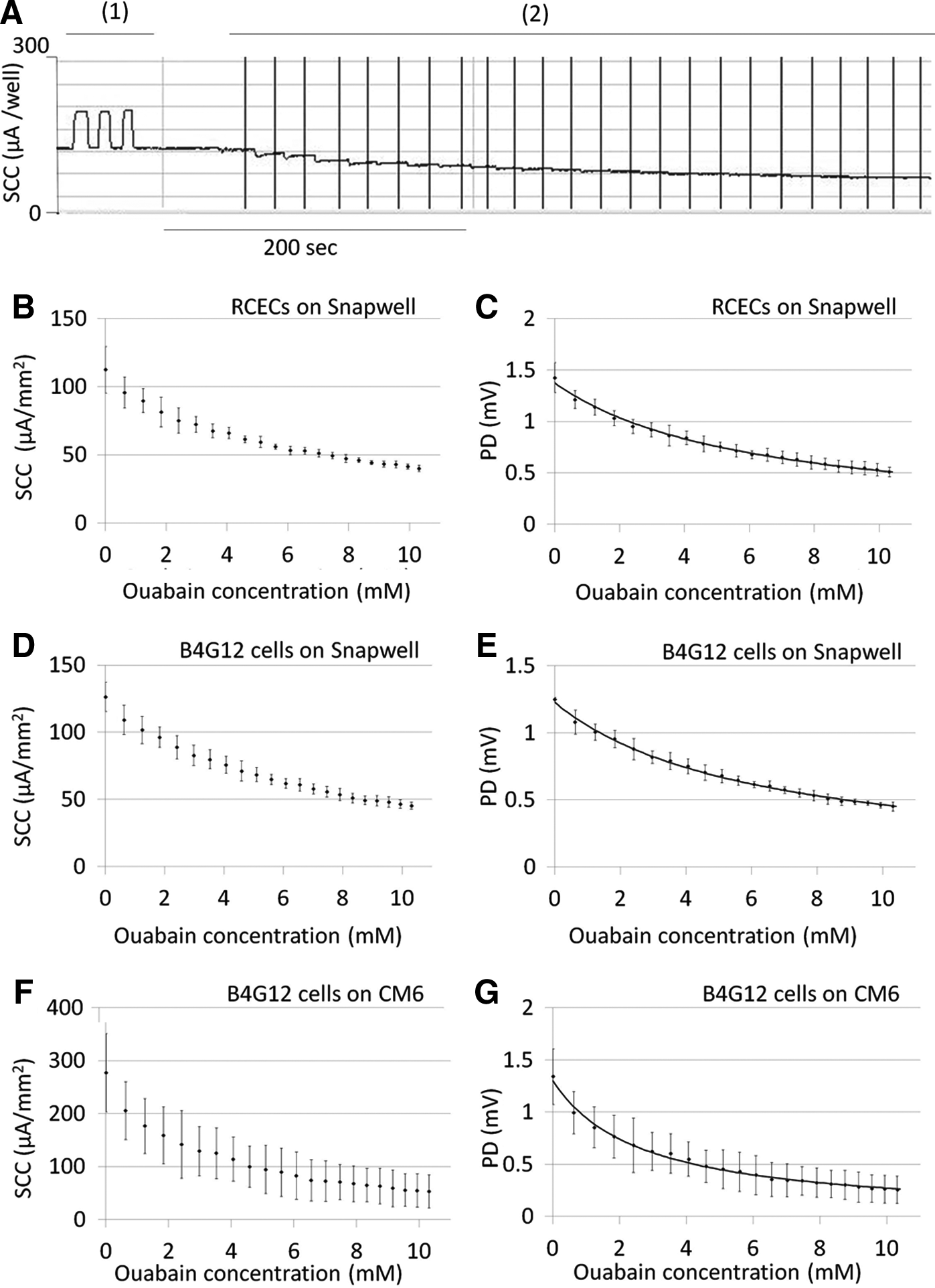
The relationship between the ouabain concentration and PD was inversely proportional. By the Gauss–Newton method, the following relationship between the ouabain concentration (X) and PD (Y) and the coefficient of determination (R2) in the RCECs or B4G12 cells were obtained;
Y=1/(0.119×X+0.728) (R2=0.952) in RCECs on Snapwell
Y=1/(0.134×X+0.815) (R2=0.968) in B4G12 cells on Snapwell
Y=1/(0.292×X+0.768) (R2=0.775) in B4G12 cells on CM6
From calculations in RCECs, 1.0, 2.0, 3.0, and 8.0 mM ouabain administration resulted in 1.2, 1.0, 0.9, and 0.6 mV pump function of RCECs, respectively. From calculations in B4G12 cells on CM6, 0.6 and 3.5 mM ouabain administration resulted in 1.0 and 0.6 mV pump function of B4G12 cells on CM6, respectively.
In vivo pump function evaluation by ouabain treatment to rabbit eye model
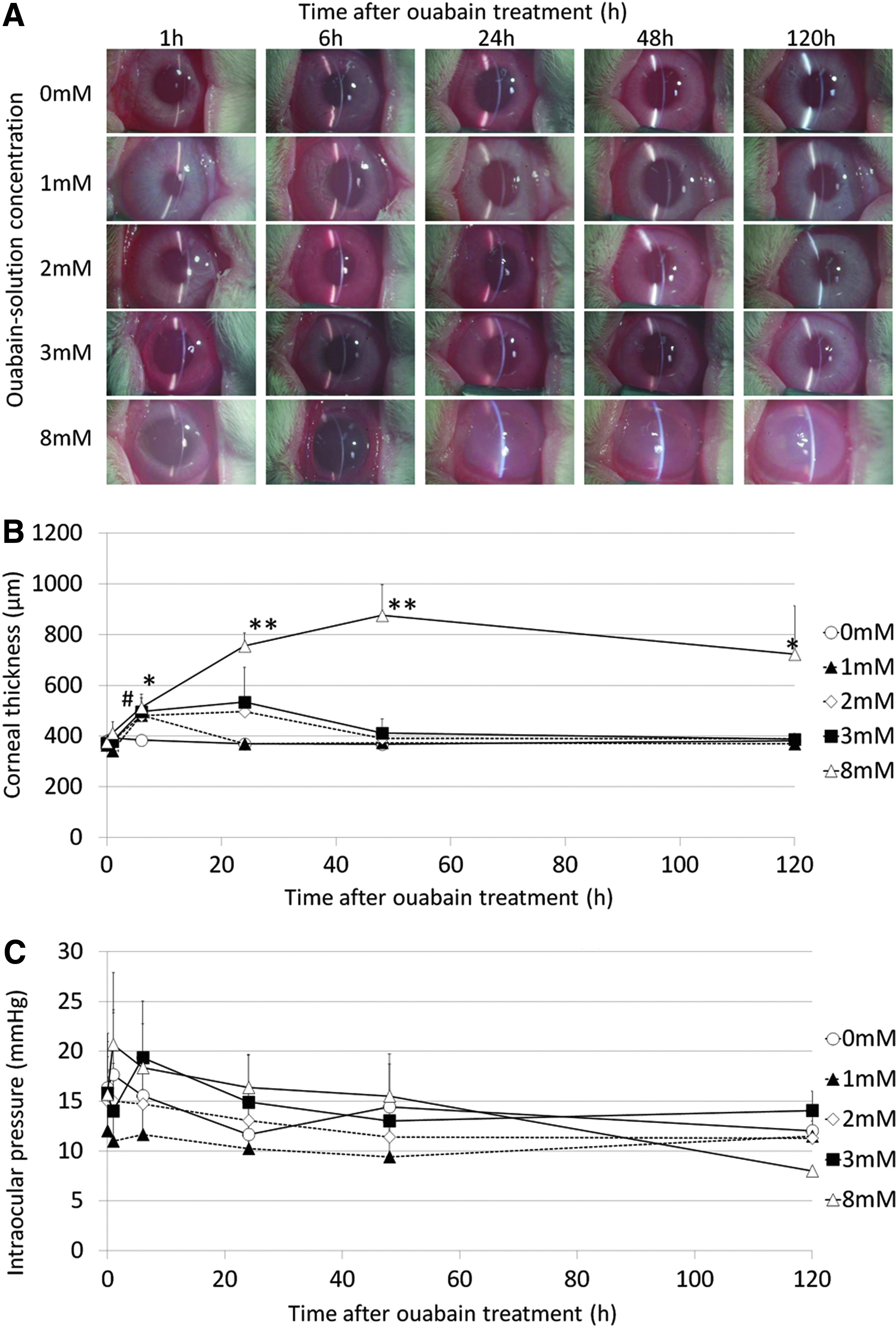
Figure 4A shows anterior segment photographs of rabbit eyes whose anterior chamber was washed with 1 mL of the Krebs-Ringer solution containing various concentrations of ouabain. In case of 0 (control), 1.0, or 2.0 mM ouabain, changes in transparency were not grossly detected, and the edematous change was detected in 3.0 mM. On the other hand, 8.0 mM ouabain resulted in marked corneal edema. The corneal thickness of 1.0 mM ouabain-treated rabbit eyes recovered close to control eyes 24 h after operation, and 2.0 or 3.0 mM-treated rabbit eyes recovered in 48 h (Fig. 4B). On the other hand, 8.0 mM-treated rabbit eyes had a significantly higher corneal thickness than control eyes during the whole observation period after surgery. Only at 6 h after the ouabain wash, 3.0 mM-treated eyes significantly increased the corneal thickness compared to control. During observation, there was no significant difference in IOP between these groups (Fig. 4C). Taken together with the ouabain concentration-PD curve of RCECs (Fig. 3C), these results suggested that the rabbit corneal thickness significantly increased when the endothelial pump function decreased below 1.0 mV.
In vivo pump function evaluation by human CEC sheet transplantation model
Figure 5A shows anterior segment photographs of rabbit eyes after transplantation of B4G12 cell sheets treated with 0 (control), 0.6, and 3.5 mM ouabain. The cell densities of these cell sheets were 3266.7±200, 3366.7±33.3, and 3288.9±69.4 cells/mm2, respectively. While B4G12 cell sheets treated with 0 and 0.6 mM ouabain maintained transparency, B4G12 cell sheets treated with 3.5 mM ouabain showed marked corneal edema. The corneal thickness of B4G12 cell sheets treated with 3.5 mM ouabain significantly increased compared to control, while there was no significance in the corneal thickness between sheets treated with 0.6 mM ouabain (Fig. 5B). During the observation, there was no significant difference in IOP between these groups (Fig. 5C). Taken together with the ouabain concentration-PD curve of B4G12 cell sheets (Fig. 3G), these results suggested that human CECs maintained the rabbit corneal thickness when the endothelial pump function was 1.0 mV, but caused an increase in thickness when the pump function was 0.6 mV.

Figure 6 shows the posterior surface and vertical section of harvested corneas 5 days after transplantation. SV40 large T-antigen-positive cells covered the posterior surface of the harvested cornea, and SV40 large T-antigen-positive monolayer cells on carrier collagen sheets also attached to the stroma of the corneal graft in vertical sections. These results confirmed that B4G12 cells covered the posterior surface of the cornea during observation.
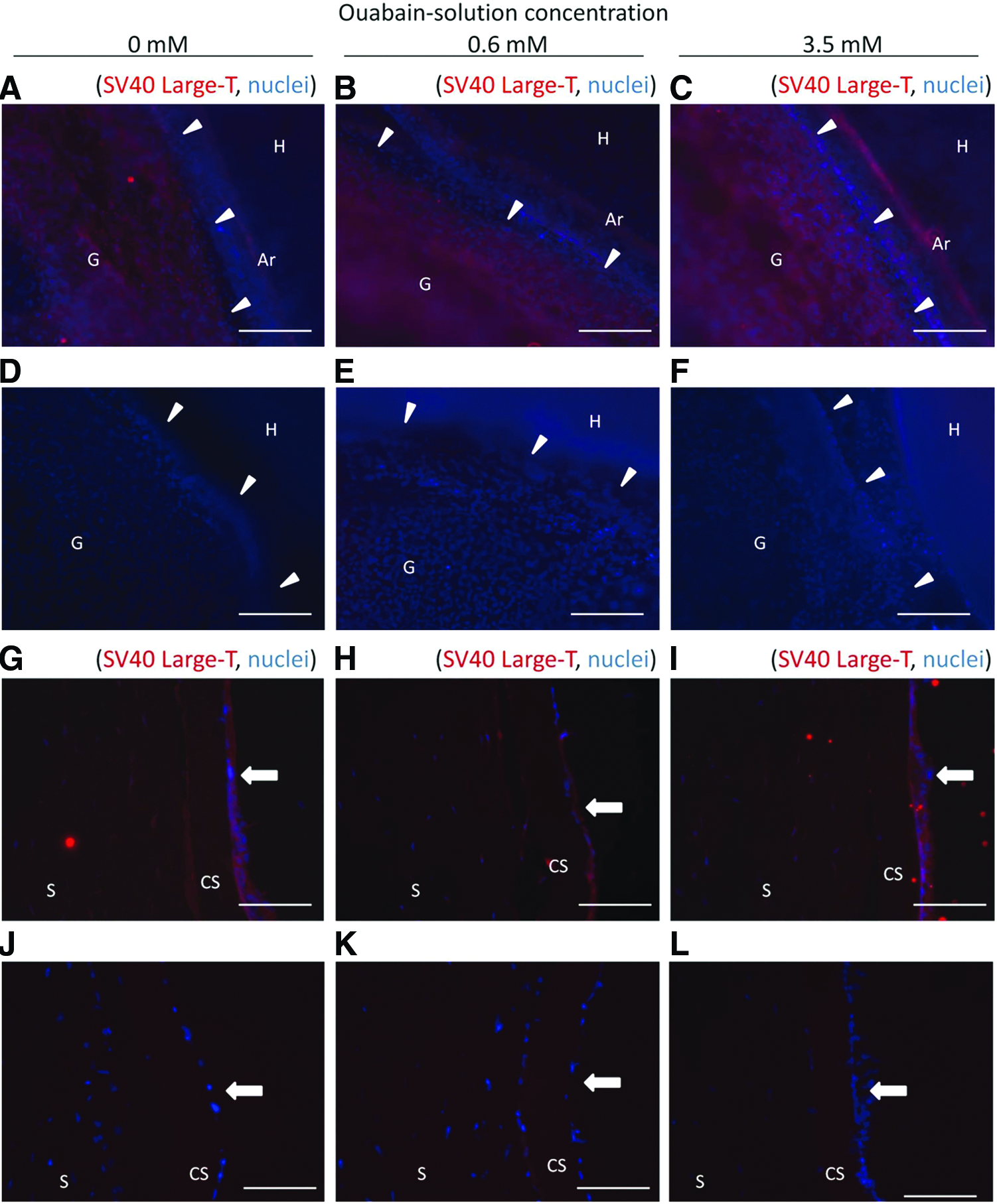
Immunocytochemistry with anti SV40 large T-antigen on host-graft junctions
Discussion
The pump function of CECs is produced by active cation transport, especially by the Na,K-ATPase of CECs. 8 When cations are actively transported to the aqueous humor side (i.e., the cell surface side in the Ussing chamber system), PD between the aqueous humor side and the stroma side (i.e., Snapwell membrane side or collagen sheets side in the Ussing chamber system) is observed. PD subsequently produces the electron flow through the endothelial cell layer and Snapwell or collagen sheet, which is represented as SCC in the Ussing chamber system. Therefore, PD directly represents the corneal endothelial function and is independent of the Snapwell membrane or collagen sheet resistance, while SCC is dependent on resistance. Since the collagen membrane absorbs the culture medium or the Krebs-Ringer solution, the membrane may be a good electrical conductor, making resistance of CM6 significantly less compared with the Snapwell polyester membrane. For this reason, even when the same B4G12 cells were analyzed by the Ussing chamber, SCC values of cells on Snapwell significantly differed from those on CM6, while PD were similar.
When pump functions as the PD value were compared between RCECs and B4G12 cells, there was no significant difference observed. The reason why TER of B4G12 cells was significantly higher compared with RCECs is unclear. This may partially be due to the difference in cell specimens, the effect of SV40 T-antigen in B4G12 cells, or the amount of basement membrane produced by each corneal endothelium. Further investigation is required to clarify this point.
The relationship between ouabain concentrations and PD was described as inversely proportional. However, it should be noted that the ethanol concentration in the chamber reached 15.5% when the ouabain concentration reached 10.3 mM. This cannot be avoided since a lower concentration of ethanol in the Krebs-Ringer solution cannot dissolve 25 mM ouabain. PD slightly decreased by administration of the Krebs-Ringer solution with only ethanol, indicating that ethanol may also have some effect on the cells. We therefore presumed that the SCC and PD decrease by the ouabain solution may be due to the net effect of ouabain and ethanol. However, this should not be a problem in terms of safety since the net effect would be an overestimation of ouabain toxicity.
The corneal thickness of ouabain-treated rabbit eyes recovered according to the concentration; however, 8.0 mM ouabain-treated eyes maintained a significantly higher corneal thickness up to 120 h after operation. The recovery of corneal thickness in lower concentrations may partially be because RCECs have proliferative capacity, or ouabain in the anterior is washed out during the long observation period. However, we believe that evaluation of the ouabain effect in a rather short time period after the ouabain wash would be acceptable to reduce the influence of such recovery mechanisms.
In vivo pump function evaluation in the rabbit corneal model revealed that 3.0 mM ouabain significantly increased the corneal thickness at 6 h compared to control, while lower concentrations did not differ significantly from control during the whole observation period. Although the difference between 2.0 and 3.0 mM ouabain concentrations seemed slight, these results suggested that the threshold of pump function may exist close to these values. In vivo pump function evaluation by B4G12 cell sheet transplantation model also revealed that the corneal thickness in 0.6 mM ouabain-treated sheets did not significantly differ from control eyes. Taken together, rabbit corneal thickness seems to significantly increase when corneal endothelial pump function decreased below 1.0 mV. Although the effect of ouabain on corneal thickness has been studied,14–17 it has not been clearly quantified as to how much pump function is needed to maintain the corneal thickness. Our data using the Ussing chamber system can be utilized as a standard of CEC pump function.
Since net pump function of RCECs before ouabain administration was 1.43 mV and that of B4G12 cells on CM6 was 1.34 mV, the threshold value of 1.0 mV pump function was equivalent to 1/1.43 of net pump function of RCECs, and 1/1.34 of net function of B4G12 cells. The 0.6 mV pump function, which induced remarkable corneal edema in this study, was equivalent to 1/2.23 (=0.6/1.34) of net pump function of RCECs, and 1/2.38 (=0.6/1.43) of net pump function of B4G12 cells. However, in general, most clinicians agree that with an endothelial cell count of <700 cells/mm2, which is 1/5 of the normal human CEC density, corneal edema becomes increasingly likely. 18 This discrepancy may suggest the existence of compensatory mechanisms of pump function in CECs when the cell number chronically decreased, which has been suggested in previous reports.19,20
As shown in Figure 1A, four cell sheets of 8-mm diameter could be obtained from one CM6 sheet. Remnant cell sheets may be used for other purposes after one sheet was used for Ussing chamber measurement. One of the sheets may be used to check tight junction by immunocytochemistry for ZO1, as shown in Figure 1D. Another sheet can be used for the validation before the clinical use of sheets from the same lot.
In conclusion, we quantified pump function of human and rabbit CECs by in vitro by the Ussing chamber system, in vivo by ouabain treatment of rabbit eyes, and in vivo pump function of human CEC sheets were analyzed using a transplantation model. The cumulative results of our study suggest that corneal endothelial pump function of more than 1.0 mV as measured by the Ussing chamber system is required to maintain the corneal thickness. This result may be utilized for standardization and validation of CEC pump function.
Footnotes
Acknowledgment
This study was supported by the Highway Program for Realization of Regenerative Medicine of the Ministry of Education, Culture, Sports, Science and Technology, Japan, (to K.N. and S.S.).
Disclosure Statement
No competing financial interests exist.
References
Supplementary Material
Please find the following supplemental material available below.
For Open Access articles published under a Creative Commons License, all supplemental material carries the same license as the article it is associated with.
For non-Open Access articles published, all supplemental material carries a non-exclusive license, and permission requests for re-use of supplemental material or any part of supplemental material shall be sent directly to the copyright owner as specified in the copyright notice associated with the article.