
Abstract
Hydrogels are widely used as three-dimensional (3D) tissue engineering scaffolds due to their tissue-like water content, as well as their tunable physical and chemical properties. Hydrogel-based scaffolds are generally associated with nanoscale porosity, whereas macroporosity is highly desirable to facilitate nutrient transfer, vascularization, cell proliferation and matrix deposition. Diverse techniques have been developed for introducing macroporosity into hydrogel-based scaffolds. However, most of these methods involve harsh fabrication conditions that are not cell friendly, result in spherical pore structure, and are not amenable for dynamic pore formation. Human tissues contain abundant microchannel-like structures, such as microvascular network and nerve bundles, yet fabricating hydrogels containing microchannel-like pore structures remains a great challenge. To overcome these limitations, here we aim to develop a facile, cell-friendly method for engineering hydrogels with microchannel-like porosity using stimuli-responsive microfibers as porogens. Microfibers with sizes ranging 150–200 μm were fabricated using a coaxial flow of alginate and calcium chloride solution. Microfibers containing human embryonic kidney (HEK) cells were encapsulated within a 3D gelatin hydrogel, and then exposed to ethylenediaminetetraacetic acid (EDTA) solution at varying doses and duration. Scanning electron microscopy confirmed effective dissolution of alginate microfibers after EDTA treatment, leaving well-defined, interconnected microchannel structures within the 3D hydrogels. Upon release from the alginate fibers, HEK cells showed high viability and enhanced colony formation along the luminal surfaces of the microchannels. In contrast, HEK cells in non-EDTA treated control exhibited isolated cells, which remained entrapped in alginate microfibers. Together, our results showed a facile, cell-friendly process for dynamic microchannel formation within hydrogels, which may simultaneously release cells in 3D hydrogels in a spatiotemporally controlled manner. This platform may be adapted to include other cell-friendly stimuli for porogen removal, such as Matrix metalloproteinase-sensitive peptides or photodegradable gels. While we used HEK cells in this study as proof of principle, the concept described in this study may also be used for releasing clinically relevant cell types, such as smooth muscle and endothelial cells that are useful for repairing tissues involving tubular structures.
Introduction
H
Recent efforts to create microchannels in 3D scaffolds have employed techniques, such as modular assembly of submillimeter-sized collagen gel rods, 3D bioprinting, layer-by-layer assembly, microfluidics, and use of cell-degradable or sacrificial template.13,33,35–40 These methods are often slow and involve complex processes, and are difficult to apply to engineer large tissues needed for repairing critical-size defects. Furthermore, most methods involve fabrication conditions that are not cell-friendly, such as the use of organic solvents, high temperature and nonphysiological salt-concentrations.24,25,41 Moreover, current techniques are mainly designed for creating hydrogels with fixed microchannel structure, and do not accommodate dynamic microchannel formation in a temporally-controlled manner. While low porosity may be preferable during initial stages to protect transplanted cells, increased microchannel formation overtime would be desirable to provide space for cell proliferation and new matrix formation.
To overcome the above limitations, here we report a facile, cell-friendly process to fabricate hydrogels with microchannel-like porosity using stimuli-responsive microfibers. These microfibers not only facilitate dynamic formation of microchannel-like pore structures, but also allow release of cells within 3D hydrogels in a stimuli-responsive manner. Specifically, calcium alginate (Ca-Alg)-based microfibers were encapsulated within photocrosslinkable hydrogels composed of methacrylated gelatin (Gel-MA). To form microchannels in Gel-MA scaffolds, the Ca-Alg microfibers were dissolved using ethylenediaminetetraacetic acid (EDTA), a calcium chelator, leaving behind cylindrical lumen spaces (Fig. 1C, D). By encapsulating cells within the microfibers, the Ca-Alg microfibers also served as effective cell delivery vehicles, which released the cells to spread on the lumen walls (Fig. 1A, B). Compared with the previously reported methods for microchannel formation,13,33,35–40 our method is fast and facile, allows dynamic formation of microchannels, and may be used to release cells in a spatiotemporally controlled manner within 3D hydrogels. To examine the effect of microchannel formation on cell viability and distribution, human embryonic kidney cells (HEKs) were encapsulated within Ca-Alg microfibers before being encapsulated within a Gel-MA scaffold. Scanning electron microscopy was used to analyze macropore morphology. The effect of EDTA exposure on cell-containing Ca-Alg microfibers and cell morphology were examined using live-dead staining and confocal microscopy at multiple time points.
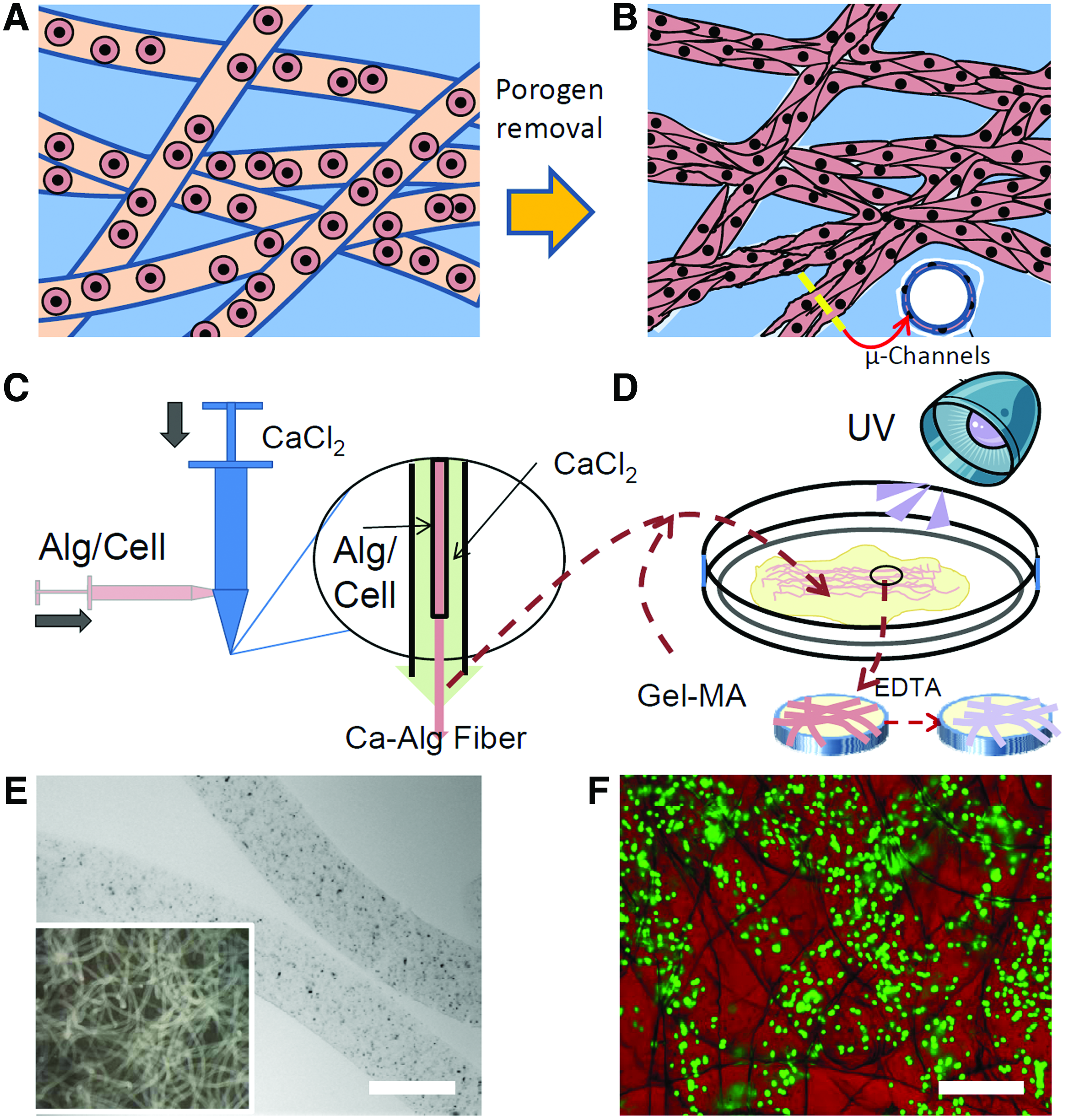
Experimental scheme for dynamic microchannel formation.
Materials and Methods
Materials
Gelatin (types A and B), glycidyl methacrylate, sodium alginate salt, dimethyl phenylphosphonite, 2,4,6-trimethylbenzoyl chloride, lithium bromide, 2-butanone, and perfluorinated oil FC-70 were purchased from Sigma-Aldrich. Disodium citrate (DSC), EDTA, sodium chloride, and calcium chloride dehydrate were purchased from Fisher Scientific.
Precursors for microfibers and hydrogels
The following methods are partially used in our previous study about spherical-macropore formation. 42 Sodium alginate, previously purified by dialysis then lyophilized, was dissolved at 2% (w/v) in Dulbecco's modified Eagle's medium (DMEM). Calcium chloride solution (CaCl2) was prepared by dissolving 1% (w/v) calcium chloride and 0.9% (w/v) sodium chloride in water. Methacrylated gelatin (Gel-MA) was synthesized as previously reported 35 using type-B gelatin (GelB), the alkali-denatured collagen that presents collagen-based binding cell sites in our hydrogel. In brief, GelB (10 g) was dissolved in 100 mL dulbecco's phosphate-buffered saline (DPBS) under 50°C, and methacrylic anhydride (20 mL) was slowly added under constant stirring at 1000 rpm. The reaction continued for 3 h at 50°C. Crude product of Gel-MA was extracted by dripping the solution into acetone (3L), which precipitated Gel-MA and removed excessive methacrylic anhydride and by products. The Gel-MA was purified by dialysis in DI water, lyophilized, and stored at −20°C until use. Photoinitiator lithium phenyl-2,4,6-trimethylbenzoylphosphinate (LAP) was prepared according to existing protocol. 43 In brief, at room temperature and under argon, 2,4,6-trimethylbenzoyl chloride (3.2 g) was added dropwise to continuously stirred dimethylphenylphosphonite (3.0 g), and the mixture was stirred for 18 h, whereupon the reaction mixture was heated to 50°C, and excess of lithium bromide (6.1 g) in 2-butanone (100 mL) was added to the mixture. After 10 min, the mixture was cooled to ambient temperature, allowed to rest for 4 h and filtered to collect precipitate. The filtrate was washed and filtered three times with 2-butanone to remove unreacted lithium bromide, and excess solvent was removed by vacuum. To prepare hydrogel precursor, Gel-MA was dissolved at 10% (w/v) with 0.05% LAP photoinitiator in phosphate-buffered saline (PBS). Type-A gelatin (GelA) was purified through dialysis then lyophilized before being dissolved at 10% (w/v) in 45°C PBS. EDTA solution was prepared at either 8 mM with 12 mM DSC or 16 mM EDTA with 24 mM DSC in DMEM. All solutions were prepared under sterile conditions.
Fabricating stimuli-responsive Ca-Alg microfibers
Ca-Alg microfibers were spun with a coaxial needle set that took advantage of shear stress generated by unequal fluid velocities between coaxial flow layers.
44
To fabricate the coaxial needle set, a 30G “core” needle was located coaxially in a 22G “sheath” needle, and inlets were built to guide separate fluids through the core and sheath needles. The needle set was fixed using acrylic resin (Fig. 1C). More details of the coaxial needle set are shown by Supplementary Figure S1 in the Supplementary Materials (Supplementary Materials are available online at
Encapsulating Ca-Alg microfibers in hydrogel scaffold
To assist microchannel formation, before encapsulation the Ca-Alg microfibers made from 2 mL sodium alginate solution were soaked in GelA solution (5 mL) at 37°C for 3 min, which forms a gelatin coating on the microfibers that prevents the diffusion of Gel-MA precursor into the microfibers. The coated microfibers were transferred to a cell strainer and were rinsed three times by warm (37°C) Gel-MA solution (200 μL each time) to remove excess GelA. To form a hydrogel, the amount of Gel-MA precursor was adjusted to make the final volume of the Gel-MA/microfiber mixture equal to 1.0 mL, and the mixture was transferred to multiple cylindrical moldings (5.6 mm in diameter and 3 mm in thickness). To crosslink the Gel-MA precursor, the moldings with Gel-MA/microfiber mixture were exposed to light (4 min, 365 nm at 2.5 mW/cm2), which turned the precursor into cylindrical hydrogels. The hydrogel with Ca-Alg microfibers was collected from the molding and incubated at 37°C and 5% CO2 in PBS for 24 h before further treatments.
Dynamic microchannel formation
Microchannel formation
To induce microchannel formation, the Gel-MA hydrogels with microfibers were incubated in EDTA solution (16 mM EDTA+24 mM DSC in DMEM) for 2 h. The treated hydrogels were rinsed by PBS and placed back into incubation. The effect of microchannel formation was studied 2 h and 2 days after the EDTA treatment.
Characterizing pore morphology
To study the efficacy of dynamic microchannel formation, variable pressure scanning electron microscopy (VP-SEM, Hitachi S-3400N) was used to monitor the changes in internal structure of hydrogel samples following EDTA treatment (on day 0 and 2). The hydrogels were cut using a razor to expose the cross-sections, and loaded into the VP-SEM chamber where they were gradually cooled from ambient temperature to −20°C, while the chamber pressure was reduced from atmospheric pressure to 50 Pa following a liquid water P/T curve. SEM images were acquired under 15 kV electron beam at ∼7 mm working distance.
Stimuli-responsive microfibers as a cell delivery mechanism
Cell encapsulation and delivery using stimuli-responsive microfibers
Trypsinized HEK cells were suspended in sodium alginate solution (5 million/mL), and the cell-laden alginate solution (2 mL) was spun into Ca-Alg microfibers following the aforementioned procedures (Fig. 1E, F). The microfibers were incubated for 24 h in HEK culture medium containing high glucose DMEM, 10% (v/v) fetal bovine serum, 100 U/mL penicillin, and 0.1 mg/mL streptomycin. After 24 h incubation, cell-laden Ca-Alg microfibers were encapsulated in Gel-MA scaffold following the aforementioned steps. However, to facilitate fluorescence imaging, in which thinner samples are preferred, the Gel-MA/microfiber mixture with cells was molded using a Teflon plate, a Petri-dish and a 500 μm-thick spacer, which shaped the mixture into a hydrogel sheet (500 μm-thick) upon exposure to UV light (Fig. 1D). To enhance the release of the hydrogel sheet, the Teflon plate was precoated with a thin layer of fluorinated oil FC-70. The hydrogel sheet was cut using a polypropylene straw into smaller samples (∼8 mm in diameter) and the samples were incubated in HEK culture medium in a standard 48-well plate for 24 h before EDTA treatments. To dissolve Ca-Alg microfibers and simultaneously release HEK cells into microchannels, the Gel-MA hydrogels were rinsed by serum-free DMEM and were exposed to EDTA at various concentrations (8 mM EDTA+12 mM DSC or 16 mM EDTA+24 mM DSC) for different durations (1 or 2 h). To remove EDTA residue, the treated samples were rinsed five times with culture medium (5 min each time).
Quantifying cell viability, morphology, and colony area
On days 1, 3, and 5, hydrogel samples from different groups (different EDTA treatments) were collected for Live/Dead and Hoechst staining (Invitrogen) following manufacture's protocols, and fluorescence images were taken with a Zeiss microscope. On the same days, the proliferation of cells in each hydrogel sample was quantified using WST-8 assays (Cayman Chemical) following manufacture's protocols (as shown by Supplementary Fig. S2 in the Supplementary Materials). On day 5, hydrogels were fixed in 4% paraformaldehyde for 2 h and stored in PBS at 4°C until processed. Fixed hydrogels were then stained by fluorescein phalloidin (Sigma-Aldrich) and Hoechst staining, and cell distribution in 3D was examined using a confocal microscope (Lesica SP5; Leisica Microsystems). Cell colony areas were quantified by analyzing the Live/Dead images using the open-source program ImageJ.
Statistical analysis
All data were expressed as mean±standard error and statistical significance was determined by analysis of variance using student's t-test with equal variance. p-values (two-tails) of less than 0.05 were considered statistically significant, and p-values less than 0.005 were considered statistically highly significant.
Results
Microchannel formation using stimuli-responsive microfibers
The effectiveness of using stimuli-responsive microfibers for microchannel formation, as illustrated by Figure 1C and D, was examined using VP-SEM. On day 0, the control group (no exposure to EDTA) presented clear structures of Ca-Alg microfibers embedded within the Gel-MA hydrogel network (Fig. 2A, B). In contrast, EDTA-treated samples showed the formation of microchannels at the place where the Ca-Alg microfibers used to be (Fig. 2E, F). There were still some noticeable microfibers residues within the microchannels at day 0, suggesting that the diffusion of alginate out of the microchannels was not yet complete (Fig. 2F). By day 2, the EDTA-treated groups displayed a network of complete microchannel formation with no noticeable microfiber residues (Fig. 2G, H), while the Ca-Alg fibers remained intact in the control group (Fig. 2C, D).
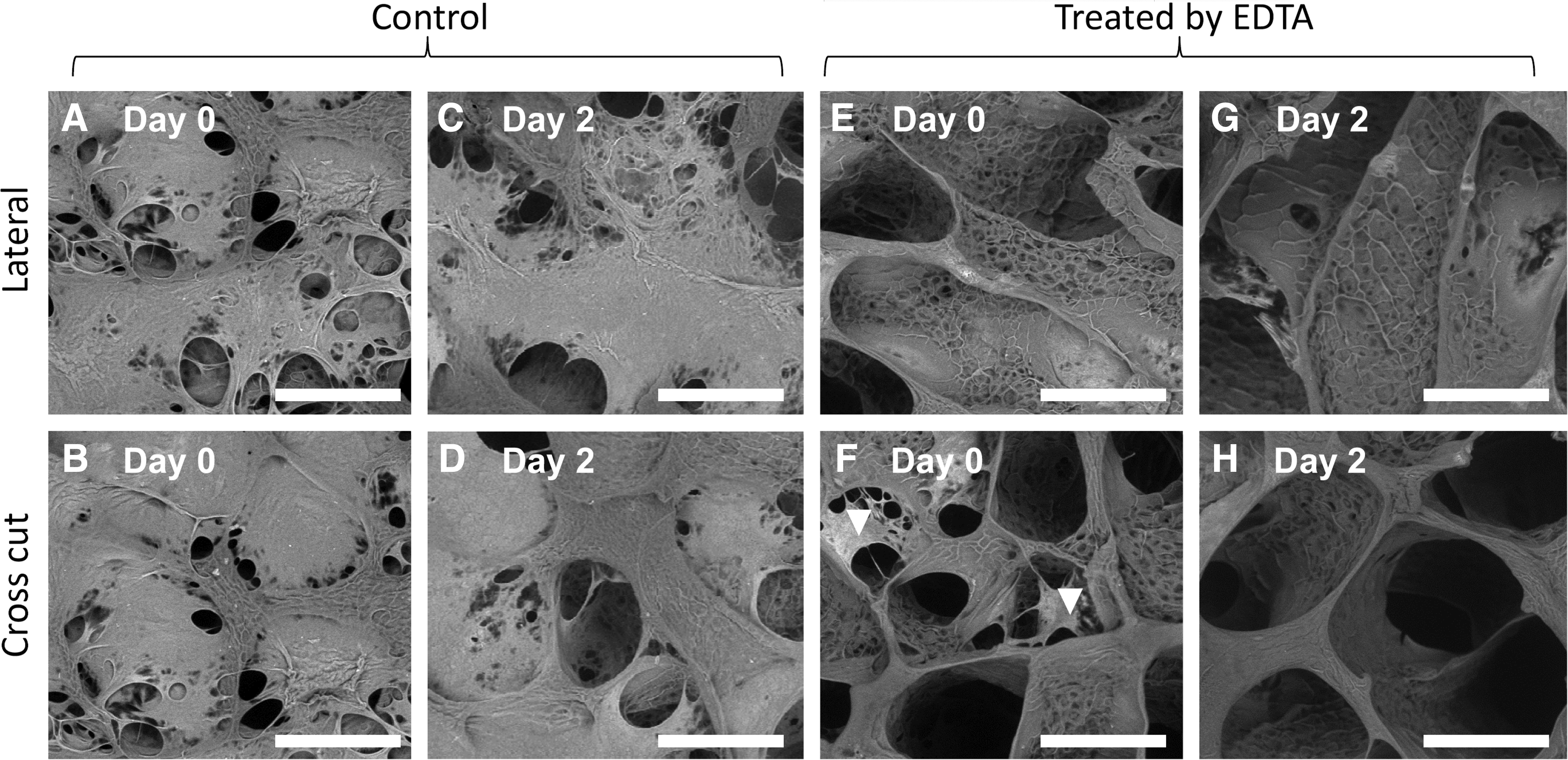
Changes of microfiber-containing hydrogel morphology by scanning electron microscopy on day 0 and 2.
Cell delivery using stimuli-responsive microfibers
To examine the efficacy of using Ca-Alg microfibers for cell delivery within hydrogels, hydrogels containing HEK-laden microfibers were exposed to EDTA at different doses (Fig. 3). Changes in cell morphology and viability in the hydrogels were monitored using Live/Dead staining in conjunction with nuclei staining (Hoechst) on days 1, 3, and 5 (Fig. 3). Cell distribution in 3D hydrogels was examined using confocal microscopy (Fig. 4). On day 1, the HEK cells in the EDTA-treated hydrogels were released from Ca-Alg fibers, spread and formed cell patches inside the lumens of the microchannels (Fig. 3B–E). Cells in the control group (nontreated) remained trapped in the Ca-Alg microfibers (Fig. 3A). By day 3, large colonies of HEK cells started to emerge in all groups treated by EDTA (Fig. 3G–J), whereas minimal cell clustering was observed in the control group (Fig. 3F). By day 5, the groups treated by EDTA showed extensive expansion of HEK colonies (Fig. 3L–O), whereas the HEK cells in the control groups increased in number but maintained small clusters in the Ca-Alg microfibers (Fig. 3K). The groups treated by EDTA presented high cell viability up to day 5 (>95%). In contrast, the control group exhibited increased cell death over time (Fig 3A, F, K). Confocal imaging also shows that the EDTA treatment resulted in cell colony formation along the lumen wall of the microchannels (Fig. 4B), while cells remain entrapped in the microfibers in the control group (Fig. 4D).
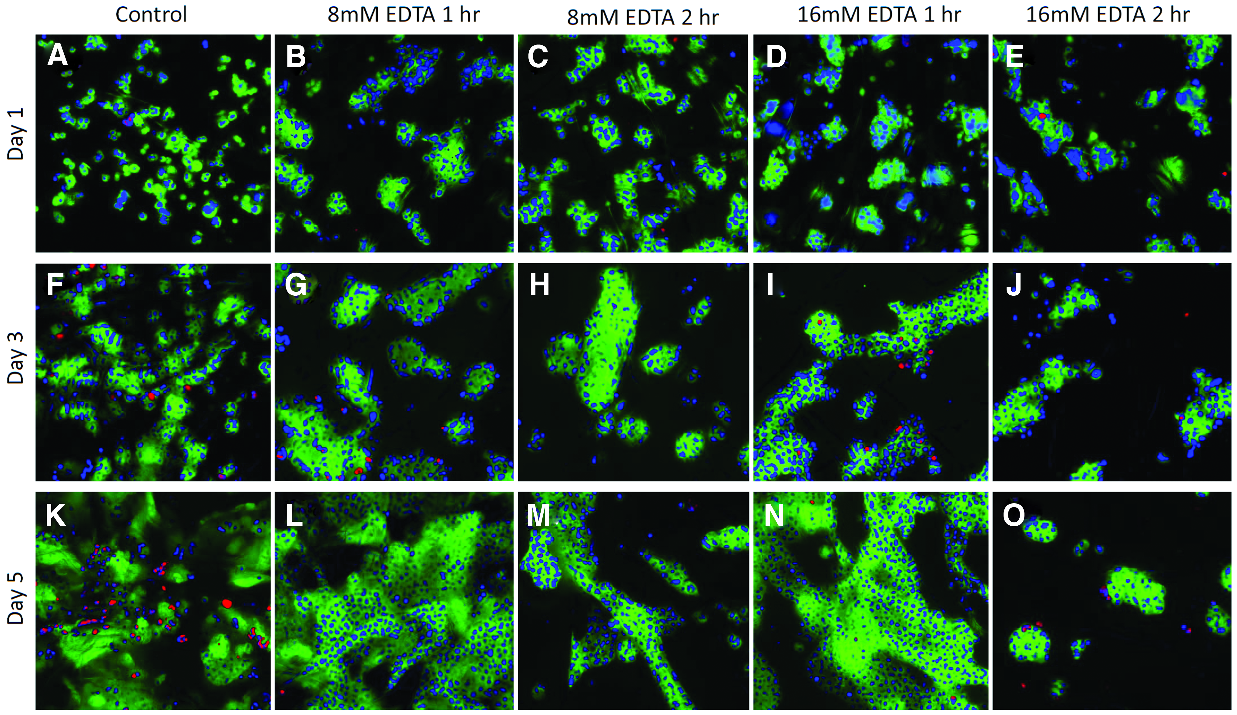
Effects of EDTA exposure at varying doses on cell viability and morphology over 5 days.

Cell morphology within hydrogels on day 5, as shown by confocal microscropy.
Histogram analysis of area covered by cell colony shows that the groups exposed to 8 and 16 mM EDTA for a shorter time (1 h) presented the highest level of colony expansion, while the control group exhibited minimal colony formation (Fig. 5). Among the EDTA-treated groups, the group exposed to the highest dose of EDTA (16 mM, 2 h) presented the lowest level of colony expansion.
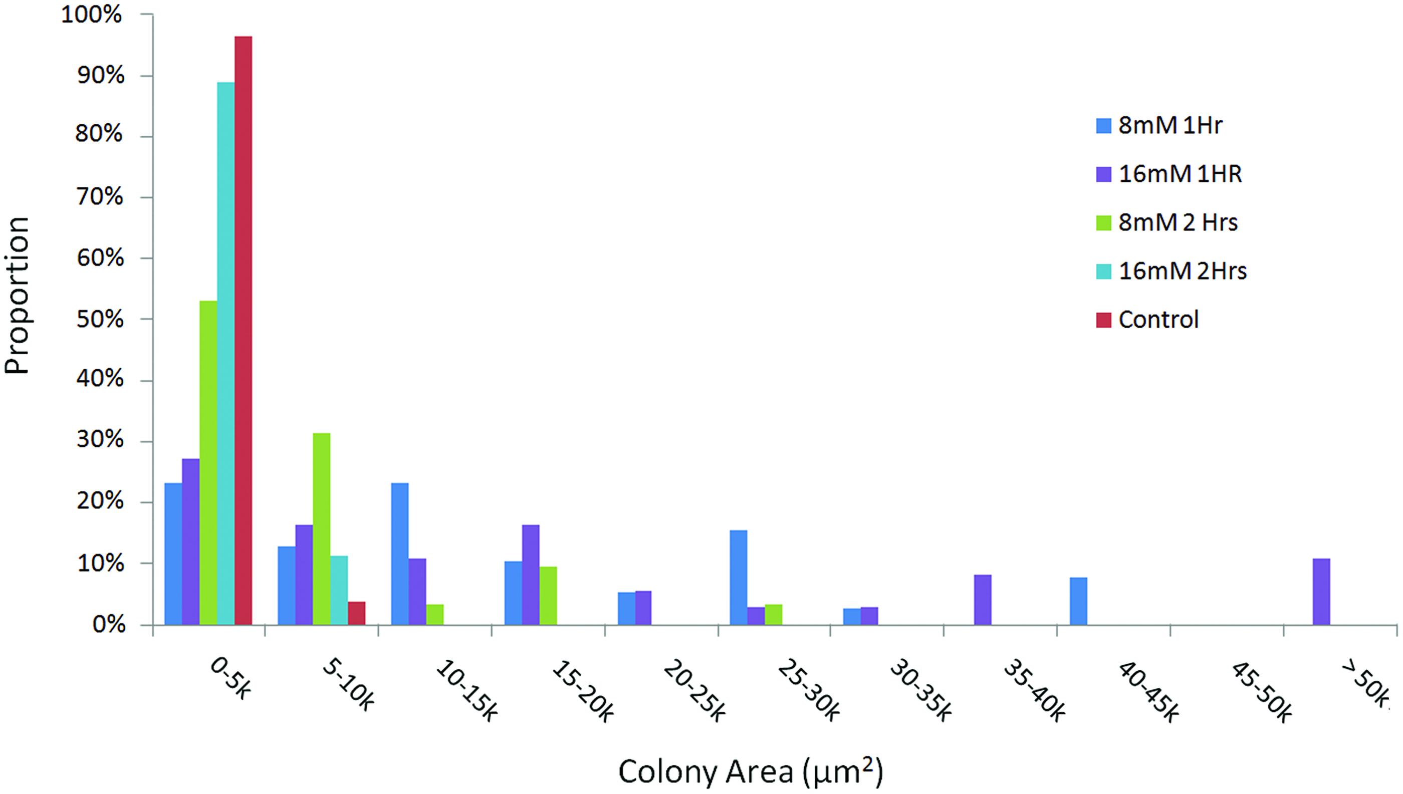
Histogram of the area of cell colony distribution on day 5. Cell number from each group is greater than 300, and EDTA (8 and 16 mM) exposure for shorter period (1 h) resulted in the formation of larger cell colonies. Color images available online at
Discussion
Here we report a facile, cell-friendly process that allows dynamic formation of microchannels within bulk hydrogels using stimuli-responsive microfibers. We further showed such microfibers can be used for delivering cells within bulk hydrogels in a spatiotemporally-controlled manner. Exposure to a chemical stimulus, in this case EDTA, caused the dissolving of the Ca-Alg microfibers, leaving behind microchannel-like porosity within bulk hydrogels (Fig. 2). The microchannels created by microfiber removal are highly interconnected and the diameter of microchannels can be tuned by modulating the diameter of the microfibers. The resulting microchannels (150–200 μm in diameter) may promote tissue formation by facilitating vascularization, cell proliferation, and production of ECM components. Given that matrix topographical cue is one important factor for regulating tissue development, we speculate that the formation of microchannel structures within hydrogel scaffolds may facilitate engineering tissue that contain tubular structures such as microvasculature network or nerve bundles. By optimize other niche cues, such as growth factors and ECM components, the microchannel structures may promote more effective formation of such tubular tissues.
To examine the effects of dynamic microchannel formation on cell proliferation and morphology in 3D, we have chosen HEK 293 cells (HEK cells), 45 a commonly used cell type in cell biology as the model cell type and encapsulated HEKs in Ca-Alg microfibers, followed by embedding the microfibers in Gel-MA hydrogels. Exposures to EDTA induced the formation of mono-layered cell colonies along the lumen wall of microchannels (Figs. 3, 4B, and 5), while cells in the control group (no EDTA) remain entrapped in 3D Ca-Alg microfibers (Figs. 3, 4D, and 5). This is consistent with the results from SEM imaging (Fig. 2), which showed that the EDTA treatment led to well-defined lumen surfaces for cell adhesion, migration, and colony formation. While groups treated by EDTA showed consistent high cell viability, increasing cell death was observed from the untreated control, which may be caused by the limited nutrient diffusion for the cells entrapped in the microfibers. Higher doses of EDTA exposure (16 mM, 2 h) resulted in more cell death, accompanied by less cell colony formation by day 5. The higher dose of EDTA might have decreased cell viability and the level of colony formation by affecting the calcium-dependent cell membrane proteins that regulate cell-cell adhesion and cell proliferation, such as cadherin proteins. Together, our results show that low EDTA exposure (8 mM, 1 h) is sufficient for microchannel formation and subsequent cell delivery, and prolonged exposure to EDTA (>2 h) may cause significant cytotoxicity to HEKs.
Our results demonstrate several advantages of the platform developed herein in comparison with previously reported microchannel-forming methods. First, we demonstrate the potential of using stimuli-responsive microfibers to control cell distribution in 3D and release cells in a temporally controlled manner, which may be particularly useful for patterning cells into microtubular structures in 3D tissue engineering constructs. Several groups have recently demonstrated the use of sacrificial materials or cell-degradable templates to create channels within a bulk scaffold, such as shellac fibers, 46 collagen gel rods 13 and carbohydrate glass framework, 31 but these materials are not capable of controlled cell release and do not allow dynamic microchannel formation. Second, at optimal EDTA doses our process for microchannel formation did not result in noticeable changes in HEK cell viability and proliferation, as shown by live-dead staining and the quantitative WST assay, and allowed more homogeneous cell distribution in microchannels, which is important to mediate cell viability and tissue-forming efficiency in 3D.
Using Ca-Alg as a model stimuli-responsive material, here we demonstrated the concept of dynamic microchannel-formation as well as the potential of controlling cell delivery in a temporal manner to facilitate tissue formation. This platform may be further extended by exploiting other materials as porogens that can be degraded using cell-friendly stimuli, such as hydrolysis, MMP-sensitive peptides 47 or photo-degradation. 48 While we used HEK cells as a model cell type to demonstrate the proof-of-principal in this study, the reported method of dynamic microchannel formation may also be applied for other cell types, such as smooth muscle cells and endothelial cells to engineer tubular tissues structures that are of clinical significance.
Conclusion
In summary, here we report a facile method to create microchannel-like structures within hydrogel scaffolds using alginate microfibers that can dissolve upon exposure to EDTA. Using optimized EDTA concentration and duration, this method also allows cell delivery in bulk hydrogels within the formed microchannel structures. Upon exposure to EDTA, a well-defined, interconnected microchannel network was created and cells were free to migrate and form colonies along the channels' internal surfaces, leading to laminar distribution of cells in 3D. The platform reported herein offers spatiotemporal control over cell seeding and distribution. Future iterations of this technique may lead to low-cost, sizable fabrication of engineered tissue grafts with enhanced cell viability and tissue formation efficiency, such as prevascularized tissue engineering building blocks.
Footnotes
Acknowledgments
The authors would like to thank the McCormick Faculty Award and Stanford Bio-X Interdisciplinary Initiative grant for funding. J.H. would like to acknowledge The Amgen Foundation for funding.
Disclosure Statement
This work has been disclosed to the Office of Technology Licensing at Stanford University.
References
Supplementary Material
Please find the following supplemental material available below.
For Open Access articles published under a Creative Commons License, all supplemental material carries the same license as the article it is associated with.
For non-Open Access articles published, all supplemental material carries a non-exclusive license, and permission requests for re-use of supplemental material or any part of supplemental material shall be sent directly to the copyright owner as specified in the copyright notice associated with the article.