
Abstract
Normal laryngeal function has a large impact on quality of life, and dysfunction can be life threatening. In general, airway obstructions arise from a reduction in neuromuscular function or a decrease in mechanical stiffness of the structures of the upper airway. These reductions decrease the ability of the airway to resist inspiratory or expiratory pressures, causing laryngeal collapse. We propose to restore airway patency through methods that replace damaged tissue and improve the stiffness of airway structures. A number of recent studies have utilized image-guided approaches to create cell-seeded constructs that reproduce the shape and size of the tissue of interest with high geometric fidelity. The objective of the present study was to establish a tissue engineering approach to the creation of viable constructs that approximate the shape and size of equine airway structures, in particular the epiglottis. Computed tomography images were used to create three-dimensional computer models of the cartilaginous structures of the larynx. Anatomically shaped injection molds were created from the three-dimensional models and were seeded with bovine auricular chondrocytes that were suspended within alginate before static culture. Constructs were then cultured for approximately 4 weeks post-seeding and evaluated for biochemical content, biomechanical properties, and histologic architecture. Results showed that the three-dimensional molded constructs had the approximate size and shape of the equine epiglottis and that it is possible to seed such constructs while maintaining 75%+ cell viability. Extracellular matrix content was observed to increase with time in culture and was accompanied by an increase in the mechanical stiffness of the construct. If successful, such an approach may represent a significant improvement on the currently available treatments for damaged airway cartilage and may provide clinical options for replacement of damaged tissue during treatment of obstructive airway disease.
Introduction
N
Tissue engineering and regenerative medicine aim at restoring function by leveraging native tissue growth and creating a dynamic living or inductive implant. In these methodologies, this restoration is achieved by mixing or seeding the cellular component of native tissue with a structural, biocompatible scaffold that enables initial retention of support and shape. The approach has been demonstrated for a number of different anatomic structures, particularly those that derive their stiffness from cartilage, including ear, 3 nose, 4 other structures of the face,5,6 as well as the trachea.7,8 Recent studies have utilized image-guided approaches to create cell-seeded constructs that reproduce the shape and size of the tissue of interest with high geometric fidelity.9–12 However, a few studies examining the use of image-guided tissue engineering for laryngeal reconstruction have been performed. Here, we describe a methodology for fabrication of upper airway cartilage using image-guided tissue engineering methods that create viable, patient-specific constructs with high geometric fidelity.
We selected an equine model, as the biochemical and mechanical properties of the equine laryngeal cartilages are well characterized. 13 In addition, the equine laryngeal cartilages are almost twice the size of the human larynx, 13 and this represents an ideal opportunity to develop tissue engineering techniques before scaling them to apply to human laryngeal reconstruction. The equine larynx is also subject to naturally occurring inflammation, infection, and degeneration of the laryngeal cartilage structure, which can result in airway obstruction.14–16 In these circumstances, removal of affected cartilage commonly results in the loss of protective swallowing mechanisms with subsequent coughing and aspiration and may also lead to laryngeal collapse due to loss of tissue support.17–20 Currently, there are a few effective treatment alternatives for remediation of abnormalities of the equine larynx. 21 These options include placement of sutures to advance the larynx or permanent placement of the tissues in an “open” position. While these treatments are effective at restoring airflow, they do not restore airway performance and can lead to aspiration of feed or water. Further, these procedures can lead to reduction or loss of airway patency. A few prosthetic or tissue engineering solutions have been suggested in the horse. An injection of Teflon into the submucosal space of the epiglottis has been suggested for restoration of structural integrity of the epiglottis.22,23 However, results of this treatment were poor, as an injection of Teflon is associated with an adverse immune response and is incapable of restoring shape or size of damaged tissues. Therefore, methods that restore the shape and strength of laryngeal cartilage would enable new therapies for reconstruction of the equine upper airway as well as provide proof-of-concept data for human applications.
The objective of the present study was to establish an image-guided tissue engineering approach to the creation of viable constructs that approximate the shape and size of clinically relevant equine airway structures, in particular the epiglottis.
Materials and Methods
Imaging and mold design
Computed tomography (CT) imaging was performed on a healthy, intact larynx harvested from a skeletally mature racehorse using a multi-slice CT scanner (Aquilion LB16; Toshiba) that was capable of sharp algorithm with a helical scanning, slice thickness, and table index of 0.5 mm. CT scans were exported in the digital imaging and communications in medicine (DICOM) format. The DICOM images of cartilage structures within the equine upper airway (arytenoid, epiglottis, cricoid, and trachea) were viewed and manually parsed using MIMICS (Materialise NV) software and three-dimensional models were created and exported as point cloud files. The point cloud files were imported to GeoMagic Studio (Geomagic, Inc.) for formatting and conversion to solid three-dimensional models. The solid model was then used for mold design in SolidWorks (Dassault Systems SolidWorks Corp.).
Briefly, the mold was designed using the three-dimensional solid model as a negative space (cavity) within a solid block. The solid block was then segmented into pieces to facilitate assembly and disassembly of the injection mold. Injection ports, as well as air exchange ports were incorporated into the design of the mold for efficient injection. Hard plastic components of the injection molds were then produced from the models using a fused deposition rapid prototyping platform (Stratasys). Figure 1 shows an overview of the mold creation process.
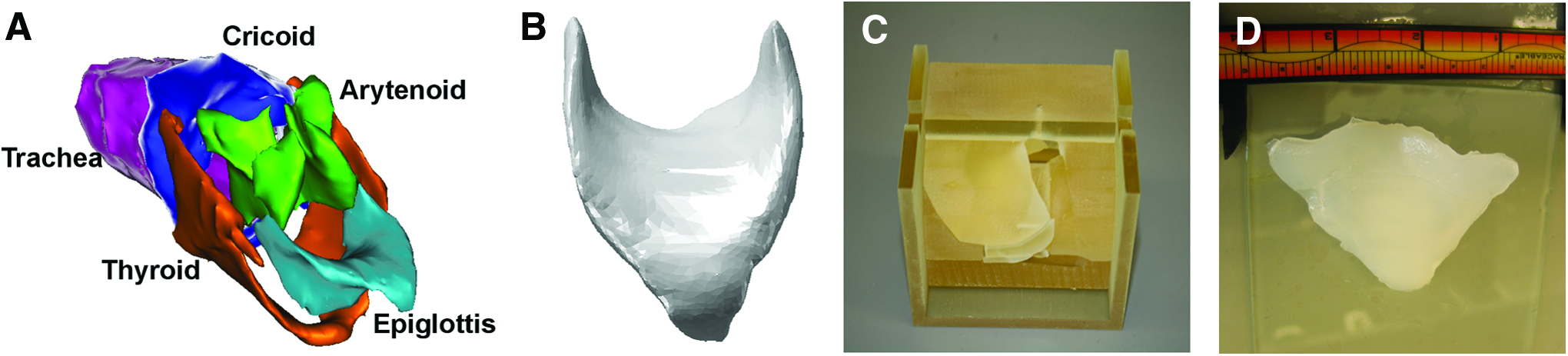
Mold design and casting. Computed tomography reconstruction of equine airway tissues
Injection molding and cell culture
Cellular harvest and construct fabrication were performed as previously described.3,10 Bovine auricular cartilage was harvested from freshly slaughtered 1–3 day-old calves (Gold Medal Packing). The ears were rinsed with soap and water before a betadine scub. The skin and the majority of the perichondrium were then removed by sharp dissection. The tissue was minced into 1 mm3 pieces, rinsed in phosphate-buffered saline (PBS; pH 7.4) containing 100 μg/mL penicillin and 100 μg/mL streptomycin, and digested overnight in a 0.3% solution of collagenase type II in Dulbecco's-modified Eagle's medium (DMEM) supplemented with 100 μg/mL penicillin and 100 μg/mL streptomycin. The cells were then harvested by centrifugation and washed in PBS containing 100 U/mL penicillin and 100 μg/mL streptomycin and counted before re-suspension in 2% w/v low viscosity high g-content alginate (FMC Biopolymer) at a concentration of 25×106 cells/mL. A total construct volume of 18 mL was used for all constructs. The alginate-cell suspension was then mixed with 0.02 g/mL CaSO4 at a ratio of 2:1 to initiate cross-linking of the alginate. The mixture was injected into the assembled mold, and the mold was allowed to sit at room temperature for 20 min before post-crosslinking in 60 mM CaCl2 for 60 min. The mold was then disassembled, and the construct was placed into static culture in DMEM supplemented with 10% fetal bovine serum, 100 U/mL penicillin, and 100 μg/mL streptomycin. Media was changed every 3 days, and constructs were harvested for evaluation after 1, 2, 3, and 4 weeks in static culture. Five constructs were harvested per time point. At each harvest point, multiple 6 mm biopsy punches were taken and frozen at −20°C until analysis. Biopsy punches were also taken from identical locations within native equine epiglottis harvested from five animals and used for testing as described next.
Viability testing
Preinjection viability of cells was determined by trypan blue exclusion. Postinjection viability was evaluated at time points of 1 and 3 days, and at 1, 2, 3, and 4 weeks using a live/dead assay kit (Life Technologies). Briefly, fresh (not frozen) 6 mm biopsy punches were taken through the thickness of the sample, and the middle portion of this biopsy was used for viability staining. Samples were submerged in PBS containing 0.15 μM calcein AM and 2 μM ethidium homodimer-1 dyes. Samples were allowed to incubate for 30 min, were sectioned using a scalpel and a portion of the sample was placed onto a glass slide for fluorescence imaging using a Nikon TE2000-S (Nikon) microscope. Three 20× objective images were captured per sample, and live (green staining) and dead (red staining) cells were counted using ImageJ image analysis software (National Institutes of Health [
Biochemical analysis
Biochemical analysis was performed on 6 mm biopsy punch samples taken at the time of construct harvest. Each sample was roughly 2 mm in thickness. Samples were weighed, then lyophilized, and, finally, re-weighed. The samples were then digested in a 1.25-mg/mL papain solution before assessment of DNA, collagen, glycosaminoglycan (GAG), and elastin content. Briefly, DNA content was assessed using a Hoescht dye assay, collagen content was assessed using a hydroxyproline assay, GAG content was assessed using a 1,9-dimethylmethylene blue assay (at pH 1.5), and elastin content was assessed using a Fastin Elastin assay (Bicolor Ltd.). All tests were performed as previously described24,25 or as per kit manufacturer instructions (Fastin assay). All biochemical assays were completed on at least two biopsy punches from each sample, and each sample was assayed in triplicate. Results are reported as content per construct wet weight.
Histologic analysis
Samples were rinsed once in PBS and then fixed in 10% neutral-buffered formalin supplemented with 1 mM CaCl2 to prevent alginate gel solubilization. Samples were embedded in paraffin, cut into 6 μm sections, and mounted on glass slides. The sections were deparaffinized by immersion in xylene, followed by a graded series of ethanol. The slides were stained using Safranin O, picosirius red, or Verhoeff's elastic stain to examine overall tissue structure, collagen architecture, distribution of GAG content, and elastin organization, respectively. Slides were dehydrated in ethanol and xylenes before cover slipping and then investigated under light microscopy using a Nikon TE2000-S.
Mechanical testing
Samples of tissue for mechanical testing were thawed in a protease inhibitor cocktail (Sigma-Aldrich) in PBS to prevent sample degradation during testing. Briefly, 6 mm biopsy punches were trimmed to a height of ∼2 mm and tested in confined compression on an ELF 3200 EnduraTec testing apparatus (Bose). Testing consisted of applying a series of 10×50 μm steps using a porous indenter, with a relaxation period between each step. The results were then fit to a poroelastic model using MATLAB as previously described5,13,26 to determine the sample aggregate modulus.
Statistical analysis
All experiments were performed on five samples per condition. One-way analysis of variance with Tukey's post hoc test and p<0.05 were used to determine statistically significant differences in variables with time. All results are expressed as mean±standard deviation.
Results
Mold creation
Gross inspection showed that the CT-based, injection molding process yielded constructs with a shape and size which approximated that of the native equine epiglottis (Fig. 2). Although quantitative measurements were not performed, constructs appeared to maintain their shape and size through 4 weeks in culture and an increase in opacity of the construct was noted with an increase in time in culture.
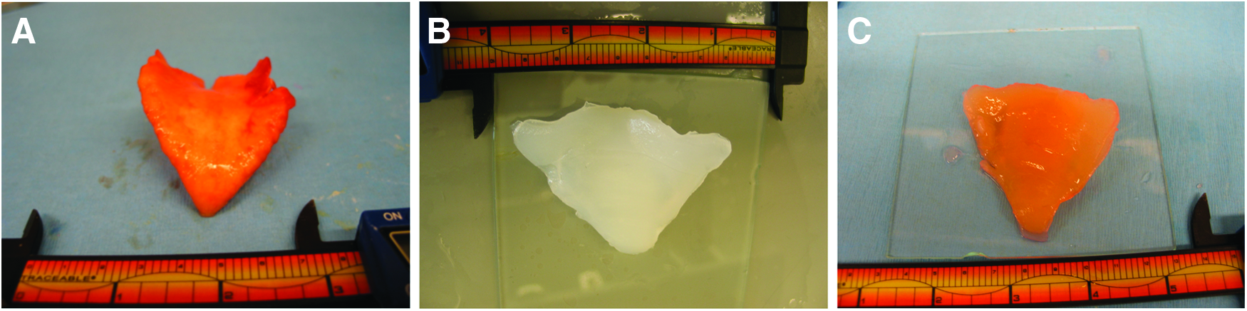
Gross morphologic appearance. Native equine epiglottis
Viability testing
Live/dead staining showed a few dead cells within the constructs at any time point during the course of the study (Fig. 3). A slight decrease in cellular viability was seen on day 1 after injection molding as compared with preinjection viability. Cellular viability, measured quantitatively, was then seen to increase at each time point thereafter. DNA content of the constructs was measured and demonstrated a drop in DNA content between weeks 1 and 2 with increasing DNA content thereafter (Fig. 3).

Live/dead staining of cultured constructs at 1 days
Histologic analysis
Similar cellular distributions were observed throughout all histology samples examined (Fig. 4). Cells had a chondrocyte-like appearance and were found within individual lacunae, which exhibited increasing maturity and matrix content in the surrounding tissue with increased time in culture. Safranin O staining indicated an increase GAG density with time in culture. No qualitative differences in staining density were observed between the bulk and periphery of the stained samples. Picosirius red staining indicated an increase in both the number and size of collagen fiber bundles within the sample from 1 to 4 week time points. However, only a small number of collagen bundles were observed. The majority of the collagen fiber bundles formed were found at the periphery of the sample at all time points. Similarly, elastin fiber formation, as demonstrated by Verhoeff's elastic stain, was found predominantly at the periphery of the samples investigated. A few, if any, elastin fibers were observed at the 1-week time point. Increased number and size of elastin fibers were noted at later time points.

Histologic architecture. Safranin O staining of constructs cultured for 1–4 weeks
Biochemical analysis
The results of the biochemical analysis correlated well with the observations of the histologic analysis. Water content of the samples was found to be similar across all time points, and greater than that of native tissues. Biochemical analysis (Fig. 5) demonstrated a general trend of increasing matrix content within the samples with time in culture. A statistically significant increase in GAG content was observed from the 1- to 4-week time points (11.5-fold). A similar trend was observed for collagen as indicated by a statistically significant increase in hydroxyproline content from the 1- to 4-week time points (25.4-fold). Elastin content was shown to increase rapidly from 1 to 2 week time points (2.0-fold), but elastin content was shown to remain relatively steady thereafter. All samples were found to be significantly lower in biochemical content than native tissues for all assays.
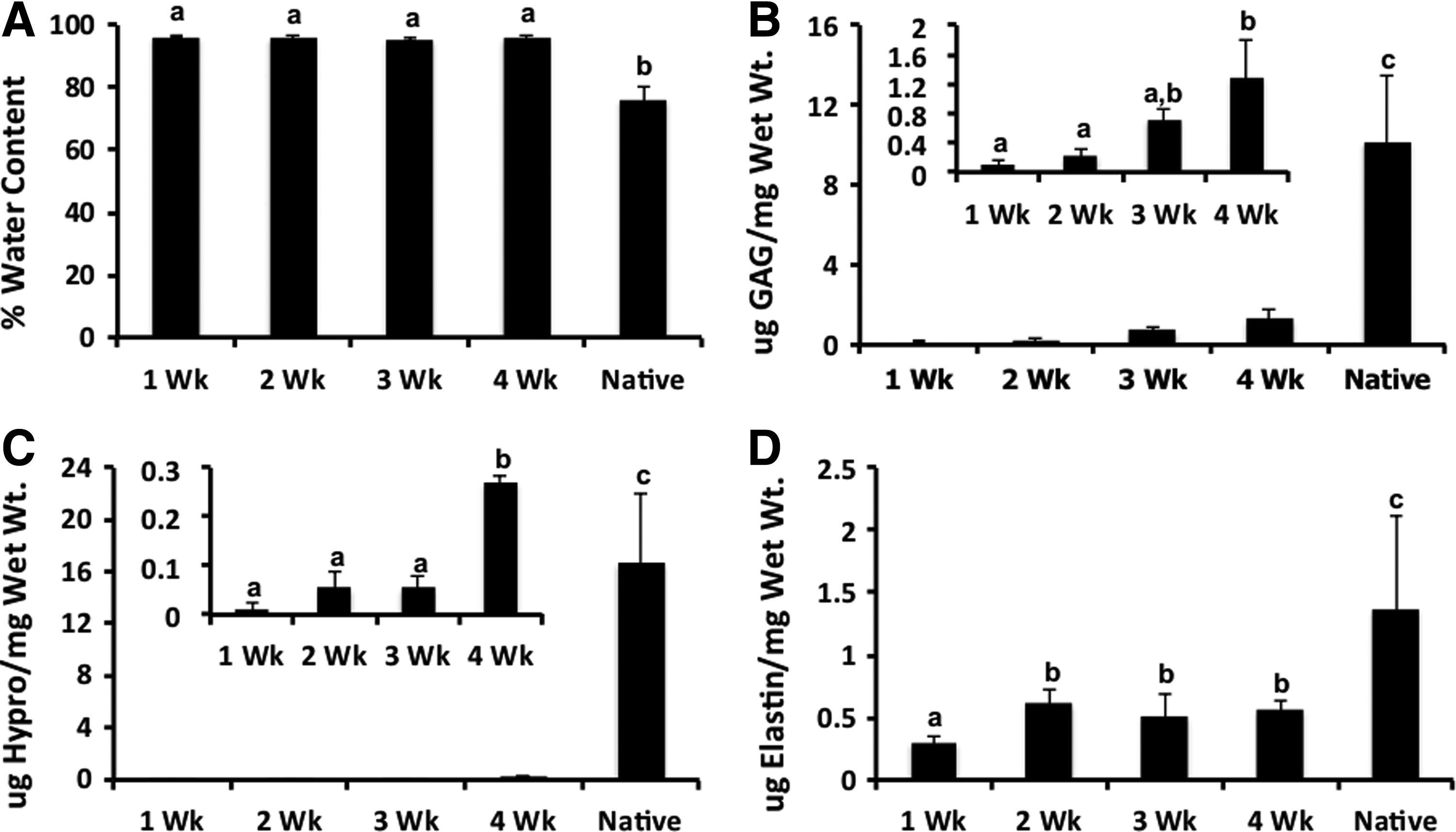
Percent water content
Mechanical testing
Increases in matrix content with time were accompanied by similar increases in the aggregate modulus of the sample with time (Fig. 6). Sample modulus increased progressively from the 1- to 4-week time points (9.1-fold). Increases in mechanical stiffness were comparable with observed increases in GAG content (9.1-fold vs. 9.7-fold, respectively).
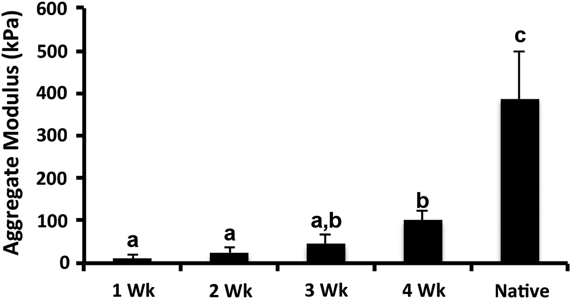
Aggregate modulus of constructs after 1, 2, 3, and 4 weeks of static culture. Aggregate modulus was observed to increase over the 4-week culture period. Native tissue is shown for reference. All values are presented as mean±standard deviation. Groups not connected by the same letter are statistically significant, p<0.05.
Discussion
The equine airway is adapted to resist high levels of airflow during exercise. During exercise, peak flow may reach 60–80 L/s resulting in turbulent flow with Reynolds numbers in excess of 100,000.27–30 These levels of flow can result in negative pressures ranging on the order of −18 to −37 mmHg. This is in contrast to the human airway, in which flow rates during exercise are ∼2 L/s with Reynolds numbers on the average of 4000.31–35 These unique environmental conditions underscore the potential effects of deformity or weakness of the cartilaginous structures on the function of the equine upper airway. At present, there are a few effective solutions for surgical reconstruction that correct such structural or mechanical defects.
An effective surgical implant for reconstruction of cartilaginous structures of the equine upper airway would need to be geometrically accurate to maintain appropriate flow through the airway, possess near-native mechanical properties, and be easily integrated with surrounding native tissues after surgical placement. The present study examined the use of a recently described CT guided approach 10 for the creation of geometrically complex three-dimensional tissue engineered constructs resembling cartilaginous components of the equine upper airway. The results of the study demonstrated that it was possible, by utilizing this image guided approach, to fabricate such implants. Specifically, these implants contained viable chondrocytes that matured in both extracellular matrix (ECM) content and mechanical properties during the course of 4 weeks in static culture without loss of geometric fidelity.
Both the spatial distribution and content of collagen, GAG, and elastin within the construct were evaluated, as they are the primary biochemical constituents of elastic cartilage of the epiglottis. The histologic and biochemical results of the present study clearly demonstrate maturation of the construct during the period of static culture. The main biochemical constituent of the constructs was observed to be GAG, although both GAG and collagen content were observed to increase throughout the course of the study. However, despite these increases, the levels of biochemical constituents were significantly lower than those observed in native tissues. The constructs were observed to possess ∼25% of the native compressive properties (101.07 kPa vs. 386.62 kPa) by the end of 4 weeks of static culture. These results were consistent with a number of other studies that have utilized chondrocytes encapsulated within alginate for the formation of three-dimensional molded cartilage constructs.36–38
In the present study, the formation of collagen and elastin were observed to be localized to the periphery of the construct, suggesting that there may be limitations in mass transport within these large-scale constructs. However, GAG formation was observed throughout the construct thickness, which suggests that GAG formation may occur more rapidly than does collagen and elastin formation. Elastin levels were found to be ∼40% of native values at the 4-week time point. This suggests that the cells are producing significant amounts of elastin, but that the soluble form is slow to assemble into fibers. Another recent study using image guided construct fabrication demonstrated that a collagen scaffold seeded with bovine auricular chondrocytes showed only limited elastin formation at 1 month post–subcutaneous implantation; however, large amounts of elastin formation were observed at 3 months post-implantation. 3 This suggests that elastin formation may occur at significantly slower rates or at later time points than collagen and GAG formation. Of note, these constructs were of a similarly large volume and thickness and GAG, collagen, and elastin formation were observed throughout the thickness of the implant, suggesting that limitations of mass transport in static culture may be overcome to some extent in an in vivo setting.
The present study represented a proof-of-concept study demonstrating the effectiveness of an image-guided tissue engineering approach to the creation of large (18 mL), anatomically accurate, viable, cell-seeded constructs. As such, culture experiments were not continued past 4 weeks, Other studies have demonstrated long-term culture of alginate-based cartilage constructs and have shown that both ECM accumulation and mechanical properties increase in cultures of approximately 8 weeks. 10 The present study examined only static culture in basal chondrocyte media; however, it is logical to assume that additional increases in matrix production and mechanical properties would have been observed with longer periods in culture. Demonstration of further matrix deposition and improvement of mechanical properties to near-native values will be required before testing in an in vivo model.
The exact amount of time required to obtain an implantable construct is likely dependent on the materials, cells, and simulating factors that are used in its construction. Ideally, the construct will match the native mechanics and ECM constituent molecules; however, an exact match is likely unnecessary as a significant amount of remodeling will occur after implantation. There is likely a minimum value for the required mechanical properties required to maintain function during remodeling; however, this has not been identified in the current study. A number of methods for increasing the deposition of ECM components within alginate constructs have been suggested.36–38 For example, dynamic mechanical stimulation of alginate constructs in the first 2 weeks of culture followed by static culture was shown to increase accumulation of both GAG and collagen with a concurrent increase in mechanical properties.36,37 However, due to the complex geometry and large size of the samples in the present study, dynamic stimulation of the image-guided constructs would likely require development of a highly complex bioreactor system. Controlled media mixing is a simple method of mechanical conditioning that has been shown in a number of studies to increase ECM production of chondrocyte-loaded constructs. 38 It is likely that such a method, when combined with growth factor stimulation or media supplementation with factors such as ascorbic acid, would significantly enhance the speed of construct maturation.
The present study evaluated alginate as an injectable hydrogel for construct fabrication. A wide array of potential injectable hydrogels exist and can be investigated for the creation of image-guided constructs in future studies.9,39,40 Alginate was chosen for the present study for its well-known ability to support chondrocyte phenotype and previous use as a hydrogel for the creation of image-guided constructs. However, the mechanical properties of alginate are not ideal at early time points after construct creation. The use of modified alginates or additional cross-linking methodologies may improve the pre-culture mechanics of the sample.41,42 Alternatively, the use of other, more mechanically robust, hydrogels may achieve improved preculture mechanics and potentially reduce culture times. For example, a recent study examined the use of high-density collagen hydrogels for the fabrication of injectable constructs for tissue engineering of whole knee menisci. 43 Menisci cultured using collagen hydrogels were found to have similar compressive moduli as those fabricated using alginate, but significantly improved tensile moduli over a 4-week culture period. Another recent study utilized high-density collagen hydrogels to create a molded scaffold, which was then implanted subcutaneously within a week of creation. 3 Results demonstrated rapid remodeling with cartilage formation and maturation to near-native values over the course of the 3-month study. This study also raises the possibility of heterotopic implantation before anatomical implantation as a strategy for construct maturation.
It should be noted that the use of alternate hydrogels may require mold redesign to account for factors related to hydrogel fabrication and the use of various cross-linking strategies and agents. In addition, mold design should allow for either swelling or shrinkage of the constructs over time, depending on the hydrogel used for fabrication. Alginate hydrogels are stable over long periods in culture, are degraded by enzymatic hydrolysis, and exhibit little swelling or shrinkage. While not quantitatively measured in the present study, the alginate constructs were observed to largely maintain their original shape and size. No signs of alginate degradation were observed from a gross morphologic or histologic perspective. Previous studies examining degradation of alginate during the culture of injection-molded constructs demonstrate minimal degradation or mass loss over 3 weeks in culture 12 .
Many engineered constructs placed into the upper airway are observed to fail despite viability and mechanical stability. There are a number of causes for these failures, including a lack of mechanical stability and/or lack of collagen and elastin. In the present study, it was demonstrated that it is possible to create a viable and anatomically accurate cell-gel construct. In future studies, and before implantation, a number of parameters should be met. First and foremost is mechanical integrity for implantation. While a significant amount of remodeling will occur post-implantation, initial minimums should be achieved to prevent airway collapse after implantation. In particular, tensile properties of the constructs should be measured in future studies, as the epiglottis undergoes bending motion. A system for testing the mechanical stability of implants for laryngoplasty under airflow in a cadaver model has been developed. 44 This system could be utilized in future studies to test the suitability of implants before implantation. It is also well known that implants within the airway should be covered by airway epithelium or they will be subject to infection and inflammatory response. Future studies should be performed to address whether this implant could be seeded with epithelial cells and/or the rate of epithelialization post-implantation within an airway environment.
Conclusions
The results of the present study proved that it is possible to create viable, cell-seeded constructs of cartilaginous structures of the equine airway. Shape, size, cellular viability, and increasing structural properties compared well with native tissue. Additional investigation is required to determine optimal hydrogel composition and culture conditions before implantation as well as subsequent host integration. If successful, such an approach would represent a significant improvement on the currently available treatments for reconstruction of damaged airway cartilage in obstructive airway diseases.
Footnotes
Acknowledgments
Funding for this study was provided by the Harry M. Zweig Memorial Fund for Equine Research. In addition, the authors would like to thank Jeffery Lipton and Hod Lipson for their assistance in the creation of the injection molds used in this study.
Disclosure Statement
No competing financial interests exist.