




Abstract
The use of multicomponent scaffolds for cell implantation has necessitated sophisticated techniques for tracking of cell survival in vivo. Bioluminescent imaging (BLI) has emerged as a noninvasive tool for evaluating the therapeutic potential of cell-based tissue engineering strategies. However, the ability to use BLI measurements to longitudinally assess large 3D cellular constructs in vivo and the effects of potential confounding factors are poorly understood. In this study, luciferase-expressing human mesenchymal stem cells (hMSCs) were delivered subcutaneously within agarose and RGD-functionalized alginate hydrogel vehicles to investigate the impact of construct composition and tissue formation on BLI signal. Results showed that alginate constructs exhibited twofold greater BLI counts than agarose constructs at comparable hMSC doses. However, each hydrogel type produced a linear correlation between BLI counts and live cell number, indicating that within a given material, relative differences in cell number could be accurately assessed at early time points. The survival efficiency of delivered hMSCs was highest for the lower cell doses embedded within alginate matrix. BLI signal remained predictive of live cell number through 1 week in vivo, although the strength of correlation decreased over time. Irrespective of hydrogel type or initial hMSC seeding dose, all constructs demonstrated a degree of vascularization and development of a fibrotic capsule after 1 week. Formation of tissue within and adjacent to the constructs was accompanied by an attenuation of BLI signal during the initial period of the image acquisition time-frame. In alginate constructs only, greater vessel volume led to a delayed rise in BLI signal following luciferin delivery. This study identified vascular and fibrotic tissue ingrowth as potential confounding variables for longitudinal BLI studies. Further investigation into the complexities of noninvasive BLI data acquisition from multicomponent constructs, following implantation and subsequent tissue formation, is warranted.
Introduction
T
Bioluminescent imaging (BLI) has emerged as a valuable tool for tracking cell populations in vivo.6,7 By measuring photon release from the luciferase (Luc)-catalyzed oxidation of luciferin substrate, BLI can noninvasively and longitudinally monitor the presence of viable cells labeled by means of constitutive Luc reporter protein expression. Alternatively, by placing Luc expression under the control of a nonconstitutive promoter, BLI can follow delivered cell function, such as the differentiation of an implanted stem cell.8,9
In addition to Luc quantity, several variables influence BLI measurement and can potentially impact its utility as an indicator of viable cell number. These variables include sample positioning, machine sensitivity, luciferin delivery technique, and measurement acquisition time. 10 The impact of confounding factors in 2D in vitro systems is relatively limited. BLI signal shows a linear relationship with viable cell number with little influence from variations in cell seeding density or time in culture.1,11 In 3D cell culture systems, BLI signal is affected by a range of additional variables such as luciferin transport kinetics and light scattering properties of the construct.10,12 BLI measured from cells seeded within constructs of significantly different material properties has been found to differ in magnitude, but to be linearly correlated to Luc content when evaluated in vitro. 13
Given the complexity of the 3D system in vitro, this evaluation becomes even more complicated when the 3D construct is implanted. The validity of BLI technique has not been rigorously evaluated for these 3D systems in vivo. While many confounding factors can be mitigated through implementation of consistent experimental practice, the development of tissue ingrowth within and around scaffolds over time is unavoidable and fundamental to tissue engineering procedures. Several studies that have demonstrated the feasibility of using BLI to track delivered cells for several weeks following implantation have also observed the development of various tissue types (including vascular, fibrotic, and mineralized) within constructs over the associated time-course.1,13–17 Thus, the impact of this tissue invasion on the accuracy of BLI measurement remains unclear.
This study constitutes an initial examination of longitudinal BLI technique applicability for use with a cell-seeded construct of large volume (>200 mm3), approaching the scale necessary to evaluate clinical applicability. 5 Luc-labeled human mesenchymal stem cells (hMSCs) were delivered subcutaneously within two hydrogel systems to investigate (1) the effects of cell dose and construct material on cell survival and (2) the development of contiguous tissues, including vascular and fibrotic. Our ultimate aim was to assess the potential influence of construct material and tissue ingrowth on in vivo BLI measurement.
Materials and Methods
Cell culture
Bone-marrow-derived hMSCs harvested from male donors 20–25 years old, with established multipotency, were purchased from the Texas A&M University Health Science Center, College of Medicine. Two individual donor cell lines were expanded at a starting density of 50 cells/cm2 on Petri dishes in minimum essential medium alpha (αMEM) containing 16.7% fetal bovine serum (Atlanta Biologicals, Lawrenceville, GA) and 100 U/mL penicillin/100 μg/mL streptomycin/2 mM
Cell labeling
Cells were cotransduced using a lentiviral vector containing green fluorescent protein (GFP) and firefly Luc downstream of the ubiquitin promoter as previously described.8,11 Briefly, hMSCs were suspended in polybrene (Sigma, St. Louis, MO) and viral vector at an MOI of 20 and incubated in flasks at a density of 10,000 cells/cm2 overnight. Culture medium was replaced daily for 3–5 days, during which time the labeling efficiency of GFP/Luc hMSCs was determined using fluorescence microscopy (Axio Observer; Carl Zeiss, Thornwood, NY) and flow cytometry (Accuri C6; BD Biosciences, San Jose, CA). Labeled hMSCs were replated at 500–700 cells/cm2 and cultured for 5–7 days prior to analysis of proliferation, luciferin exposure, or in vivo construct preparation. In situ proliferation of labeled and unlabeled hMSCs (n=5) was measured using uptake of bromo-2′-deoxy-uridine (BrdU; Roche Applied Science, Indianapolis, IN). Cells, plated at a density of 8000 cells/cm2, were incubated with BrdU for 90 min following 48 h of culture. Subsequent steps were carried out according to manufacturer's instructions, using a DAPI nuclear counterstain (Invitrogen) prior to image acquisition. ImageJ (U.S. National Institutes of Health, Bethesda, MD) was used to quantify BrdU+ and DAPI+ events in triplicate for each sample. To investigate potential luciferin toxicity on cell viability, hMSCs (n=6) were plated at 2000 cells/cm2 and exposed to either continual (31.5 μg/mL) or pulsed (31.5 μg/mL for two 20-min sessions) beetle luciferin (Fisher Scientific, Hampton, NH) or vehicle only. After 48 h, samples were harvested for DNA content using the Quant-iT PicoGreen dsDNA Kit (Molecular Probes, Eugene, OR).
Construct preparation
A dual-syringe protocol, similar to that previously described by Kolambkar et al.
18
for production of acellular constructs, was adapted to embed the GFP/Luc hMSCs within hydrogels (final weight percentage 2% w/v). Cells were embedded in low-molecular-weight–irradiated RGD-functionalized alginate (FMC BioPolymer, Ewing, NJ)19,20 or SeaPlaque Agarose (Lonza, Basel, Switzerland) at densities of 0, 0.25, 0.5, 1.0, and 2.0×10⁶ cells/150 μL of gelled volume (Supplementary Fig. S1a; Supplementary Data are available online at
Surgical procedures
All animal procedures were conducted in accordance with the Georgia Institute of Technology Institutional Animal Care and Use Committee protocol No. A10021. Eighteen 11-week-old female, athymic nude rats (Charles River Labs, Wilmington, MA) were anesthetized using isoflurane. Two incisions were made slightly lateral to the spine of each animal and a custom-made tunneling device was used to create four subcutaneous pockets. One construct was placed in each pocket. Alginate or agarose constructs containing 0, 0.25, 0.5, 1.0, or 2.0×10⁶ hMSCs (n=4) were implanted in a balanced manner, such that every group contained an implant placed at each of the subcutaneous pocket locations and samples were randomly distributed across the operated animals. Following construct implantation, incisions were closed using suture and wound clips and rats were maintained under anesthesia to perform day 0 BLI. After imaging, a 0.03-mg/kg subcutaneous dose of buprenorphine was administered to each animal. On day 7, these samples were assessed for cell survival, construct vasculature, and histology.
Bioluminescent imaging
BLI was performed on each animal at days 0 and 7. Rats were anesthetized using isoflurane and 300 μL of luciferin (21 mg/mL in saline) was injected subcutaneously at a distance of 2–4 mm from the construct site. Using an IVIS Lumina machine (Caliper Life Sciences, Hopkinton, MA), animals were positioned with lateral side facing up and scanned at 10, 20, and 30 min postluciferin injection (10-s exposure; 12.5-cm field of view). The animals were then scanned on their other side and maintained in the dorsal position when not actively being scanned. BLI images were evaluated by demarcation of a 4-cm2 elliptical region of interest (ROI) centered on each construct using Living Image software version 3.2 (Caliper Life Sciences). BLI counts were normalized first by exposure time and ROI area and then to the corresponding day-0 value for each sample. BLI signal profile values were calculated by normalizing the difference between two same-day BLI measures by the BLI value obtained at 30 min.
Vasculature analysis
Following day-7 BLI acquisition, 10 rats underwent a vascular perfusion protocol modified from Duvall et al.21,22 Briefly, after induction of anesthesia, rats were maintained at 4% isoflurane and the thoracic cavity was opened to insert an 18-gauge catheter (SURFLO Teflon IV catheter; Terumuo Medical, Somerset, NJ) through the left ventricle of the heart into the ascending aorta. The inferior vena cava was cut and 0.9% saline was perfused through the vasculature using a peristaltic pump (Masterflex; Cole Parmer, Vernon Hills, IL) until complete clearance was achieved. A solution of 0.9% saline containing 0.4% (w/v) papaverin hydrochloride was then perfused followed by 10% neutral buffered formalin (NBF) for 5 min. Animals received a final perfusion of 20–25 mL of the radiopaque contrast agent Microfil (Flow Tech, Carver, MA) and were left to cure at 4°C overnight. In this way, animals were euthanized by the combined effects of isoflurane overdose and exsanguination. Explants were then excised, incubated in NBF for 24 h, and imaged via microcomputed tomography (μCT) scans on a MicroCT42 (Scanco Medical, Brüttisellen, Switzerland) at 55 kVp, 145 μA, and a 12-μm voxel size. Acquired slices were contoured to define a volume of interest of tissue within the construct interior alone. To segment perfused vasculature from the surrounding soft tissue within each construct, a range of global thresholds were applied and evaluated by visual inspection to determine a minimum threshold value that best reflected the 2D grayscale tomograms. This threshold and a low-pass Gaussian filter (sigma=1.2, support=2) were then applied to all explants evaluated.
Histology
Perfused samples were prepared for histological analysis following μCT scans. Constructs were paraffin embedded and 5-μm-thick sections were cut using a microtome (Microm, Walldorf, Germany). Masson's trichrome and immunostaining for von Willebrand factor (vWF) were performed and images were collected using an Axio Observer fluorescent microscope (Zeiss). vWF staining was conducted using overnight incubation with rabbit anti-rat vWF primary (Abcam, Cambridge, MA) at 1:200 dilution, followed by an incubation of Alexa Fluor 568 donkey anti-rabbit secondary antibody (Invitrogen) and DAPI counterstain. Incubation in serum-free protein block (Dako, Carpinteria, CA) preceded application of each antibody.
Explant digest and flow cytometry
In a parallel study to evaluate the applicability of day-7 BLI measurement, alginate constructs containing a range of cell densities up to 1.0×10⁶ hMSCs were implanted using the previously described technique. Following day-7 BLI, these constructs were analyzed using ex vivo flow cytometry. Rats were euthanized by CO2 asphyxiation and constructs were removed by careful dissection. Each explant was cut into 10 pieces, placed in a digest solution of 1 mg/mL collagenase 1A (≥125 U/mg; Sigma) in Hank's balanced salt solution (Invitrogen), and incubated on a rocker plate at 37°C for 30–40 min. After this time, digest solutions were analyzed by flow cytometry until 20,000 live cell events had been collected or 2 min had expired. The remaining solution was then diluted in 10 mL of isotonic solution and analyzed using a Multisizer 3 Coulter Counter (Beckman Coulter, Brea, CA) that measures cell size and number distribution. A Flow Cytometry Size Calibration Kit (Invitrogen) was used to equate forward scatter values from flow cytometry with diameter data from the Multisizer to obtain estimates of the live cell events per construct.
Data analysis
For flow cytometry analysis of labeling efficiency on cell cultures, a GFP+ threshold was set using an unlabeled hMSC population with the assumption that 95% of the events were subthreshold. Data were analyzed using one-way and two-way analyses of variance (ANOVAs) with Tukey post hoc analyses and Minitab software (State College, PA) unless otherwise stated. Linear regressions were conducted using GraphPad Prism 5 software (GraphPad Software, La Jolla, CA). For flow cytometry analysis of digested explants, the GFP+ event threshold was set at 20-fold higher than the mean GFP emission value for each sample to negate interference from surrounding tissue debris. A linear regression was performed on the day-7 BLI versus GFP+ events data for all samples. Correlations between BLI and vascular data were assessed using GraphPad software (two-tailed analysis; assumed Gaussian distribution) and data from cell-seeded constructs only. After performing a Grubbs' test on the acquired vascular data for each hydrogel type, one sample in the alginate construct data set was found to be a mathematical outlier and excluded from the analysis. Data are displayed as mean±standard error of mean with corresponding values of n and statistical significance defined by p<0.05, if not otherwise specified.
Results
BLI protocol development
GFP/Luc-labeled hMSCs retained a fusiform morphology and expressed GFP as early as 24 h following lentiviral cotransduction, as indicated by fluorescent microscopy (Fig. 1A). Flow cytometry determined that the labeling efficiency was sufficient for in vivo tracking, with ∼92% of hMSCs expressing GFP (Fig. 1B). To investigate any impact of labeling on cell phenotype, proliferation and viability assays were conducted in vitro. Labeling did not impact hMSC proliferation and frequency of luciferin exposure had no effect on hMSC viability in culture (Fig. 1C, D).
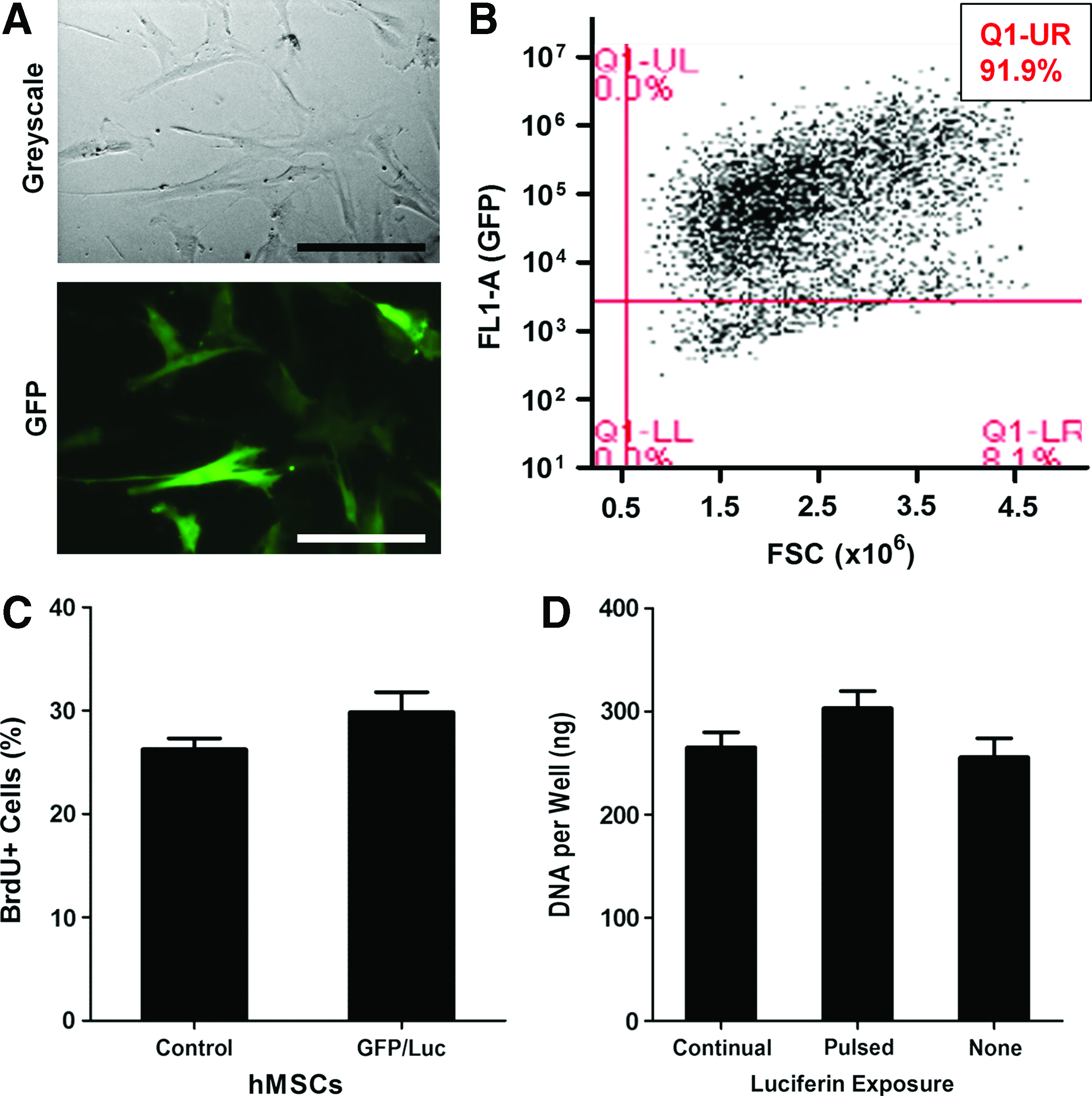
Characterization of green fluorescent protein (GFP)/luciferase (Luc)-labeled human mesenchymal stem cells (hMSCs).
In preparation for in vivo delivery, the dual-syringe protocol used to embed hMSCs within hydrogel constructs was evaluated for resulting cell viability and seeding accuracy. It was determined that sufficient hMSC viability, GFP expression, and spatial distribution were achieved for both hydrogel types (Fig. 2A). Spatial distribution of the embedded hMSCs was also confirmed using confocal microscopy (Supplementary Fig. S2). hMSC seeding density was validated for both agarose and alginate hydrogels (Fig. 2B). Embedded cell dose was linearly related to metabolic activity for agarose (r2=0.992) and alginate (r2=0.977) gels.

Evaluation of dual-syringe hydrogel/cell delivery. Labeled-hMSCs were incorporated into 2% w/v hydrogels using a dual-syringe technique.
hMSC-seeded constructs ranging in cell dose up to 2.0×10⁶ were subcutaneously implanted and day-0 BLI was conducted at 10-, 20-, and 30-min timepoints following injection of luciferin substrate (Fig. 3A). Linear regression analysis demonstrated a strong relationship between BLI signal and cell number for each hydrogel type irrespective of BLI measurement time. The highest correlation coefficients for agarose (r2=0.71) and alginate (r2=0.91) constructs were observed using the 30-min acquisition time (Fig. 3B, C). Although a similar BLI protocol was effective for both hydrogel types, the magnitude of BLI signal differed with hydrogel type such that alginate constructs exhibited approximately twofold greater counts than agarose constructs at comparable hMSC doses.

Development of bioluminescent imaging (BLI) protocol and determination of hMSC survival. Agarose and RGD-alginate constructs were used to deliver a range of hMSC doses: 0–2.0×10⁶. Positive correlation between BLI signal and implanted cell number was observed at all imaging acquisition time points (10, 20, and 30 min) following luciferin delivery.
To evaluate the effects of hydrogel type and cell dose on hMSC survival, BLI data obtained using the 30-min acquisition protocol on days 0 and 7 in vivo were compared. Although all agarose groups showed a decrease in live cell number over 1 week, the 0.25×10⁶ cell dose displayed a significantly higher cell survival ratio (67%) compared with all other doses (Fig. 3D). For the alginate delivery vehicles, only the highest two cell doses exhibited a significant decrease in cell number (Fig. 3E). By day 7, a larger percentage of hMSCs remained viable for the 0.25×10⁶ cell dose group (61% survival) compared with the 2.0×10⁶ dose (3.8% survival).
BLI longitudinal assessment
To investigate the applicability of BLI measurement following implantation in vivo, hMSC-seeded alginate constructs were explanted at 1 week and subjected to ex vivo analysis using flow cytometry. While day-7 BLI counts were significantly correlated to GFP+ events under flow (Fig. 4A), goodness of fit (r2=0.422) was less than that observed on day 0 (Fig. 3C). In addition to examining the BLI signal for each construct on day 7, BLI signal profile (defined as the change in BLI signal over the 30 min following luciferin delivery) was evaluated. The BLI signal profiles for alginate constructs on day 7 suggested a delay in signal rise over the 30-min imaging span compared with day 0 (Fig. 4B). This observation was supported quantitatively by evaluating the BLI signal at 10 min (BLI10min/BLI30min) for each construct group. This value was significantly decreased at day 7 compared with day 0 for both agarose and alginate constructs (Fig. 4C, D). There was no apparent effect of cell dose on BLI signal kinetics.
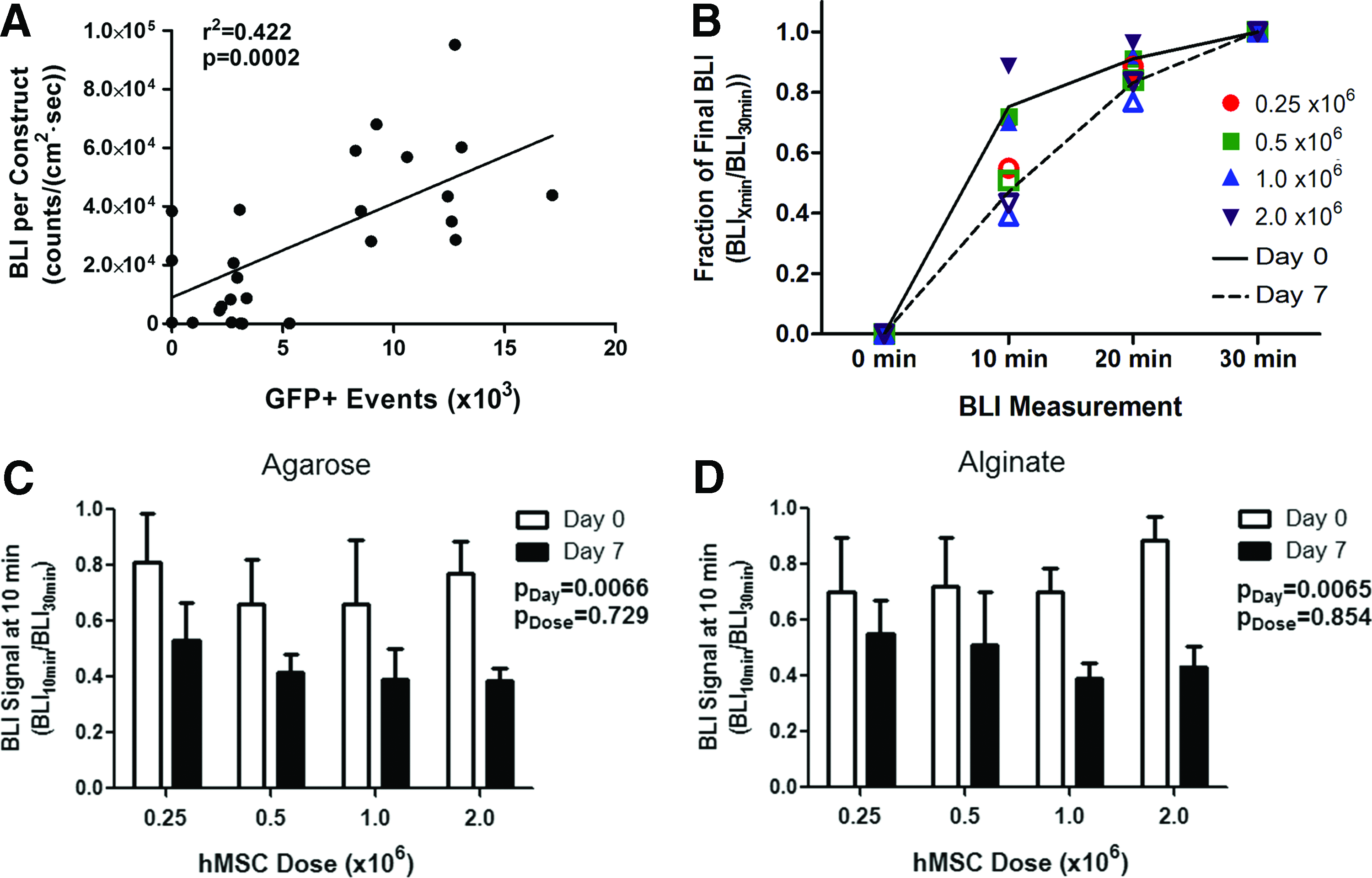
Shift in BLI profile after 1 week in vivo.
Tissue development within and exterior to the constructs on day 7 was assessed using Masson's trichrome staining for fibrotic tissue. Tissue growth was localized predominantly to the construct exterior (Fig. 5A). Irrespective of hydrogel type and cell dose, the construct mesh was lined with a layer of cells and fibrotic tissue. Construct interiors were composed of hydrogel islands containing vacant and cell-occupied pockets (Fig. 5B). Qualitatively, pocket density appeared to be dependent on the initial cell dose, but independent of hydrogel type. The extent of tissue infiltration through macroscopic mesh pores and tube ends of each construct was variable and unrelated to hydrogel type, cell dose, or day-7 BLI signal attenuation (Fig. 5C, D). Although all samples contained a fibrous capsule, capsule thickness and vascularization were inconsistent and did not appear related to experimental group, interior construct vasculature, or day-7 BLI profile behavior.

Construct encapsulation and tissue ingrowth at 1 week postimplantation in vivo.
Construct vasculature
μCT reconstruction of the explant vasculature illustrated the presence of blood vessels surrounding the nanofiber mesh exterior as well as infiltrating the construct through 1-mm-diameter macropores in the tube (Fig. 6A). Quantification of vessel volume within the construct interior determined that all groups contained vasculature and there was no difference between cell dose groups for either hydrogel type (Fig. 6B, C). Histological analysis was used to corroborate vascular perfusion data (Fig. 6D). Both agarose and alginate groups showed pockets of perfusion contrast agent contained within immature vasculature localized near the construct periphery. Specificity of the perfusion technique was supported by colocalization of the contrast agent and vWF immunostaining.
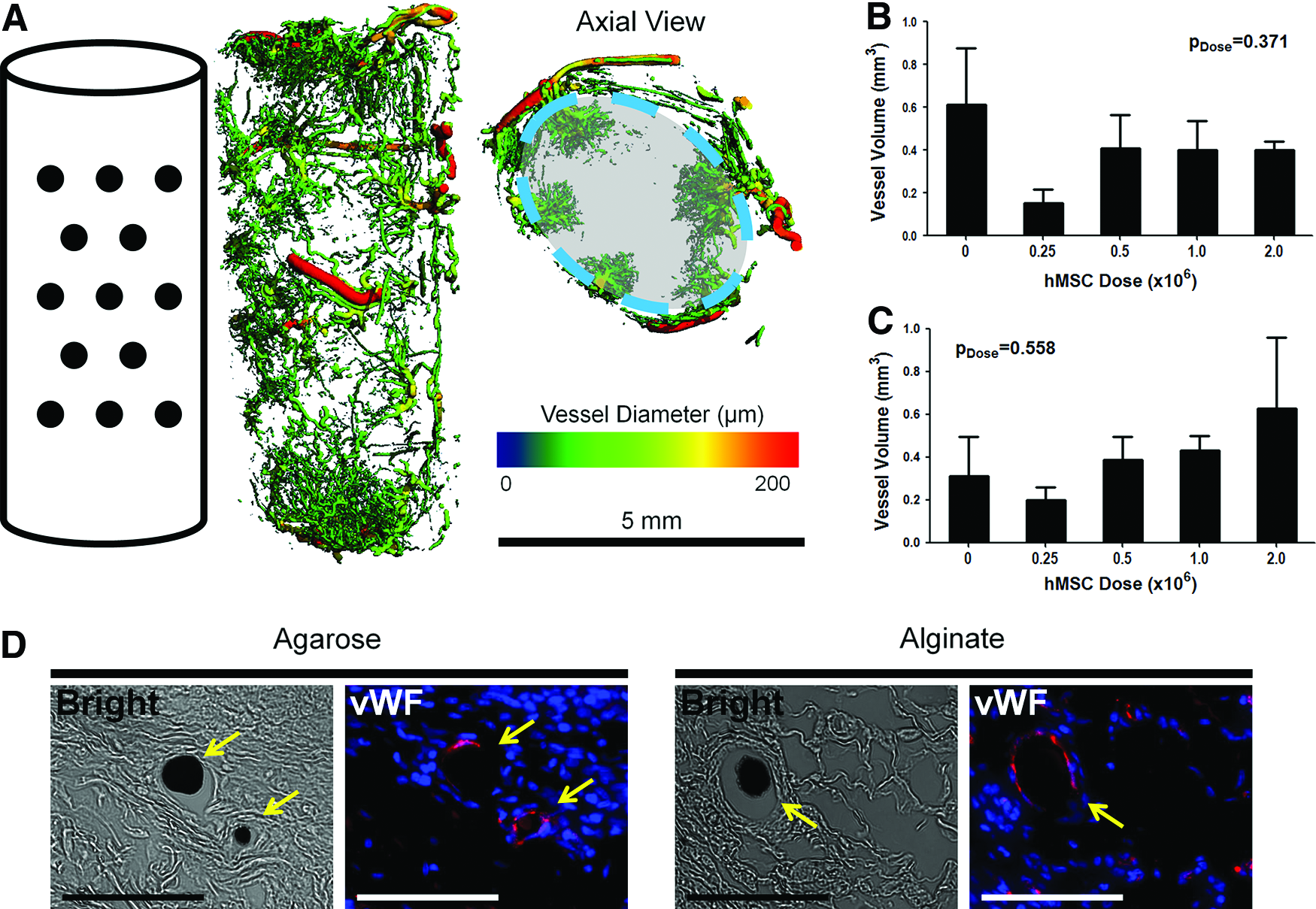
Construct vascularization at 1 week postimplantation in vivo.
The association between development of vasculature and live cell number (BLI signal) on day 7 was examined. For agarose constructs, there was no correlation between vascular volume and either BLI signal (30-min measurement) or BLI signal profile (Fig. 7A, B). Alginate constructs showed no correlation between live cell number (BLI signal) and vascular volume on day 7 (Fig. 7C). However, alginate construct vasculature was significantly correlated to BLI signal profile (Fig. 7D). Higher vascular volumes were associated with a smaller rise in BLI signal during the first 10 min following luciferin delivery, but accelerated BLI signal increase thereafter.

Construct vascularization and BLI signal profile with hydrogel type.
Discussion
This study investigated the impact of hydrogel type on BLI measurement, cell survival, and tissue ingrowth in cell-seeded agarose/mesh and alginate/mesh constructs implanted in a subcutaneous rat model. Based on previous unpublished data, we elected to use nude rats for these studies as the model presented fewer technical challenges, such as cross-over of BLI signal between constructs, compared with the mouse model. We used a hydrogel/mesh hybrid delivery vehicle, previously characterized in an acellular capacity for treatment of a critically sized femoral defect.18,23 Within this challenging bone injury model, the alginate/mesh hybrid delivery system has facilitated effective restoration of limb function upon codelivery with osteogenic growth factor.
At day 0, we found a strong correlation between BLI signal and viable cell number within each hydrogel type. The implementation of a 30-min window between luciferin injection and BLI acquisition provided a good representation of live cell number contained within these systems while operating within the constraints of an in vivo model. Although both hydrogel types showed a linear correlation between BLI signal and live cell number using the 30-min imaging protocol, there was a difference in the magnitude of BLI signal measured between the agarose and alginate constructs. Previous work has noted a difference in BLI signal magnitude for materials of significantly distinct opacity and physiochemical properties. 13 However, the agarose and alginate constructs examined in this study each consist of translucent hydrogels reconstituted at similar density. Within these materials, diffusive transport is shown to be limited for molecules >6.5–70 kDa, a size roughly 200-fold larger than that of luciferin.24–26 Therefore, differences in luciferin transport kinetics are unlikely to account for the material-dependent magnitude of BLI signal. The observed phenomenon could be due to a difference in construct light-scattering properties resulting from dissimilar hydrogel rearrangement behavior. Following syringe-injected construct preparation, the ionically crosslinked alginate is capable of reforming under physiological conditions whereas agarose hydrogel would require a temperature increase in order to reassemble.24,27
Irrespective of hydrogel type, the lower cell doses were found to survive 1 week in vivo proportionally better than the high cell doses. Although the presence of a necrotic core is not seen in vitro, it is possible that cells compete for nutrients and only a fraction of the implanted cells could be supported in vivo. This is suggested by the fact that, in contrast to the proportional viability just discussed, there were no differences in absolute live cell number between groups on day 7. This might indicate the attainment of an equilibrium live cell number that can be initially supported or sustained in the absence of an integrated vasculature. The alginate constructs supported improved hMSC viability in comparison to the agarose vehicles. This may be due to functionalization of the alginate hydrogel with the RGD peptide in order to facilitate cell attachment and manipulation. 27 In the absence of RGD modification, agarose and alginate hydrogels lack adhesive sequences, resulting in abnormal morphology and reduced viability of encapsulated cells. 28
Despite its widespread employment for tracking viability and location of injected cell in vivo, BLI has yet to be validated for large 3D cellular constructs. Variations in tissue ingrowth over time following implantation lead to development of an inherently noisy system for optical imaging analysis. 5 Even in vitro, transport properties of cell-seeded agarose and alginate hydrogels change over time in culture, presumably due to cell-mediated matrix synthesis. 29 Previous work has corroborated in vivo BLI measurement to ex vivo construct analysis of Luc content or histological staining.4,13,15,30 In this report, we evaluated BLI measurement of large constructs subsequent to tissue ingrowth in vivo by directly quantifying labeled cell number on a whole-construct basis. BLI signal correlated linearly to GFP+ flow cytometry events with a coefficient lower than that observed on day 0. This reduction in correlation is suggestive of confounding variables present within the system on day 7.
An examination of the factors that could affect the BLI signal on day 7 reveals the dependency of BLI signal on oxygen and ATP levels within the tissue-engineered constructs. 10 This is generally viewed as a positive feature, as it constrains measurement to the presence of live cells. However, in exceptionally hypoxic environments, such as those simulating a tumor necrotic core, oxygen can become rate limiting within the bioluminescent system. 31 Aside from the Luc enzyme, photon release is highly dependent on the availability of luciferin substrate. Luciferin is a small molecule that readily diffuses through tissue. 32 However, endogenous cell populations may deplete luciferin en route to the labeled cells of interest through uptake of substrate across the cell membrane. 33 Adding to the complexity of luciferin availability, vascular networks have the potential to impact substrate delivery by providing a means of convective transport.
For both agarose and alginate constructs, increase of BLI signal over the 30-min imaging window was attenuated on day 7 in comparison to day 0. To investigate whether the modified BLI profile might be attributed to hindered luciferin transport kinetics, tissue within and proximal to the explant on day 7 was examined by histology. Neither fibrous capsule thickness nor degree of the peripheral vascular network exhibited a relationship to hydrogel type, initial cell dose, or day-7 BLI profile. However, the presence of a fibrous capsule across all experimental groups may be responsible for the attenuated BLI profile observed on day 7, as a logical consequence of such tissue development would be restricted luciferin availability.
Although MSC delivery has been shown to promote angiogenic and vasculogenic processes, this effect was not significant in the current study, perhaps due to particulars of the vehicle or timescale examined.34–36 Vasculature did weakly correlate to BLI signal behavior in the alginate constructs over the 30-min imaging window, suggesting an impact of vascular volume on luciferin availability within the construct. Interestingly, greater vasculature correlated to a delayed rise in BLI signal following luciferin delivery initially, but then accelerated BLI signal increase between 10 and 30 min. No correlations were observed in the agarose constructs. These results provide further insight on the temporal- and matrix-dependent effects of vasculature on BLI measurements. 37
Noninvasive cell tracking strategies include alternatives to BLI, such as fluorescence or magnetic resonance imaging.38–41 However, techniques that rely on the monitoring of cell-contained particles are limited in their utility for longer durations in vivo. For example, particles may be phagocytized upon cell death, leading to the tracking of host macrophages.42–44 Further, although particle-based labels are conferred to daughter cells upon proliferation, per cell signal magnitude is altered.45–47 This limitation disrupts the ability to detect cell number increase, of particular importance when tracking rapidly dividing stem cells. In contrast, BLI specifically monitors viable cells of interest as signal is tied exclusively to labeled cell viability (Supplementary Fig. S3) and is passed through genetic code upon cell division. 48 Due to these significant advantages, BLI has become an invaluable optical imaging technique for the development of cancer therapies and cell-based regenerative medicine strategies within experimental models.7,10
Earlier studies have established that BLI signal is dependent on variables, including construct composition, luciferin delivery method, animal model, sample positioning, and progression of tissue development in vivo.6,10,12 Our work draws attention to the impact of tissue and vascular ingrowth on the accuracy of longitudinal BLI measurement. As BLI is frequently used to visualize systems containing a dynamic vascular network, we believe this validation is critical.49,50 Logeart-Avramoglou et al. previously demonstrated a statistically significant correlation between in vivo BLI signal and ex vivo Luc activity for constructs explanted up to 3 weeks following implantation. 13 In the present study, we performed novel validation of BLI for evaluating live cell count within implanted, multicomponent constructs of large size (>200 mm3). We conclude that, while BLI may prove advantageous for informing large volume cell-based therapeutic design, care should be taken when implementing and drawing quantitative conclusions using this assessment technique. Further, our work highlights that two hydrogels of relatively similar material properties can exhibit divergent behavior under bioluminescent analysis. Validation of BLI protocol and investigation of potentially confounding variables, such as vascular ingrowth and fibrosis, should be conducted on a system-by-system basis in order to draw representative conclusions.
Footnotes
Acknowledgments
This work was supported by NIH grant R01-AR 056694. Wafa Tawackoli assisted with the cell labeling technique. Emily Butts, Albert Cheng, Christopher Dosier, Chelsea Fechter, Angela Lin, Lauren Priddy, Brennan Torstrick, Brent Uhrig, and Jason Wang assisted with the surgical procedures.
Disclosure Statement
No competing financial interests exist.
References
Supplementary Material
Please find the following supplemental material available below.
For Open Access articles published under a Creative Commons License, all supplemental material carries the same license as the article it is associated with.
For non-Open Access articles published, all supplemental material carries a non-exclusive license, and permission requests for re-use of supplemental material or any part of supplemental material shall be sent directly to the copyright owner as specified in the copyright notice associated with the article.
