
Abstract
It is well documented that cryopreservation and resuscitation of human embryonic stem cells (hESCs) is complex and ill-defined, and often suffers poor cell recovery and increased levels of undesirable cell differentiation. In this study we have applied Quality-by-Design (QbD) concepts to the critical processes of slow-freeze cryopreservation and resuscitation of hESC colony cultures. Optimized subprocesses were linked together to deliver a controlled complete process. We have demonstrated a rapid, high-throughput, and stable system for measurement of cell adherence and viability as robust markers of in-process and postrecovery cell state. We observed that measurement of adherence and viability of adhered cells at 1 h postseeding was predictive of cell proliferative ability up to 96 h in this system. Application of factorial design defined the operating spaces for cryopreservation and resuscitation, critically linking the performance of these two processes. Optimization of both processes resulted in enhanced reattachment and post-thaw viability, resulting in substantially greater recovery of cryopreserved, pluripotent cell colonies. This study demonstrates the importance of QbD concepts and tools for rapid, robust, and low-risk process design that can inform manufacturing controls and logistics.
Introduction
T
Complex processes require a systematic approach to develop understanding of key controls that reduce the risk of process failure. Quality by Design (QbD) is the current paradigm for pharmaceutical product and bioprocess design, incorporating tools to address these issues, including risk assessment, process mapping, and factorial design of experiments (DoE).4–6 Risk assessment ensures that capital is allocated efficiently to high-risk areas. Process mapping ensures a consensual understanding of process issues and variables. DoE enables efficient understanding of key variables identified in process mapping. 7 Such tools are central to product development and manufacturing systems for delivering consistent quality.8–10
Applying QbD approaches to cell therapy products have additional challenges compared with conventional pharmaceuticals. QbD relies on an understanding of critical quality attributes (CQAs) that determine the product performance. A scientific understanding of process and other operational risks that may detrimentally affect these attributes can then be developed. However, cell therapy products do not currently have complete measureable CQA profiles that guarantee function through mechanistically understood relationships. Further, processes typically involve numerous biological input materials that are equally poorly defined and have limited in-process monitoring and control of key variables. 1 Resultant statistically uncontrolled or high intrinsic variation processes are very challenging to characterize at a level expected for manufacturing and have led to production processes with poor control and significant endpoint quality testing and wastage. This results in high-risk product manufacture from a regulatory and economic perspective, a situation exacerbated by the restricted ability to purify the end product,11,12 which increases the criticality of the entire bioprocess to the integrity of the end-product relative to molecular therapies. 13
Our process risk assessments, both with industrial partners and in-house processes, have repeatedly highlighted product cryopreservation as a highly variable and high-risk process step for hESC bioprocesses. It is well documented that cryopreservation and resuscitation of hESCs is complex and ill-defined leading to recovery rates as low as 5% and undesirable cell differentiation. 14 We therefore selected it as the unit operation focus to explore the potential for a QbD-driven approach for process improvement.
There are two primary techniques used for cryopreservation of hESCs: slow freezing15–17 and vitrification.18–20 Although vitrification can result in high viability and improved resuscitation survival compared to most slow-freezing protocols,18,21 the technique is significantly limited for current good manufacturing practice (cGMP) of RM cellular therapies. The limitations pertain to achievable scale and process complexity, and potential regulatory concerns regarding the use of very high concentrations of cryoprotective agents (CPAs), LN2 contact, and potential for contamination and cross-contamination. 18 Slow-freezing processes confer advantages for cGMP in terms of increased achievable scale (large numbers of cells or colonies per cryovial), process simplification, lower CPA concentrations, alternatives to direct LN2 contact, and more appropriate storage systems for mitigation of contamination and cross-contamination risks. 21 Slow freezing is currently widely employed in the cell therapy industry and was therefore selected as the focus for this study where we have applied robust process design and QbD principles to define a low-risk, high-quality process for the unit operation of hESC colony culture cryopreservation and resuscitation.
Materials and Methods
A set of in-house standard operating procedures and process maps of the culture, cryopreservation, and resuscitation substeps were used to reduce experimental noise. These maps showed high-level points of control (e.g., timed substeps and temperatures), points of measurement, and the measurement systems used.
hESC culture and maintenance
The hESC line H9 (WiCell Research Institute; p25) was cultured on growth-factor-reduced, hESC-qualified Matrigel™ (BD Biosciences) in mTeSR1 medium (StemCell Technologies). Working cell banks (p36) for these studies were cryopreserved in this format in mFreSR medium (StemCell Technologies), using a Nalgene Mr Frosty (Thermo Scientific) held at −80°C for 18 h before being transferred to LN2 vapor phase storage. Experimental cultures (p37–42) were maintained in six-well plates at 37°C and 5% CO2 in a humidified atmosphere with medium change every 24 h. Intact colony dissociation for passage was performed with Dispase (StemCell Technologies; 1 mg/mL). Cultures were morphologically assessed for ∼70% confluence and <20% differentiation at passage (∼4–5 days between each passage) with manual removal of excessive differentiation. Temperature control was monitored throughout the study using MSR®145 dataloggers (MSR Electronics GmbH). Assessment of cell pluripotency was performed using the Guava EasyCyte 8HT Flow Cytometer (Millipore) and the following conjugated antibodies and appropriate isotype controls, used according to manufacturer's instructions: NANOG-Alexafluor 488 (AF488), OCT4A-AF488, and SOX2-AF488 (BD Biosciences).
Cryopreservation protocol
Detached colonies were transferred to a 50-mL tube and centrifuged at 300 g for 5 min. The supernatant was gently aspirated and mFreSR was slowly added to the cell pellet (1 mL/dissociated well) and mixed gently. The cell solution was aliquoted into Nunc cryovials at 1 mL/vial using a 5-mL stripette. Cryovials were cooled to −80°C at experimental freeze rates using an Asymptote EF600 controlled-rate freezer (Cell Cryogenics Ltd.) or Nalgene Mr Frosty and held at −80°C for 12 h; cells were then transferred to LN2 vapor phase storage.
Resuscitation protocol
mTeSR1 was prewarmed to 37°C, and matrigel-coated 24-well plates were warmed to room temperature. Cryovials were thawed at experimental temperatures and times in a waterbath. One microliter of mTeSR1 was added slowly to each cryovial and gently mixed. The cell suspension was transferred to a 15-mL tube (separate tube per cryovial) and an additional 4 mL of mTeSR1 was gently added. Colonies were pelleted by centrifugation (300 g for 5 min) and gently resuspended in 2 mL mTeSR1. Using a 5-mL stripette, the 2 mL of cell suspension was used to seed wells in triplicate at 0.5 mL/well. Cells were incubated at 37°C and 5% CO2 for 1 h and then removed for analysis of adherence and viability.
Cell adherence and viability assay
Culture supernatant was transferred to individual eppendorf tubes. Wells were gently washed with 0.5 mL DMEM/F12 (Life Technologies) and added to the supernatant samples. To dissociate colonies into a single-cell suspension, 0.5 mL of Accutase (StemCell Technologies) was added per well and the cells were incubated at 37°C for 15 min. Dissociated samples were transferred to separate eppendorfs, the wells were washed with 0.5 mL DMEM/F12, and the wash solution was added to the dissociated samples. The supernatant and dissociated samples were centrifuged at 300 g for 5 min, resuspended in 700 μL of 0.01 μM calcein AM live cell stain (Life Technologies), and incubated at 37°C for 7 min. Samples were then plated into a 96-well round-bottomed plate at 200 μL/well (each sample in triplicate) and analyzed for Calcein AM expression using a Guava EasyCyte 8HT Flow Cytometer. Adherence was calculated by dividing the number of cells in the dissociated sample by the total number of cells in both the supernatant and dissociated samples. Viability was measured by calculating the percentage of cells expressing over a set level of fluorescence (Fig. 1). Controls included nonfrozen cells and unstained cells.
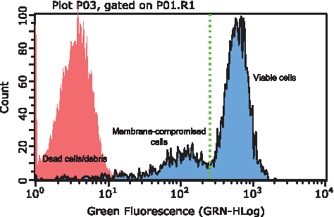
Histogram depicting the regions observed in populations of human embryonic stem cells (hESCs) stained with Calcein AM. The viable region was utilized for determining the recovery of cells from cryopreservation.
Experimental designs
A systematic approach was used to evaluate the effect of various formulation and process variables on the health of cells during the cryopreservation and resuscitation subprocesses. Experimental factors for investigation were selected from risk assessments and review of process maps. The adherence-based viability assay described previously was used as an experimental response. All experimental designs were created and analyzed using Design Expert 8.0.7.1 (Stat-Ease, Inc.). ANOVA was applied to establish a prediction model for each response. A two-replicate, two-level, central composite design (24) was employed to investigate the cryopreservation subprocesses. Experimental parameter levels were set around normal process levels within limits of practicality (i.e., avoiding excessive confluence or cryopreservation time [≥3 h]). Center points for each variable were included to make the design more robust and enhance the predictive power of the model. Experiments were randomized as far as possible within operational constraints. A 2-level, fractional factorial design with eight center points was used to investigate the resuscitation subprocess. Input culture quality was varied using data from the cryopreservation subprocess experiment to select a suboptimal and optimal set of freeze conditions. The selected models were evaluated according to a range of adequacy tests. Data were transformed if required prior to modeling and insignificant terms were removed using backward selection. Diagnostics data were used to determine best fit for model selection.
Results
The objective of this work was to develop a systematic approach, in line with QbD concepts, to increase quality and recovery of cryopreserved hESC colonies and to reduce process risk. A screening exercise identified a suitable assay for detecting damage to cells postresuscitation with the speed, throughput, and sensitivity to enable a DoE approach. A process map identified key variables for a series of designed experiments to identify critical determinants of hESC quality during the cryopreservation and resuscitation process.
Establishing a cell assay for recovery from cryopreservation
Application of multifactorial experiments for efficient and robust process design depends upon high-throughput, rapid, and accurate postresuscitation measurement to predict long-term recovery (a key CQA). Using two different cryopreservation protocols (controlled rate freeze at 1°C/min and 2°C/min), we analyzed multiple cell attributes up to 96 h postresuscitation for their ability to predict recovery. These attributes included adherence, cell esterase activity (Calcein AM), apoptosis (Caspase 3, 8, and 9), membrane integrity (CellROX), pluripotency (Nanog, Oct4, Tra-1-60, Tra-1-81, SSEA1, and SSEA4), and proliferation (Ki67). Early measurement of cellular adherence and viability of adhered cells (by cell esterase activity; Calcein AM, 1 h postseeding) correlated with 4-day proliferation data and allowed early discrimination between cell recovery from different freeze conditions (Fig. 2A, B). Cells would continue to adhere and recover esterase activity beyond the first hour, but the extent of recovery after 1 h was good enough to predict long-term recovery. However, due to measurement as early as 1 h postseeding, values for adherence and viability appeared relatively low. Adherence was selected as a primary quality output due to highest sensitivity to long-term recovery in response to process variation; cell esterase activity was used as a secondary quality output as it provided a further layer of information on the health of the adhered cells. Instability in the Calcein AM viability assay was observed over time (Fig. 2C); removal of a wash step after staining resulted in prolonged signal stability as cells were able to continue cleaving Calcein AM, thus maintaining viable cell fluorescence (Fig. 2D). This modified method had the throughput and stability required to assess hES recovery from cryopreservation experiments and develop models of the cryopreservation process.
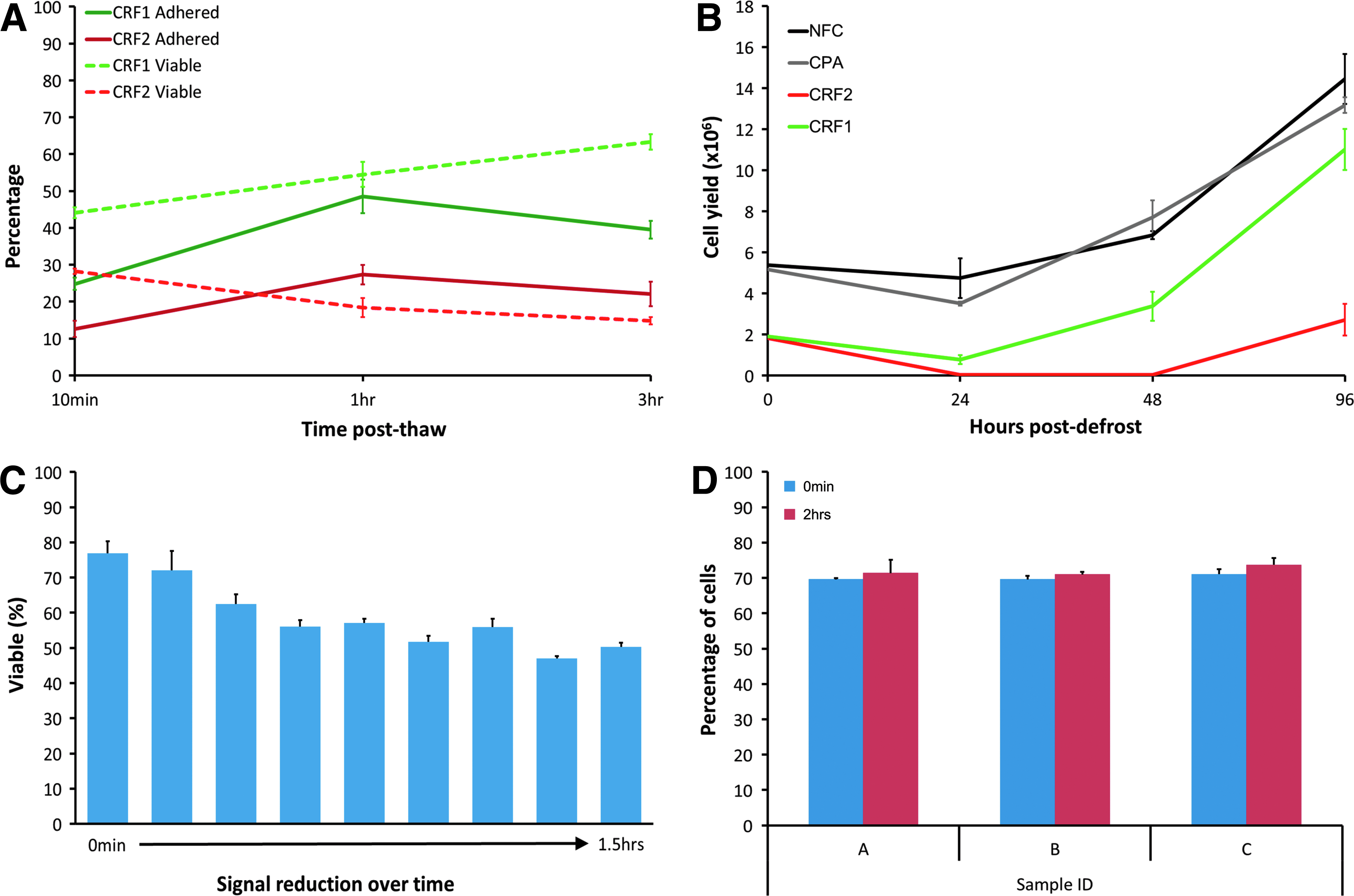
A difference in protocols that produced healthy (CRF1) or poor (CRF2) cell populations could be detected as early as 1 h postresuscitation by analyzing the adherence and viability of the adhered cells
Establishing cryopreservation experimental parameters and analysis
A risk assessment of hESC cryopreservation and resuscitation was produced following a systematic review of existing protocols and literature. This enabled identification of process parameters amenable to experimental optimization and control (Table 1). Parameter levels for each prioritized factor listed in Table 1 were set at low, medium (where possible), or high for the experimental designs and models (see Table 2). This identified the necessity to split experimental planning into two subunits (cryopreservation and resuscitation) to pragmatically restrict experiment size. Experimental designs were conducted in triplicate and the cell recovery was measured with the method established in “Establishing a cell assay for recovery from cryopreservation” section. This data was used to generate statistical models of the effect of the experimental parameters on the cell recovery in terms of both cell adherence and viability of adhered cells. All models passed standard regression diagnostics and lack-of-fit tests, and correlation plots of predicted versus experimental values (as well as R2 values) indicated that models should be predictive. The signal-to-noise ratio of the data was adequate (>4), and analysis via ANOVA found the experimental models to be significant (p<0.001). See Supplementary Tables S1–S4 (Supplementary Data are available online at
hESC, human embryonic stem cell.
DoE, design of experiments.
Effect of cryopreservation parameters on cell quality and definition of the optimal process-operating space
Cell recovery positively correlated with longer preculture duration and slower freeze rates (Fig. 3A), but was insensitive to cryoprotectant volume and temperature (within the experimental range). The model of adherence predicted that optimization of significant factors could improve adherence from 27.0% to 62.2%. Minor model curvature was observed within both preculture duration and freeze rate, indicating that the factor ranges used may be near optimal for adherence. The model describing the viability of adhered cells was reassuringly similar to that of total cell adherence, that is, increasing with longer preculture duration. However, when using this quality measure, freeze rate only had a minor impact and was not independent of preculture duration; a peak in viability (55–58%) was observed across all freeze rates at 5.8–6.8 days in preculture (Fig. 3B).
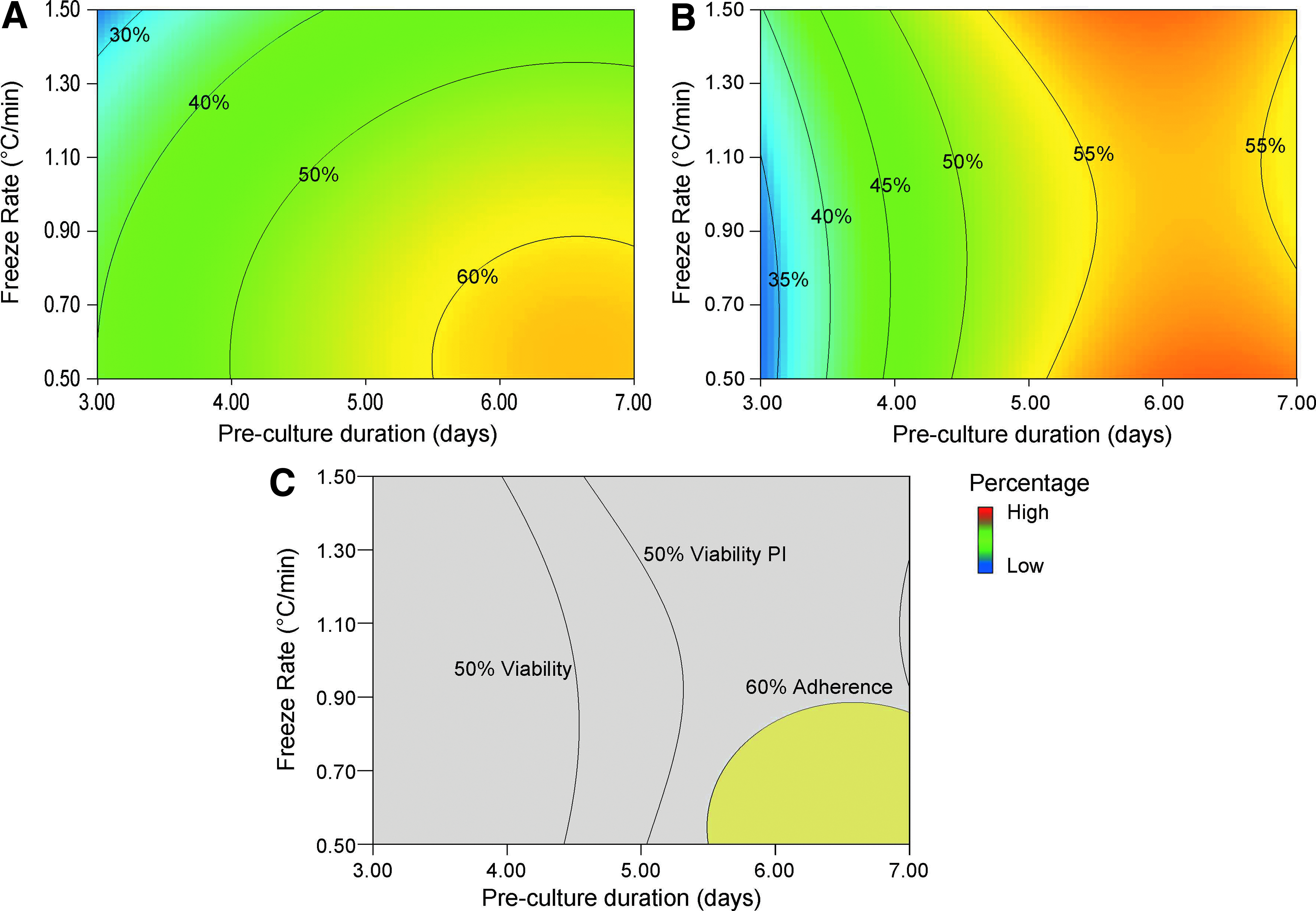
Contour heat plots demonstrating adherence and viability (combined together to provide a definition of cell health) within the cryopreservation DoE. High adherence/viability regions are red; low adherence/viability regions are blue. Adherence plots demonstrate that a longer preculture time (time since previous passage) and slower freeze rate promote best cell recovery
Process design space optimization was performed using minimum desired values of 60% adherence and 50% viability of adhered cells at 1 h postseed. This is a software function that uses the process models to calculate a parameter operating tolerance in order to consistently deliver product within specification. Minimum desired values must be selected from within the experimental data range. Cryoprotectant volume and temperature were set at 0.5 mL and 4°C, respectively, as the models predicted no effect on cell quality and these settings improved economy and ease of control. The optimal experimental design space (see Fig. 3C) confirmed that longer preculture times coupled with lower freeze rates are optimal for cell recovery.
Establishing resuscitation experimental parameters and analysis
To determine the relationship between the cryopreservation subprocess described earlier (“Effect of cryopreservation parameters on cell quality and definition of the optimal process operating space” section) and the linked resuscitation subprocess, we created two cryopreserved cell populations using optimal and significantly suboptimal freeze processes (based on the output of the previous cryopreservation models). These populations were used as alternative (“good” and “bad”) inputs to screen the resuscitation process parameters identified in Table 2. Importantly, this highlighted how the ability of resuscitation parameters to affect the cell recovery, either positively or negatively, is dependent on the cryopreservation of the resuscitated sample.
Effect of parameters on cell adherence
Thaw bath temperature, exposure time of cryovial in thaw bath, and temperature of the dilution media added to the thawed cells had significant effects on cell adherence, with the latter two factors being nonindependent. Seeding density did not have a significant effect. Cryopreserved input cell quality (optimal vs. suboptimal) was significant but nonindependent of all other factors. Thaw bath temperature was only significant for the suboptimally cryopreserved input cells, where a higher temperature increased cell adherence by up to 11%. Also, longer exposure in the thaw bath and a higher dilution media temperature significantly improved recovery of suboptimally cryopreserved cells (up to 9% increase in adherence) regardless of other factors. Optimally cryopreserved cells were less sensitive to the resuscitation process. Thaw bath exposure time and dilution media temperature interacted such that they only affected cell adherence when the other factor was set at a low level; if exposure time of cells was low, then adherence increased up to 20% at higher dilution media temperatures. Optimization of exposure time or dilution media temperature increased robustness of the process to variation in the other factor; thus, longer exposure times resulted in improved adherence and increased robustness to variations in dilution media temperature. The adherence model indicates that high levels of each significant factor would produce the most robust, optimal resuscitation process for both optimal and suboptimal cryopreserved cells.
Effect of parameters on adhered cell viability (Calcein AM)
The Calcein AM viability assay provided limited additional information on the effect of resuscitation parameters beyond the adherence assay. It confirmed that a longer exposure time in the thaw bath improved the cell recovery (by up to 10.3%) and that parameters affected the suboptimally cryopreserved cells more than the optimally cryopreserved cells. In addition to the adherence data, a higher seeding density had a minor beneficial effect to cell viability in both inputs. The viability model indicated that high levels for all significant factors improved the resuscitation process for both optimally and suboptimally cryopreserved cells.
Design spaces
The details of the models described in “Effect of parameters on cell adherence” and “Effect of parameters on adhered cell viability (Calcein AM)” sections can be combined to generate process design (Fig. 4A, B) and desirability (Fig. 4C, D) charts using both cell adherence and cell viability responses to predict parameter levels that will deliver cells above minimum selected specification. Optimal regions for both cell quality inputs were found at high levels of thaw temperature, exposure time, and dilution media temperature. Optimization of both the cryopreservation and resuscitation subprocesses predicted significant improved adherence and recovery of cryopreserved cells (Table 3).
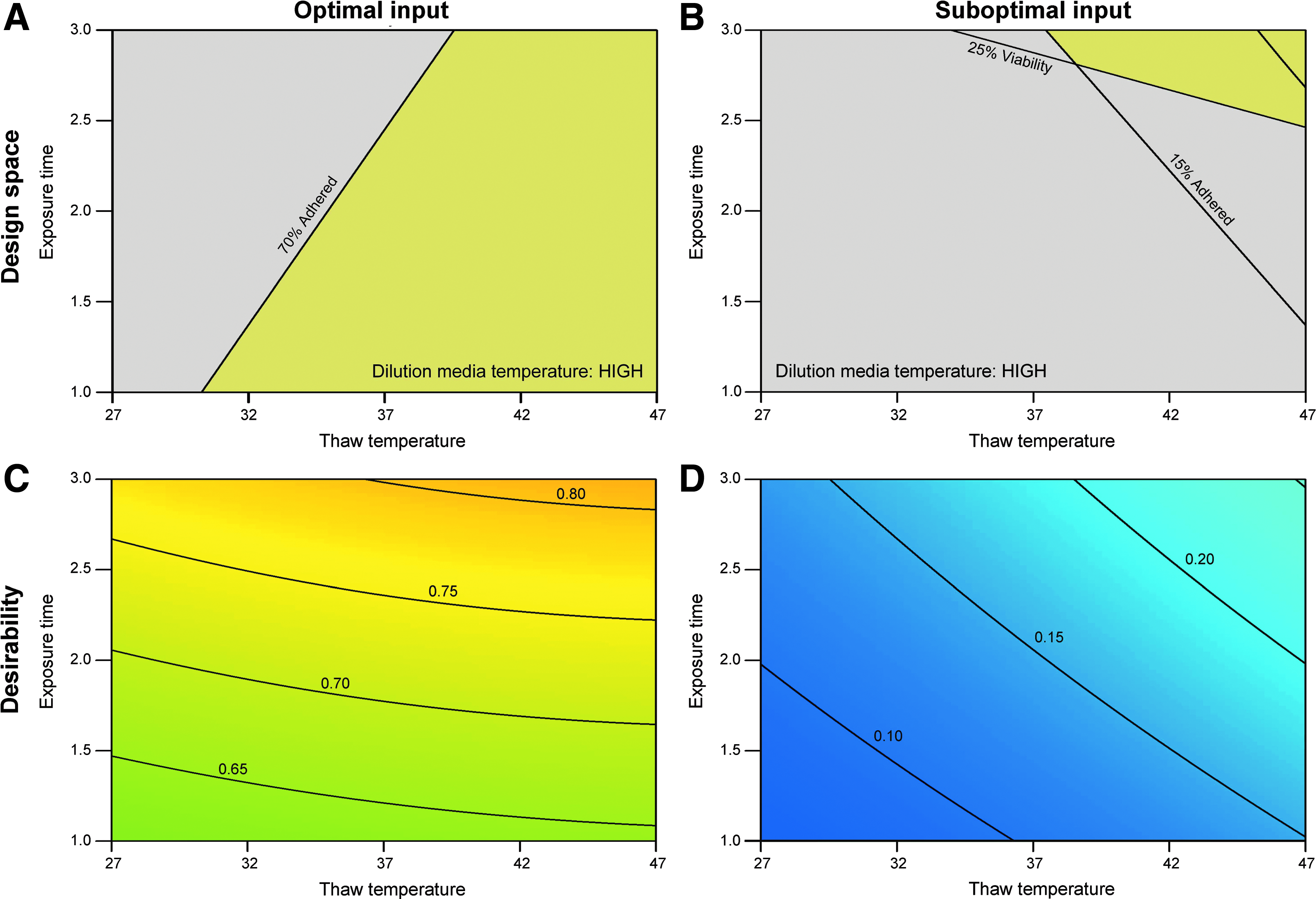
A combination of both the adherence and viability data was utilized to produce design spaces for optimum cell health and recovery from defrost for each cryopreserved input cell quality. The % adherence and % viability lines demonstrate the parameter levels at which the average response will meet the minimum desired values. The yellow regions depict the region that parameters must be within to produce the minimum desired values for both adherence and viability
Values were predicted using the DOE models based on raw data.
Model validation
Due to the inherent interaction of the cryopreservation and resuscitation subprocesses, model validation was performed simultaneously as a single complete process to allow measurement of cell quality postcryopreservation. All values observed in the validation runs were within an acceptable range of the predicted values, thus validating the models (Fig. 5A). Analysis of typical pluripotency markers Nanog, Oct4a, and Sox2 showed no detectable differences pre- and postcryopreservation (Fig. 5B). Extended culture (96 h postseeding) of optimally cryopreserved inputs demonstrated a rapid recovery of proliferation after a lag phase of 24 h (Fig. 5C); suboptimal inputs demonstrated a lag phase of up to 48 h (Fig. 5C, inset).

Validation plots proving the predictive ability of the models for both subprocesses are shown in
Discussion
This study demonstrates the application of a QbD approach to gain further understanding of the processes involved in cryopreservation and resuscitation of therapeutic cells. Tools used include risk assessment, CQA selection, and multifactorial statistically designed experiments and models. These methods would be utilized by a manufacturer to develop models and validate the operating conditions for their specific cell bank. This QbD-type approach offers an improvement over less systematic and one-factor experimental methods. It increases experimental efficiency, enables detection of interactions between processing variables, and allows a risk-driven quantitation of process sensitivities, therefore enabling development of scientifically informed process control strategies. It is also a model for development of cell manufacturing processes through division into manageable subprocesses that facilitates systematic characterization and engineering control. hESC colony culture cryopreservation is explored as an exemplar due to its complexity, common poor cell recovery, and associated process risk. 14
There are multiple steps required to design the cryopreservation and resuscitation operating spaces to reduce process risk. These include identifying subprocess outputs that are directly predictive of the subsequent process (and ultimately product) performance (a critical process or quality attribute). Correct identification ensures that successfully controlling output from one subprocess will determine the performance of the next. Some of these outputs, such as cell viability, are intuitive; a wide distribution of viability will clearly impact cell quality further down the process. 2 It is therefore a good initial target relative to pluripotency factors or surface marker measurements where a quantitative and timely link to cell quality is poorly defined. 32 Further, detecting physical damage from cryopreservation is unlikely to immediately manifest in pluripotency markers. This does not mean that these attributes are not important; they just may not be relevant for rapid detection of a changed output from this specific subprocess. For this same reason, the common method of testing cells for karyotypic change after cryopreservation was not performed because the nature of karyotypic change suggests that a prolonged growth period would be required to detect the emergence of a karyotypically abnormal population. Further, the stochastic nature of karyotypic change would require a very large number of experimental runs to statistically determine the relationship between cryopreservation conditions and karyotypic stability.
Many current assays are based on cell viability; however, these are often invasive and open to interpretation. For example, membrane integrity methods are not sensitive to cells that succumb to delayed onset cell death and do not give information on proliferating cells, 33 and measures of metabolic activity 34 that can fluctuate independent of viability. We investigated the performance of conventional viability tests both at product defrost and at other points in the manufacturing process to determine those that are most predictive of longer term cell health; many were insensitive with regard to predicting cell recovery. The cell adherence system we report was a rapid, high-throughput, and stable measurement of postcryopreservation cell recovery. We observed that measurement of adherence and viability of adhered cells at 1 h postseeding was predictive of cell proliferative ability after 96 h in this system.
In a limited number of experimental conditions, the cell adherence and cell viability by esterase activity were contradictory; this resulted in a dilemma whereby adherence was optimal at certain factor levels, but viability was optimal at others. In this study our design spaces selected for the weighting of adherence and viability that resulted in the greatest numbers of viable recovered cells for manufacturing. This is an example of the care required in meaningfully interpreting CQAs and model outputs. The complexity of therapeutic cell responses suggests that using multiple-modeled outputs to generate agreement in the effect of processing parameters will be important to reduce risk of misinterpreting findings.
The factors selected in the models for cryopreservation and resuscitation were chosen based on both their prominence in literature and our previous studies suggesting influence on cryopreserved cell quality. Although the models allow substantial process improvement, the noise in the data indicates that there are one or more factors that introduce variation that are not fully controlled. This variation may come from the existing variation in the cell lines used, operator variation, or from environmental effects, such as variation in room temperature. QbD methodology, as a risk-driven process, should continue to target variables until adequate control and process knowledge can be demonstrated commensurate with process risk. Response surface models can be used to provide higher resolution models of relationships if required to predict process performance. However, selection of both further variables and more intensive experimental designs require significant resource and require risk-based justification to allocate capital.
The models used for cryopreservation and resuscitation steps indicate that both subprocesses are critical to cell recovery. Preculture duration and freeze rate were identified as critical parameters affecting the cryopreservation subprocess. This data agrees with the current trends observed in literature, stating that hESCs require sufficient time postpassage to recover E-cadherin, essential for their survival. 22 It is clear that cell state as a function of culture time is a critical indicator of cell robustness to cryopreservation. However, it is currently unclear how this could be accurately measured (we noted a decrease in EdU incorporation with longer culture, which may be a candidate predictive assay.). Evidence suggests that a faster freeze rate is beneficial with DMSO-based cryoprotectants as it protects cells from toxicity. However, we found that a slower freeze rate was optimal, potentially due to increased time for equilibration of DMSO-throughout colonies, reducing intracellular ice formation.
All factors tested in the resuscitation model had an effect, either individually or in interaction with one another. The effect of seeding density on cell recovery showed substantial noise, most likely due to the challenges of controlling colony seeding density based on the dilution factor of a vial rather than specific cell counts. Cells treated with a short exposure time had increased adherence when resuspended in warmer media. Use of warm resuspension media increased robustness of cell viability to variations in thaw temperature, this is likely due to it aiding rapid defrost of still-frozen cell solutions. 29 In samples with longer exposure times, cell solutions were already defrosted and therefore are less affected by lower dilution media temperatures. This data suggests that a rapid solid-to-liquid transition is the most critical part of the resuscitation process to maintain cell health.
The interdependence of cryopreservation and resuscitation indicates that although poorly frozen cells will not recover to levels observed with optimal freezing protocols, it is possible to significantly improve adherence and viability of the cells through controlled defrost, thus increasing the total yield available for manufacture. The ability to improve the robustness of the defrost process to variations in cryopreservation or vice versa potentially reduces one of the subprocesses that needs stringent control during manufacturing and improves logistical flexibility. This is exemplified in the resuscitation model where many parameters had a greater effect on poorly cryopreserved input samples relative to optimally cryopreserved samples. The process confidence delivered by such systematic modeling methods has the potential to greatly facilitate regulatory acceptance of new cell products, define operational boundaries (and therefore logistics and costs), and reduce risk for product developers.
Footnotes
Acknowledgments
The authors gratefully acknowledge the financial support from Pfizer Neusentis and the EPSRC center for regenerative medicine. The authors acknowledge Miss Kirsty-Louise Marrow for her laboratory support and technical input. The authors also acknowledge Dr. Andrew Picken and Dr. Katie Glen at the Centre for Biological Engineering, Loughborough University, for their constructive discussions and useful input.
Disclosure Statement
No competing financial interests exist.
References
Supplementary Material
Please find the following supplemental material available below.
For Open Access articles published under a Creative Commons License, all supplemental material carries the same license as the article it is associated with.
For non-Open Access articles published, all supplemental material carries a non-exclusive license, and permission requests for re-use of supplemental material or any part of supplemental material shall be sent directly to the copyright owner as specified in the copyright notice associated with the article.