




Abstract
Current protocols for chondrocyte isolation are inconsistent, resulting in suboptimal cell yield and compromised cell quality. Thus, there is a need for an improved isolation protocol that is able to give a maximum yield with optimal cell viability while preserving the chondrocyte phenotype. In light of this, we developed an improved isolation protocol based on enzymatic digestion using 0.1% (w/v) collagenase II. Different from existing methods of digesting minced cartilage for a prolonged period (usually 14–16 h), we performed two additional digestions, with a 5- and 3-h interval in between. The results showed that this multiple digestion method was able to yield a total number of cells that are more than a fivefold increase as compared to any of the common isolation protocols. More importantly, a high percentage of the isolated cells remained viable. Furthermore, an evaluation of the effect of additional digestions on chondrocyte phenotype indicated that cells harvested from the second and third digestion showed a comparable or higher proliferative capacity than the first digestion and all the cells expressed chondrocyte-specific markers tested, with cells from the third digestion showing exceptionally high gene expression levels for collagen type II (Col II), aggrecan, and COMP. Additionally, their ability to produce collagen type II as well as their morphology were not affected by the two additional digestions. Taken together, the results suggested that the use of this isolation protocol resulted in a higher cell yield and the quality of the isolated cells was maintained. Hence, we recommend this isolation protocol to be employed for more efficient cell harvesting especially from limited biopsied cartilage tissue samples.
Introduction
D
The commonly used protocols for harvesting these cells are based on enzymatic digestion of the extracellular matrix (ECM) with different proteinases, mainly targeting the extensive collagen network by using collagenases.2,6,7 According to protocols published, the concentrations and incubation times of digestion enzymes vary significantly. A combination of trypsin and collagenase or just collagenase alone has been used; in addition, the concentration of the digestion enzyme such as collagenase may also differ from 0.05% to 2% w/v depending on the incubation length used. 2 Despite variations in chondrocyte isolation protocols, chondrocyte isolation efficiencies remained unsatisfactory.2,8 Typically, only 1–5×10⁶ cells out of 1×10⁸ of the total chondrocytes present in each gram of cartilage were successfully isolated. A large proportion of cells remained entrapped within the cartilaginous matrix, suggesting an incomplete digestion of the ECM, leading to suboptimal isolation efficiency. Thus, there is a need for an improved isolation protocol to optimize harvesting of viable cells.
In this study, we developed a protocol for more efficient primary chondrocyte isolation from articular cartilage tissue, focusing on maximizing the cell yield while maintaining the quality of the isolated cells. It is well known that serum is a necessary medium component to ensure cell viability during the isolation process. However, it also contains factors that would inhibit the collagenase activity, thereby reducing cell isolation efficiency. Hence, instead of employing a single long digestion time of 10–22 h, as seen in many current protocols, multiple short incubation periods are used. The fresh collagenase solution would be replenished at the end of each digestion round to ensure the continuous activity of collagenase throughout the entire isolation process.
Materials and Methods
Chondrocyte isolation and culture
Unless stated otherwise, all cell culture-related items used were supplied by Invitrogen. Cartilage was extracted from the patellar articular surface of the femur in 5-month-old domestic pigs (∼3.5 g each, n=3). All cartilage pieces were washed with phosphate-buffered saline (PBS) before they were cut into smaller pieces (∼2×2 mm in size) in a 100-mm Petri dish and immersed in 15 mL of filtered collagenase-containing medium for digestion. The collagenase-containing medium was prepared by dissolving collagenase type II at the concentration of 1 mg/mL in Dulbecco's modified Eagle's medium glutamax and 10% fetal bovine serum. A total of three rounds of digestions were carried out and their duration were 14 h (first digestion), 5 h (second digestion), and 3 h (third digestion), respectively. The remaining cartilage pieces were rinsed with PBS before addition of collagenase-containing media for subsequent rounds of digestion. Additionally, a single digestion period of 22 h was also carried out as a comparison.
Cell-harvesting efficiency
After each digestion, supernatants were collected and the total cell number was determined using a hemocytometer before centrifuging at 1100 rpm to remove collagenase. To calculate the efficacy of cell harvesting during each digestion round, the total cell number was counted and normalized by the weight of cartilage digested.
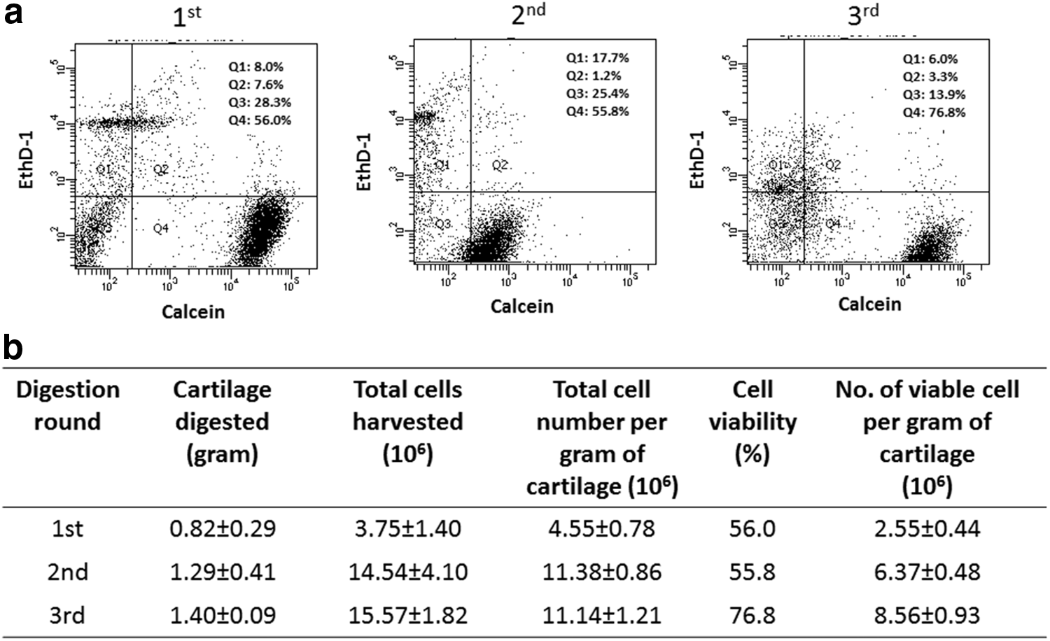
To determine the cell viability of the harvested chondrocytes, live/dead staining was carried out immediately after digestion. Chondrocytes from the same round of collagenase digestion were pooled together to be used for Live/Dead dye staining (Molecular Probes, Invitrogen). Briefly, 0.5 μL of calcein-AM and 2 μL of ethidium homodimer were added to every milliliter of cell suspension. After 30 min of incubation at 37°C, the cells were loaded into the flow cytometry (BD LSR II) for analysis. The result presented is based on a record of 10,000 events.
Cell proliferation assay
Cell proliferation capacity was evaluated using the WST-1 assay. Briefly, 40,000 cells from each sample were seeded into each well in a 96-well plate. At designated time points, media were removed and replaced with a mixture containing 100 μL of media and 10 μL of WST-1 reagent. The mixture was incubated with cells for 1.5 h before reading absorbance at wavelength 450 nm with reference wavelength at 620 nm.
Gene expression
At designated time points, RNA from chondrocytes was extracted using TRIzol® (Invitrogen). The acquired RNA samples were reverse transcribed to cDNA by M-MLV reverse transcriptase (Promega) before the subsequent polymerase chain reaction (PCR). For semiquantitative PCR, cDNA was amplified by respective primers using the following PCR parameters: 94°C for 60 s followed by 35 cycles of 94°C for 30 s, 58°C for 30 s, 72°C for 30 s, and finally, 72°C for 5 min. The amplified product was loaded into a 1.5% agarose gel and a constant voltage of 100 V was applied for 50 min. The gel was then stained with the ethidium bromide solution before viewing. Real-time quantitative PCR (qPCR) was performed using iQ SYBR Green Supermix (Bio-Rad), and gene expression was calculated with the comparative 2−ΔΔCT method, using β-actin and TATA box protein (TBP) as the reference genes and the values were further normalized to the first digestion group of respective time points. All the primers used in PCR and qPCR were synthesized by AIT Biotech and their sequences are presented in Tables 1 and 2, respectively.
AT, annealing temperature; PS, product size.
Immunohistology staining for collagen type II and F-actin
Primary cells harvested were seeded directly in a 24-well plate at a cell density of 200,000 cells/well. Cells at each time point were fixed in 4% (w/v) paraformaldehyde for 1 day. They were gently rinsed with PBS before permeabilized with 1% Triton-X for 10 min. To prevent nonspecifc binding of IgG, incubation with 2% bovine serum albumin was carried out for 30 min at room temperature before staining. Cells were incubated with the Col II (2 μg/mL in PBS; Chemicon) primary antibody at 4°C overnight and washed with PBS. Subsequently, they were incubated with Anti-IgG (5 μg/mL in PBS; AlexaFluor 488; Invitrogen) at room temperature for 1 h in the dark. Nuclei were then counterstained by 4′,6-diamidino-2-phenylindole (DAPI). The samples were rinsed with PBS washing for three times before viewing using a fluorescent microscope.
Statistical analysis
Where appropriate, ANOVA is used to analyze results using Tukey or Games-Howell as the post hoc test. Data are presented as mean±standard deviation, calculated from three specimens from each sample group, and any statistical significance is denoted in the figures.
Results
Cell-harvesting efficiency
We investigated the effect of multiple enzymatic digestions on cartilage digestion. After each digestion, the volume of cartilage pieces decreased and most of them were completely digested after the third round (Fig. 1). To evaluate the cell-harvesting efficiency for each digestion, the yield of chondrocytes after each digestion was determined and expressed as the total number of cells harvested per gram of cartilage. As seen in Figure 2b, the total number of cells per gram of cartilage collected from both second and third digestion was ∼2.5-fold higher than the first digestion. Additionally, we also evaluated cell viability through flow cytometry analysis: ethidium homodimer (EthD-1) was used to stain for dead cells, whereas calcein was used to stain for live cells (Fig. 2a). Similarly, the second and third digestion yielded a much higher number of viable cells as compared to the initial round of digestion (2.5- and 3.4-fold, respectively). It is interesting to note that although the total number of cells collected during the additional two digestions was similar, the third digestion yielded the highest percentage of isolated viable cells (as high as 76.8%). Although a single 22-h digestion was shown to digest 2.22 g of cartilage, only about 4 million cells were harvested, and of which 2.34 million of them were viable from each gram of cartilage (Supplementary Fig. S1; Supplementary Data are available online at

Cartilage fragments before and after each round of digestion showing a progressive decrease in cartilage volume with increasing digestion. Color images available online at
Cell proliferation
Besides determining the cell-harvesting efficiency, it is also important to characterize the harvested cells to ensure that the introduction of two more digestion steps does not affect the cells, both structurally and functionally. We first examined the cell proliferative capacity after the cells were plated and cultured for 1, 4, and 7 days using the WST-1 proliferation assay (Fig. 3). As compared to the first digestion, the two additional digestions did not affect the capacity of the cells to proliferate. In fact, cells harvested from the second digestion consistently showed the highest cell proliferation as indicated by the highest absorbance value among the three, but the difference was especially significant on day 4 and 7. At day 4, cells from the third digestion also showed a significantly higher cellular proliferative activity as compared with the first digestion. Although the same phenomenon was not observed on day 7, their proliferative activity still remained comparable to that of the control group.

Cell proliferation assay using WST-1. *p<0.05 while **p<0.01 when compared to first round of digestion.
Gene expression
To evaluate the effect of multiple digestions on cartilage-specific phenotype, both PCR and qPCR were performed to investigate the expression of chondrocyte markers. As shown by the PCR results in Figure 4, all important chondrocyte markers tested were expressed, except for some irregularities observed for collagen type II on early time points (day 1 and 4). Of particular importance was the expression of collagen type I, which was not observed throughout the entire experimental period. It is also interesting to note that the expression of collagen type X was high initially, but its expression was attenuated on day 7, as suggested by the decrease in band intensity. As for the qPCR results (Fig. 5), cells from the third digestion generally showed an upregulation for all the anabolic genes tested (col II, Agg, COMP, and Sox 9). The increase in gene expression levels was most significant on day 4 for all the important cartilage matrix components tested, namely, Col II, aggrecan, and COMP (p<0.01). The cells isolated from the second digestion also showed a marked increase in all their anabolic gene expressions at day 4, except for COMP. Besides the anabolic genes, we also analyzed the expression of Rho A and collagen type I. The expression levels of Rho A for both the second and third digestion were comparable to, if not higher, the control group. As for collagen type I, we observed that cells from the two additional digestions showed significantly lower expressions for this gene as compared with the first digestion toward the end of the experimental period (p<0.05).
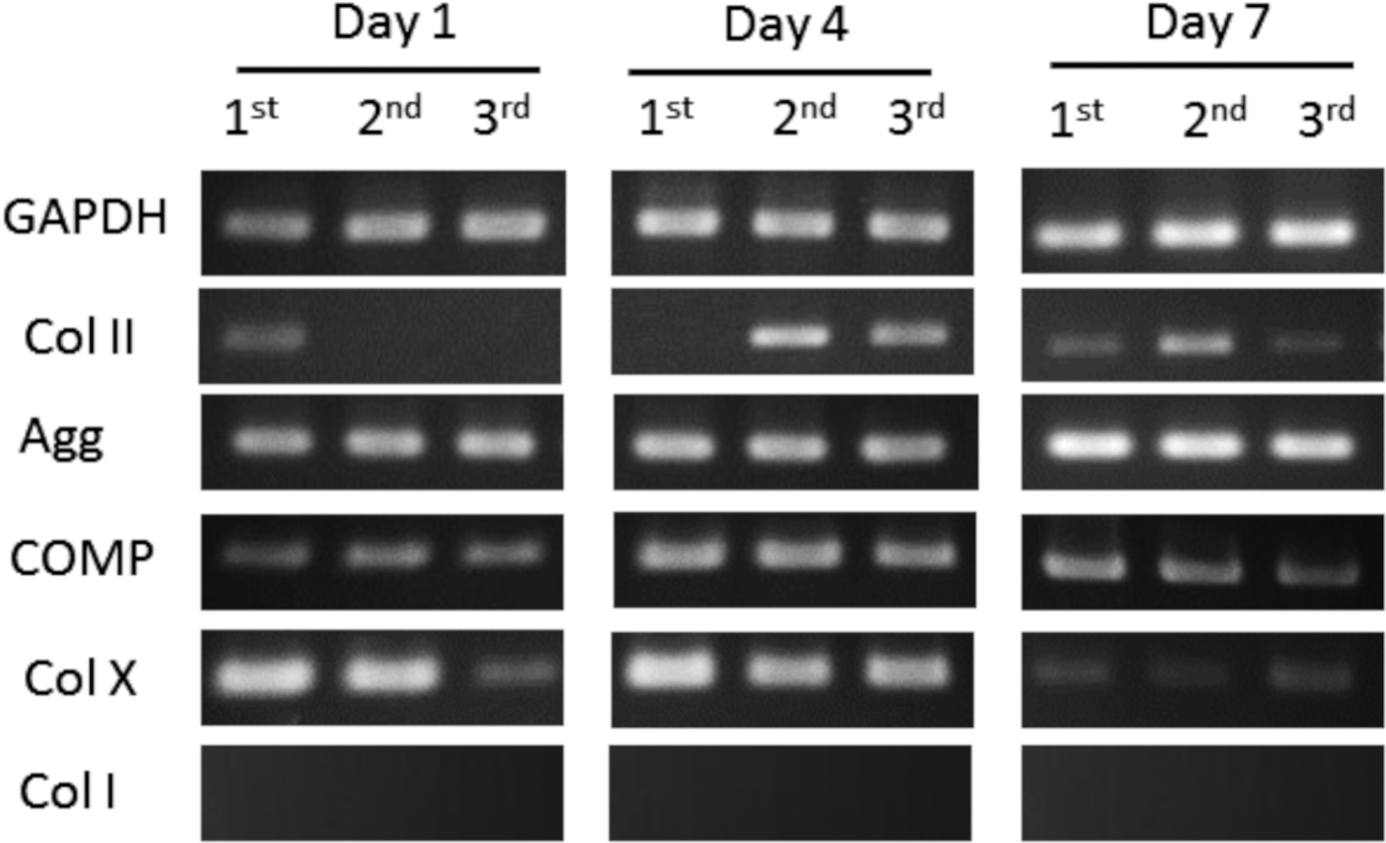
Gel electrophoresis of the chondrocyte markers in cells isolated from various rounds of digestion.

Quantitative polymerase chain reaction analysis of the chondrocyte markers in the cells isolated from different rounds of digestion. *p<0.05 while **p<0.01 when compared to the first round of digestion at each respective time point.
ECM synthesis and cell morphology
After examining the transcriptional levels of collagen type II, immunofluorescent staining was also performed on the harvested cells for this protein (Fig. 6). Regardless of the digestion round, all three groups were shown to produce a comparable amount of collagen type II. Apart from protein expression, the cell morphology was also analyzed by phalloidin staining. Over the 7-day culture period, the cell appearance took on the usual polygonal shape as seen in typical monolayer-cultured chondrocytes. There was no observable difference in the cell spreading area among the three groups.

Discussion
Chondrocyte isolation from cartilage is one of the most critical steps in determining the success of cell-based repair procedures such as ACI and cartilage tissue engineering. To date, the efficiency of cell isolation from native cartilage still remains suboptimal. As reported by previous studies, common isolation protocols only yield about 1–5 million viable chondrocytes per gram of cartilage, which is well below the total number of available cells in the native cartilage.2,6 In light of this, an improved protocol with enhanced cell isolation efficiency was developed. Instead of performing the usual one round of digestion in collagenase for a prolonged period of time (10–22 h),9–12 this new protocol essentially involves three consecutive digestions that comprises 14, 5, and 3 h respectively. For each round of digestion, a freshly prepared digestion medium containing 0.1% (w/v) of collagenase type II is employed.
The chondrocyte isolation efficiency in this proposed protocol is evaluated based on two criteria: cell yield and cell quality, the two major concerns in the development of any cell isolation protocol. From the results obtained in this study, we improved cell yield by more than fivefold; we successfully isolated a total of 1.75×10⁷ cells/g of viable chondrocytes as compared with the 2.34×10⁶ cells/g of cartilage in a single 22-h digestion (Supplementary Fig. S1). A higher cell yield is the direct result of the multiple digestion method employed, allowing more complete cartilage digestion, as evidenced by the greater reduction in volume of the digested cartilage fragments (Fig. 1). Furthermore, the use of fresh digestion medium at each digestion also helped to ensure a consistently high proteolytic activity from the collagenase, thus resulting in the release of more cells from the collagen fibril network. 2 Interestingly, it is also noted that the two additional digestions yielded much more viable cells than the first digestion even though the digestion periods are much shorter. This observation can be attributed to the first digestion that acted as a priming step for the initial disruption of the extensive collagen networks in cartilage ECM, thereby facilitating subsequent cell retrieval. Another possible reason is that the collection of cells at each round of digestion reduced the collagenase exposure in the isolated chondrocytes, leading to higher cell viability. This is evident from the 22-h digestion data where substantial amounts of small cell debris were observed (Supplementary Fig. S1c). It is hypothesized that this is a result of apoptosis of the isolated cells due to long hours of continuous collagenase digestion.
Besides cell viability, successful cell-based repair procedures also require high cell quality.13,14 In other words, isolated cells must retain their proliferative capacity and tissue-specific phenotype when cultured in vitro. Therefore, the effect of additional digestions on the proliferative capacity, gene expression, ECM synthetic activity, and cell morphology was assessed. The WST-1 proliferation assay and gene expression analysis of Rho A collectively showed that the newly developed protocol had no adverse effects on the proliferative capacity of the isolated cells. In fact, we observed that cells obtained from the additional rounds of digestion generally proliferated at a higher rate.
To evaluate the maintenance of phenotype in the chondrocytes isolated from all three digestion rounds, gene expression of various hyaline cartilage markers was examined. The gel electrophoresis results confirmed that cells from all three digestions strongly expressed collagen type II, aggrecan, and COMP. As mentioned, there were some irregular expressions of collagen type II. We believed that such irregularities are due to the constant exposure to a high collagenase activity. Chondrocytes from the second and third digestions experienced a total of 19 and 22 h of collagenase exposure, respectively, and require time for recovery 8 and thus did not express any Col II on day 1, but only on day 4. Collagen type I, a typical dedifferentiation marker for chondrocytes, remained undetected. To quantify the gene expression among different sample groups, qPCR was carried out. From the results obtained, the cells harvested from the third digestion were observed to have the highest expression levels for the anabolic genes examined. The zonal structures and depth dependence on collagen crosslinking extensiveness were previously mentioned,15–17 which may be a possible explanation for the results observed. It is hypothesized that the chondrocytes retrieved from the third digestion may contain a higher proportion of cells collected from the deeper zones of the native cartilage, which were reported to have greater synthetic capabilities for the main ECM components of cartilage than the superficial zone cells. 18
Although collagenase digestion may cause severe disruptions to the cellular structure, especially after prolonged treatment, 8 cells harvested from the additional digestion protocol have no observable morphological difference for the cells collected as compared to those from the first digestion, suggesting that the additional rounds of digestion did not cause any cell behavioral change. The results of this study generally suggested that the cells retrieved using this new protocol retained their chondrocytic phenotype and are manifested in their ability to synthesize the cartilage-specific ECM. This is particularly important for the construction of functional tissue-engineered constructs since the production of cartilaginous materials is very much dependent on the cells seeded in the construct. 8
Collectively, this study suggests that a more efficient protocol for isolating chondrocytes is established. This protocol has greatly improved cell yield for primary chondrocyte isolation by more than fivefold and also yielded cells with good quality. Therefore, we suggest that neither a longer digestion time nor a higher collagenase concentration is necessary; instead, multiple short digestions with freshly prepared digestion medium (0.1% w/v of collagenase) may be the technique to be used. The use of this isolation protocol is recommended especially for isolating cells from biopsied cartilage tissue samples in adult species for efficient utilization of the limited sample source.
Footnotes
Acknowledgment
This work was supported by AcRF Tier 1 RG 36/12 and AcRF Tier 2 ARC 1/13, Ministry of Education, Singapore.
Disclosure Statement
No competing financial interests exist.
References
Supplementary Material
Please find the following supplemental material available below.
For Open Access articles published under a Creative Commons License, all supplemental material carries the same license as the article it is associated with.
For non-Open Access articles published, all supplemental material carries a non-exclusive license, and permission requests for re-use of supplemental material or any part of supplemental material shall be sent directly to the copyright owner as specified in the copyright notice associated with the article.
