




Abstract
Enzymatic isolation of chondrocytes from a cartilage biopsy is the first step to establish in vitro models of chondrogenesis or to generate cell-based grafts for cartilage repair. Such process is based on manually operated procedures and typically results in yields lower than 20% of the total available cells. In this study, we hypothesized that, as compared to conventionally used protocols, the enzymatic digestion of human articular cartilage in the presence of ascorbic acid 2-phosphate (AscA2P) or of sodium chloride (NaCl), in combination with the use of a perfusion bioreactor system, leads to a higher and more reproducible yield of cell populations with high proliferation and chondrogenic capacity. The addition of AscA2P within the enzymatic digestion medium did not significantly increase the cell yield, but resulted in a significant decrease of the intradonor variability in cell yield (−17.8%±10.7%, p=0.0247) and in a significant increase of the proliferation rate of the isolated chondrocytes (+19.0%±1.4%, p<0.05) with respect to the control group. The addition of NaCl during cartilage digestion did not modulate cell yield. When the cartilage digestion was further performed under direct perfusion flow, beneficial synergistic effects were achieved, with an overall increase of 34.7%±6.8% (p<0.001) in the cell yield and an average decrease of 57.8%±11.2% (p<0.01) in the coefficient of variation with respect to the control group. Importantly, by implementing this strategy it was possible to retrieve clonal subpopulations more efficiently capable of undergoing chondrogenesis, both in vitro and in vivo. Our findings bear relevance for the preparation of human chondrocytes for laboratory investigations, and in the perspective of efficient and streamlined manufacturing of cell/tissue grafts for articular cartilage repair.
Introduction
A
A high cell density is known to be critical to initiate chondrogenic redifferentiation,4,5 which prompts for the generation of large numbers of chondrocytes. Thus, despite the associated costly, labor intensive, and time-consuming processes, isolated hAC are generally expanded in vitro prior to being transplanted at the defect site. During this expansion phase, hAC undergo a process defined as de-differentiation, losing the original phenotype characterized by round morphology and collagen type II production, and acquiring fibroblastic-like features as elongated shape and collagen type I deposition.6–10 Since de-differentiation may negatively affect the cell chondrogenic capacity and consequently the outcome of chondrocyte-based techniques for cartilage regeneration, cell expansion should be limited in time and scale, possibly maximizing initial cell yield while addressing standardization.
To our knowledge, the relation between specific digestion conditions and functional characteristics of the isolated AC, such as adhesion, proliferation rate, cell phenotype, and chondrogenic potential has been studied only in rabbits, 11 pigs, 12 ovines, 13 and only preliminarily by our group by one decade ago. 3
In this study, we aimed at improving the process of hAC isolation from a cartilage biopsy to (1) reduce their ex vivo expansion, (2) increase their proliferation and chondrogenic capacity, and (3) enhance the standardization across different preparations. These objectives were addressed by targeting different strategies. In particular, the differentiation medium was supplemented with (1) ascorbic acid 2-phosphate (AscA2P), to protect cells from oxidative stresses associated with the switch from hypoxic environment of the native matrix to the super physiological oxygen conditions during explantation14,15 or (2) sodium chloride (NaCl), to balance osmotic properties between native milieu and in vitro medium conditions. 16 Moreover, direct perfusion was introduced to enhance mass transport and to combine enzymatic with mechanical digestion, and to limit manual operations.3,17
Materials and Methods
Experimental design
A schematic diagram of the experimental design is displayed in Figure 1 and described in details in the sections below.
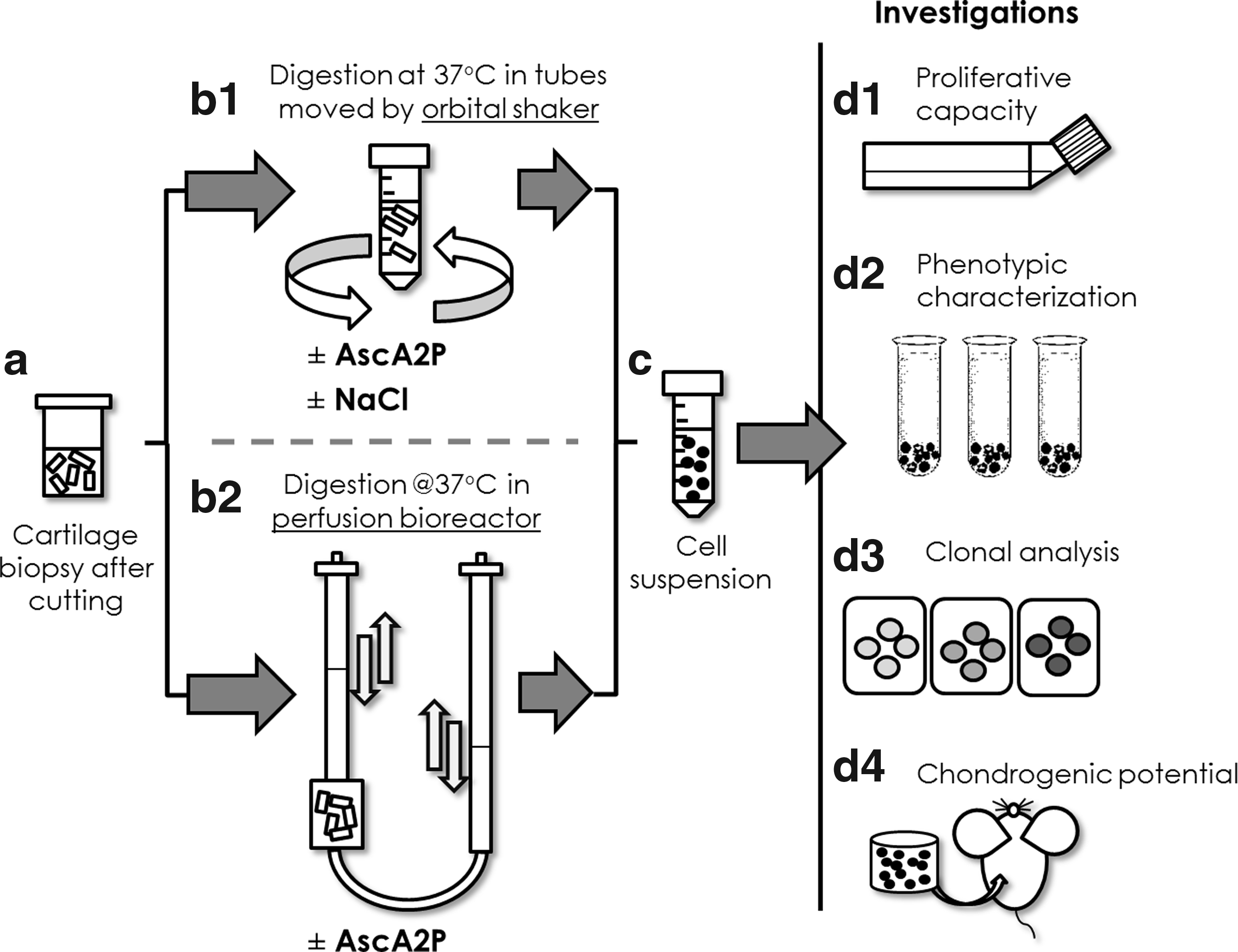
A schematic of the experimental design. We tested both the effect of ascorbic acid 2-phosphate (AscA2P) and sodium chloride (NaCl) added to the standard digestion solution in contact with cartilage pieces
Cartilage harvesting, cutting, and incubation
Full-thickness human articular cartilage samples were collected from the femoral lateral condyles of seven cadavers and 22 patients undergoing partial/total knee replacement (mean age 67.1±2.1, range 47–85) after informed consent by relatives was obtained and in accordance to the local ethical commissions. From such specimens, the macroscopically normal cartilage tissues (i.e., relatively smooth and whitish parts) were cut in 1–2 mm thick pieces with surgical blades (Fig. 1a) and kept in an a humidified 37°C/5% CO2 incubator between 24 and 72 h in Dulbecco's modified Eagle's medium containing 4.5 mg/mL D-glucose, 0.1 mM nonessential amino acids, 1 mM sodium pyruvate, 100 mM HEPES buffer, 100 U/mL penicillin, 100 μg/mL streptomycin, and 0.29 mg/mL L-glutamine (basic medium) supplemented with 10% fetal bovine serum (FBS; complete medium, all the reagents just mentioned were purchased from Gibco, Life Technologies, Zug, Switzerland). Some of the minced cartilage pieces were sampled and either stored at −20°C for further biochemical analyses or fixed and embedded in paraffin for histological evaluations.
We are aware that the cartilage specimens analyzed in our study, mainly derived from individuals relatively old and thus with possible early degenerative changes, may have different properties compared with the cartilage biopsies normally considered for ACI/MACI applications. However, the latter samples could not be considered in our study due to the difficulty of procurement (also for ethical reasons) and to their anyway limited size (that could not have allowed performing all the described investigations).
Cartilage digestion on orbital shakers
Chondrocytes were isolated (n=17 donors) by digestion of the cartilage pieces with 0.15% type II collagenase (355 U/mg; Worthington Biochemical Corp., Lakewood, NJ) in basic medium (CTR), or further supplemented with AscA2P (200 or 500 μM; Sigma-Aldrich, Buchs SG, Switzerland). These two AscA2P concentrations were chosen after a preliminary initial screening (data not shown). The digestion was performed in 50 mL tubes, considering 10 mL of digestion solution per gram of cartilage pieces, sealed with Parafilm for 22 h on an orbital shaker (40 rpm) in a humidified 37°C/5% CO2 incubator (Fig. 1b1). At least three independent replicates were evaluated per each experimental condition and donor. Upon digestion, chondrocytes were resuspended in complete medium, passed through a 100-μm cell strainer, and washed twice with complete medium via centrifugation at 1100 rpm for 5 min. Cells were then resuspended in complete medium (Fig. 1c) and counted using trypan blue. Cell yield was calculated as 106 cells/g of cartilage pieces or 106 retrieved cells/106 cells in native biopsy (estimated by DNA quantification, as described below). Freshly isolated chondrocytes were then plated at the initial density of 104 cells/cm2 and cultured for proliferation rate assessment (Fig. 1d1). An aliquot of cell suspension was cryopreserved in FBS supplemented with 10% (v/v) dimethyl sulfoxide (Sigma, St. Louis, MO) for following fluorescence-assisted cell sorting (FACS) analyses (Fig. 1d2).
The osmolality of digestion media was adjusted to match the value of the native tissue. A freezing point osmometer (Advanced Instrument, Inc., Norwood, MA) was used to calculate that 25 mM NaCl solution had to be added to the collagenase-based media (with or without AscA2P 200 μM, Fig. 1b1) to increase the osmolality from the original value of the digestion solutions (335 osm) to the value of articular cartilage (380 osm). 18
Cartilage digestion using perfusion bioreactors
Perfusion bioreactor systems—previously developed for cell seeding and cultivation into 3D scaffolds 17 —were also used for cartilage digestion (n=15 donors). Cartilage pieces (total weight: ∼1 g) were allocated in each chamber unit of a single closed perfusion bioreactor (Cellec Biotek AG, Basel, Switzerland), confined by two grids allowing liquid perfusion but not excessive movement of the cartilage pieces. The different digestion solutions (10 mL per bioreactor unit), prepared as described in the previous section, were injected in each bioreactor system (Fig. 1b2). At least three independent replicates were included per each experimental condition and donor. Bidirectional flow of digestion medium was applied at different rates, ranging from 390 to 1300 μm/s (which corresponds to 3.6–12 mL/min) by using a syringe pump, allowing perfusion of digestion medium through cartilage pieces. The bioreactor systems were placed inside a humidified 37°C/5% CO2 incubator, and the perfused digestion was prolonged for 22 h. Upon digestion, chondrocytes were treated as described below.
Chondrocyte proliferation
Isolated chondrocytes (n=13) were expanded in complete medium. After ∼14 days from the initial plating, when about 80% confluence was reached, cells (first passage cells, P1) were rinsed with phosphate-buffered saline (PBS), detached using 0.05% trypsin/0.53 mM EDTA (Gibco), and replated at 5×10³ cells/cm2. After 1 more week, when cells were again about 80% confluent, second passage cells (P2) were detached and induced to redifferentiate in pellet cultures as described below. At each passage the proliferation index was calculated as the ratio of log2(N/N0) to T, where N0 and N are the numbers of cells respectively at the beginning and the end of the expansion phase, log2(N/N0) is the number of cell doublings, and T is the number of days required for the expansion.
Clonal cultures of freshly isolated chondrocytes
Cell cloning was performed by limiting dilution as previously described, 19 starting from freshly isolated chondrocytes obtained using the different digestion conditions (Fig. 1d3). Briefly, the cell suspension was serially diluted to plate cells at a density of 1.0 cells/well in 96-well plates. Based on meticulous microscopic observations, cell populations were considered clonal if derived from wells where not more than one cell had been originally seeded. Clonal cell populations were cultured in complete medium supplemented with FGF2 (5 ng/mL) and TGFβ1 (1 ng/mL) (R&D Systems, Abingdon, United Kingdom) until confluence and then passaged in 24-well plates. Upon reaching a second confluence, cells were detached and induced to chondro-differentiate as described below.
Colony forming efficiency
Freshly isolated chondrocytes (n=8) were plated in 100 mm diameter Petri dishes at the clonal cell density of three cells/cm2 and cultured for 14 days. Colony forming efficiency (CFE) was calculated as the number of colonies observed following Crystal violet staining (at least three dishes per experimental condition), and normalized to the total number of seeded cells.
In vitro chondrogenic differentiation
Chondrogenic differentiation was induced in 3D micromass pellet cultures using a defined serum-free medium, as previously described. 20 Single cell-derived colonies were suspended in chondrogenic medium, consisting of basic medium supplemented with insulin 10 μg/mL, transferrin 5.5 μg/mL, selenium 5 ng/mL, human serum albumin 0.5 mg/mL, linoleic acid 4.7 μg/mL, AscA2P 0.1 mM (Sigma), dexamethasone 10−7 M (R&D Systems, Minneapolis, MN), and TGFβ1 10 ng/mL. Cell suspensions were diluted at 106 cells/mL, distributed (0.5 mL per tube) into 1.5 mL polypropylene conical tubes (Sarstedt, Numbrecht, Germany) and centrifuged at 1100 rpm for 3 min to form spherical pellets. Pellets were cultured for 2 weeks, with medium changed twice weekly and finally assessed histologically and biochemically.
In vivo chondrogenic differentiation
Freshly isolated chondrocytes from three donors were seeded into collagen scaffolds (1.6×10⁶ cells per 6 mm in diameter and 3 mm in thickness disks of Ultrafoam®; Davol, Providence, RI). Static seeding on the biomaterial was performed as previously described. 17 Cell-seeded scaffolds (n=4 per each experimental condition per each donor, three donors) were let in a humidified 37°C/5% CO2 incubator overnight in complete medium (Fig. 1d4) and then directly implanted in subcutaneous pouches on the back of nude mice (four scaffolds/animal). 21 Mice were sacrificed 8 weeks after surgery and the constructs were harvested for histological and biochemical analyses. All animal studies were approved by the responsible ethical authorities and by the Swiss Federal Veterinary Office.
Histology
Cultured micromass pellets and in vivo constructs were washed in PBS after harvesting, fixed in 4% formalin overnight at 4°C, embedded in paraffin, cross-sectioned (5 μm thick), and stained with Safranin-O for sulfated glycosaminoglycans (GAG). The Bern score was used to evaluate the quality of engineered chondrogenic samples 22 : scoring >3 (possible maximum value: 9) was considered corresponding to chondrogenic samples (assessment performed by three independent and expert evaluators). When possible, a full-thickness biopsy from native tissue was harvested and treated with the same protocols to assess the quality of the starting material and possibly correlate it with isolation yield data. The quality of the cartilage was assessed using the modified Mankin's score in Safranin-O-stained sections. 23
GAG and DNA assays
To measure total GAG and DNA amounts of native cartilage, micromass pellets, and in vivo constructs each sample was washed in PBS, digested with protease K (0.5 mL of 1 mg/mL protease K in 50 mM Tris with 1 mM EDTA, 1 mM iodoacetamide, and 10 μg/mL pepstatin-A for 15 h at 56°C). GAG amounts were spectrophotometrically measured using dimethylmethylene blue, 24 with chondroitin sulfate as a standard, and normalized to the DNA amounts, spectrofluorometrically measured using the CyQUANT Kit (Molecular Probes, Eugene, OR) and with calf thymus DNA as a standard. 20 GAG contents are reported either as μg GAG/μg DNA for in vitro samples and native tissues or μg GAG/μg wet weight for in vivo samples.
FACS analysis
Frozen cells, separately stored per each experimental condition after isolation (n=13), were thawed, washed, blocked in 1% bovine serum albumin for 20 min at room temperature, and then incubated in the dark at 4°C for 15 min with a fluorochrome-conjugated antibody for CD29, CD44, CD49c, CD49e, CD49f, CD90, CD105, CD166, and s100 (Becton Dickinson, Franklin Lakes, NJ) or the correspondent IgG control, using propidium iodide to exclude dead cells. Cells were analyzed using a FACSCalibur flow cytometer (Becton Dickinson). Data analysis was performed with FlowJo software (version 3.4; Tree Star, Inc., San Carlos, CA). 25
Statistical analysis
Data are presented as mean±standard error. The number of donors used is specified for each experiment. For each donor and experimental condition, at least triplicate samples were used for each assessment. Statistical analysis was performed using two-tailed unpaired t-test or the nonparametric Mann–Whitney U-test, after assessing the normality of distribution of the collected data by means of Prism® software (GraphPad Software, La Jolla, CA). One-way analysis of variance (ANOVA), in combination with a Tukey's range post hoc test, was used when comparing more than two groups. p≤0.05 was selected to define statistically significant differences.
Results
AscA2P addition during cartilage digestion results in a positive trend in the cell yield
We first assessed the effect of AscA2P supplemented to the standard digestion solution (CTR) at two different concentrations (200 and 500 μM). The time for complete digestion was maintained constant (22 h), based on a macroscopic evaluation of the state of tissue digestion and previously established protocols. 3 Upon retrieval of single cell suspensions, a trend toward an enhancement of the cell yield was observed when AscA2P was added at 200 μM (8/11 donors with cell yield higher than CTR), whereas AscA2P 500 μM induced a cell yield higher than CTR only in 2 out of 6 donors, as shown in Figure 2a. However, none of these differences were statistically significant (p=0.158 for AscA2P 200 μm vs. CTR comparison, p=0.357 for AscA2P 500 μm vs. CTR comparison).

Despite the large deviations in the measurements due to the normal inter- and intradonor variability when isolating hAC, we could observe a clear positive, though not statistically significant, trend in the cell yield when 200 μM of AscA2P were added to the collagenase-based digestion medium. Thus, further analyses were focused on a collagenase-based digestion medium supplemented with 200 μM AscA2P, defined as the condition more consistently increasing the cell yield.
Since the density of chondrocytes can vary between cartilage specimens, the cell yield was also calculated as a fraction of the total number of cells available in the tissue (Fig. 2b). The cellular content in cartilage tissue was determined biochemically by measuring the DNA content of tissue digests, 26 considering a DNA content per human chondrocyte of 11.6 pg. 3 The cell yield normalized to the DNA content for each specific donor displayed a nonsignificant trend similar to the one of the absolute values.
When adding AscA2P 200 μM, the coefficient of variation between the replicates of the different conditions per each donor (i.e., intradonor variability) was significantly reduced with respect to the CTR group in 12 out of 17 cases, with an overall reduction of −17.8%±10.7% (Fig. 2c, p=0.0247). In the remainder of the study, the chondrocyte yield will be expressed as a percentage of the total cell number and not normalized to the tissue weight.
Aiming at further improving the hAC isolation yield, the osmolality of digestion media was adjusted by adding 25 mM NaCl solution to the collagenase-based media (with or without AscA2P 200 μM) to match the value of the native tissue. However, no significant effects were found when NaCl was added either alone or in combination with 200 μM AscA2P (Fig. 2d).
AscA2P increases 2D cell proliferation rate and the clonogenicity upon digestion while modulating key cellular surface markers
The proliferation index was higher for cells isolated in the presence of AscA2P with respect to the CTR group for the majority of the donors (10/13; Fig. 3a), with an overall increase of 19.0%±1.4% (p=0.0324). The proliferative capacity was enhanced also at clonal level, as observed by an evident increase of colony diameter (Fig. 3b). The CFE was not significantly higher in the AscA2P group versus the CTR group (+26.6%±2.9%, p=0.0651; Fig. 3c). We then assessed the chondrocyte phenotype upon isolation (n=13 donors). We focused on mesenchymal markers, such as CD29, CD105, and CD166, and putative predictive markers for chondrogenic differentiation capacity, that is, CD49e, 27 CD44 and CD49f, 28 s100, 29 and CD90. 30 Statistically significant higher percentages of CD44-, CD49e-, CD49f-, s100-, CD90-, and CD105-positive cells were found in AscA2P condition. CD166, CD49c, and CD29 were not modulated with respect to the CTR condition (Fig. 3d).
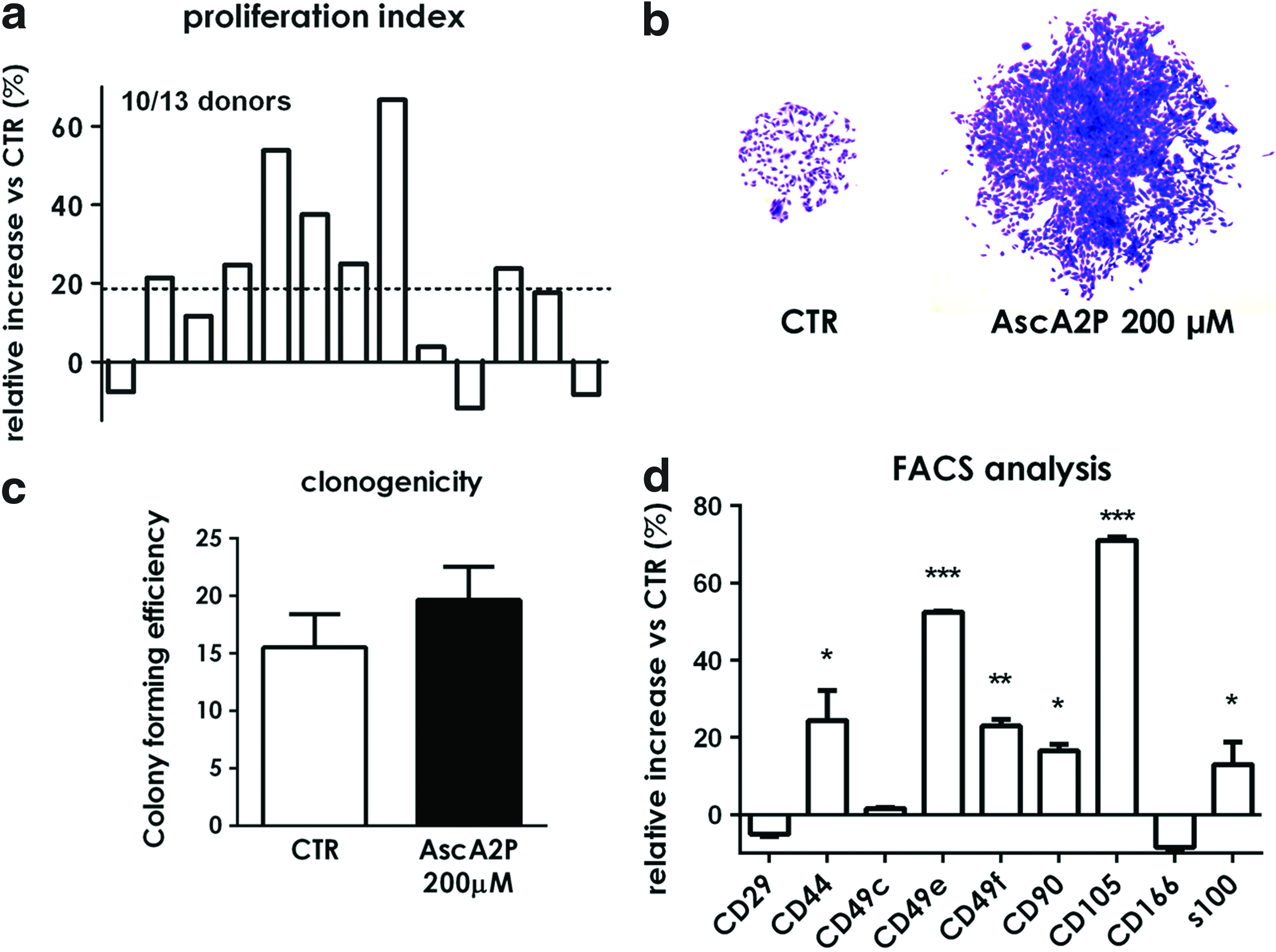
AscA2P increased 2D cell proliferation rate upon digestion while modulating key cellular surface markers. The proliferation index was higher in the AscA2P with respect to the CTR group in 10 out of 13 donors, both at a multiclonal
Bioreactor-based digestion improves cell yield and standardizes the isolation procedure
We then investigated whether mechanical forces under the form of fluid-induced shear could be coupled to the conventional enzymatic-based cell retrieval process to further increase the hAC isolation yield and quality. A perfusion-based bioreactor system (Fig. 1b), previously used for cell seeding and tissue development into 3D scaffolds,17,31 was thus introduced (n=15 independent runs). Initially, different flow rates were screened using the standard collagenase-based digestion medium. Looking at the trend (Fig. 4a), 650 μm/s (corresponding to 6 mL/min) was selected as the one providing both the highest cell yield and proliferation rate of the isolated cells. Another set of experiments was carried out to compare the standard shaker- and the bioreactor-based digestion without (CTR-shaker; CTR-bioreactor) or with AscA2P 200 μM (AscA2P-shaker; AscA2P-bioreactor). Digestion under perfusion did not result in an increased the cell yield (+7.5%±6.0%, p=0.16) (Fig. 4b) but significantly decreased the coefficient of variation (−41.1%±10.9%, p=0.0043) (Fig. 4c) as compared to the standard shaker-based method (i.e., CTR-shaker condition). When the bioreactor-based digestion process was combined with the use of AscA2P (AscA2P-bioreactor condition), an overall increase of 34.7%±6.8% (p<0.001) in the cell yield and decrease of −57.8%±11.2% (p<0.001) in the coefficient of variation with respect to the CTR-shaker condition were observed (Fig. 4b, c). Such percentages are larger than the sum of the changes introduced by the AscA2P supplementation or bioreactor introduction, indicating a beneficial synergistic role of AscA2P and perfusion.
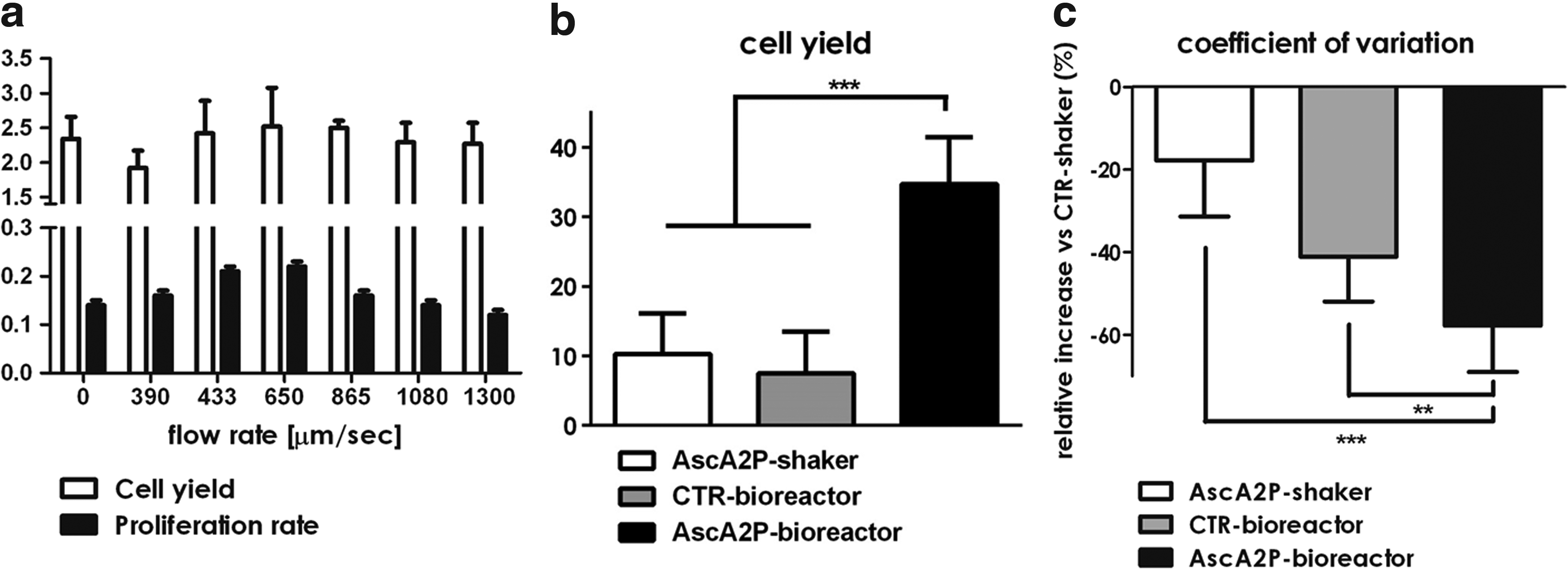
Bioreactor-based digestion in combination with the addition of AscA2P 200 μM in the medium improved the cell yield and standardizes the isolation procedure (n=15 donors).
No correlations were found between the enhanced cell isolation yield and the tested donor-related parameters, including donor age, modified Mankin Score, cellularity, and GAG content of the corresponding native tissue (data not shown).
Bioreactor-based digestion improves the chondrogenic potential of hAC at clonal level
To investigate whether the digestion method can alter the ratios of subpopulations with different chondrogenic potential, a clonal study upon digestion for three experimental groups (i.e., CTR-shaker, AscA2P-shaker, and AscA2P-bioreactor) was performed. Compared with the CTR-shaker group, in the two AscA2P-containing digestion group 10.7% more clones with highly proliferative features (defined as the clones with a proliferation index higher than 0.7) were derived. Upon expansion, each single cell-derived progeny was tested for chondrogenic potential in vitro by micromass culture. Safranin-O pictures of representative clones qualitatively display the higher chondrogenic potential of the clones in the AscA2P group (Fig. 5a–c). The AscA2P-bioreactor was the only experimental group showing an average Bern score higher than 3 (threshold of chondrogenic potential for a Bern Score)—even in the low proliferating clones—and a higher percentage of chondrogenic clones—namely 66.7% as compared to 20.0% in the CTR-shaker and 33.3% in the AscA2P-shaker groups (Table 1 and Fig. 5d, e).
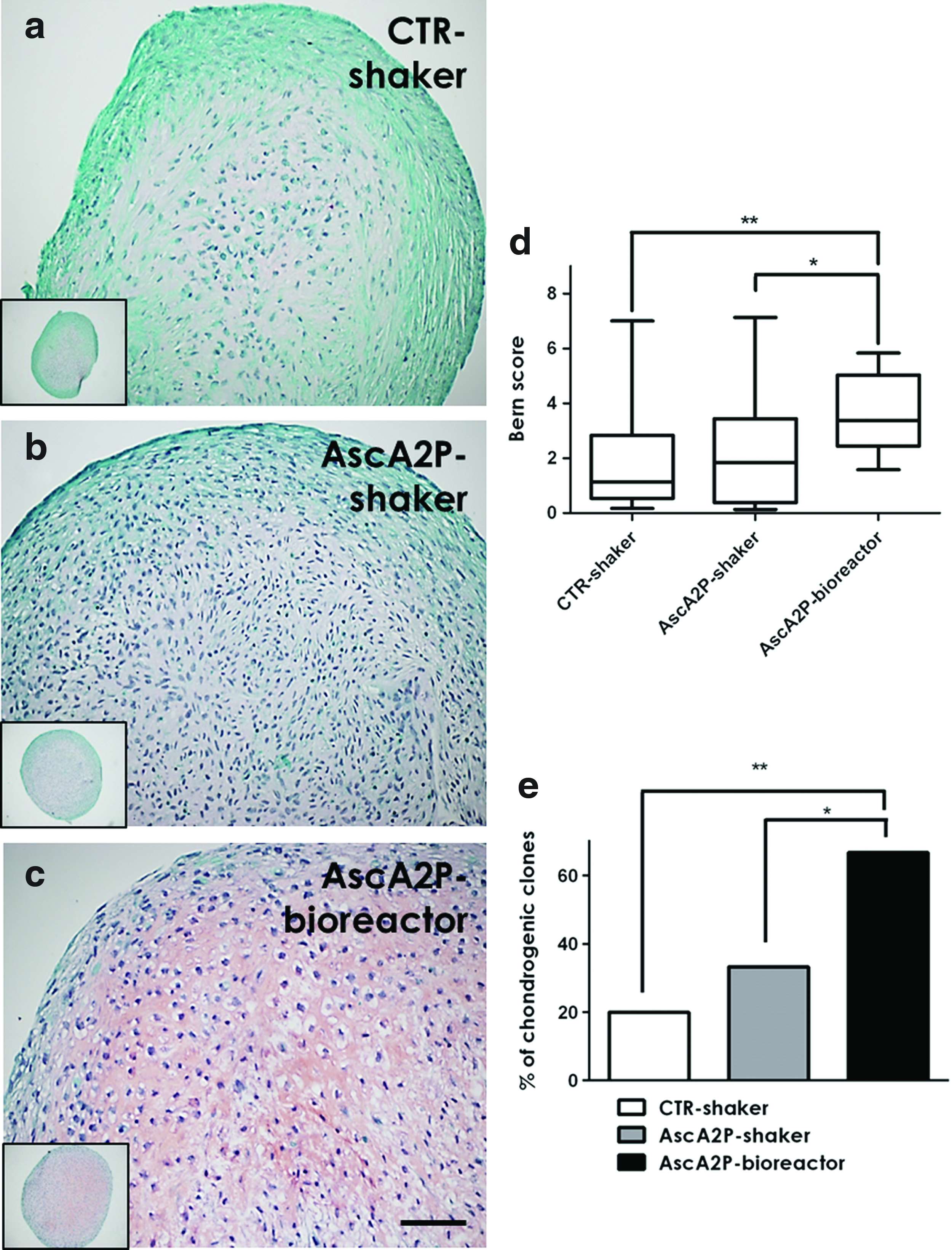
Bioreactor-based digestion improved the chondrogenic potential of hAC at clonal level. Safranin-O pictures of representative clones, corresponding to the clones that better match the average Bern score, for the CTR-shaker
Chondrocyte preparations were divided into low and high proliferative clones, generated using the three digestion protocols.
Bioreactor-based digestion improves the in vivo chondrogenic potential of hAC
We then tested the in vivo cartilage forming capacity of hAC isolated using either the standard shaker-based protocols without (CTR-shaker) and with AscA2P (AscA2P-shaker) or the bioreactor-based digestion method in the presence of AscA2P (AscA2P-bioreactor).
Immediately after digestion, chondrocytes were statically seeded on 3D collagen-based scaffolds (Ultrafoam®); cell-seeded constructs were then subcutaneously implanted in nude mice. Despite the normal inter- and intradonor variability evidenced by the differences in the size of harvested constructs, hAC isolated with the AscA2P-bioreactor protocol displayed a higher chondrogenic differentiation capacity with the respect to the other two experimental groups in all the three donors tested. The finding was confirmed both qualitatively, as shown by the Safranin-O staining (Fig. 6a–i), and quantitatively, as assessed by the statistically significant higher amount of deposited GAGs and by the 43% higher Bern Score (Table 2).
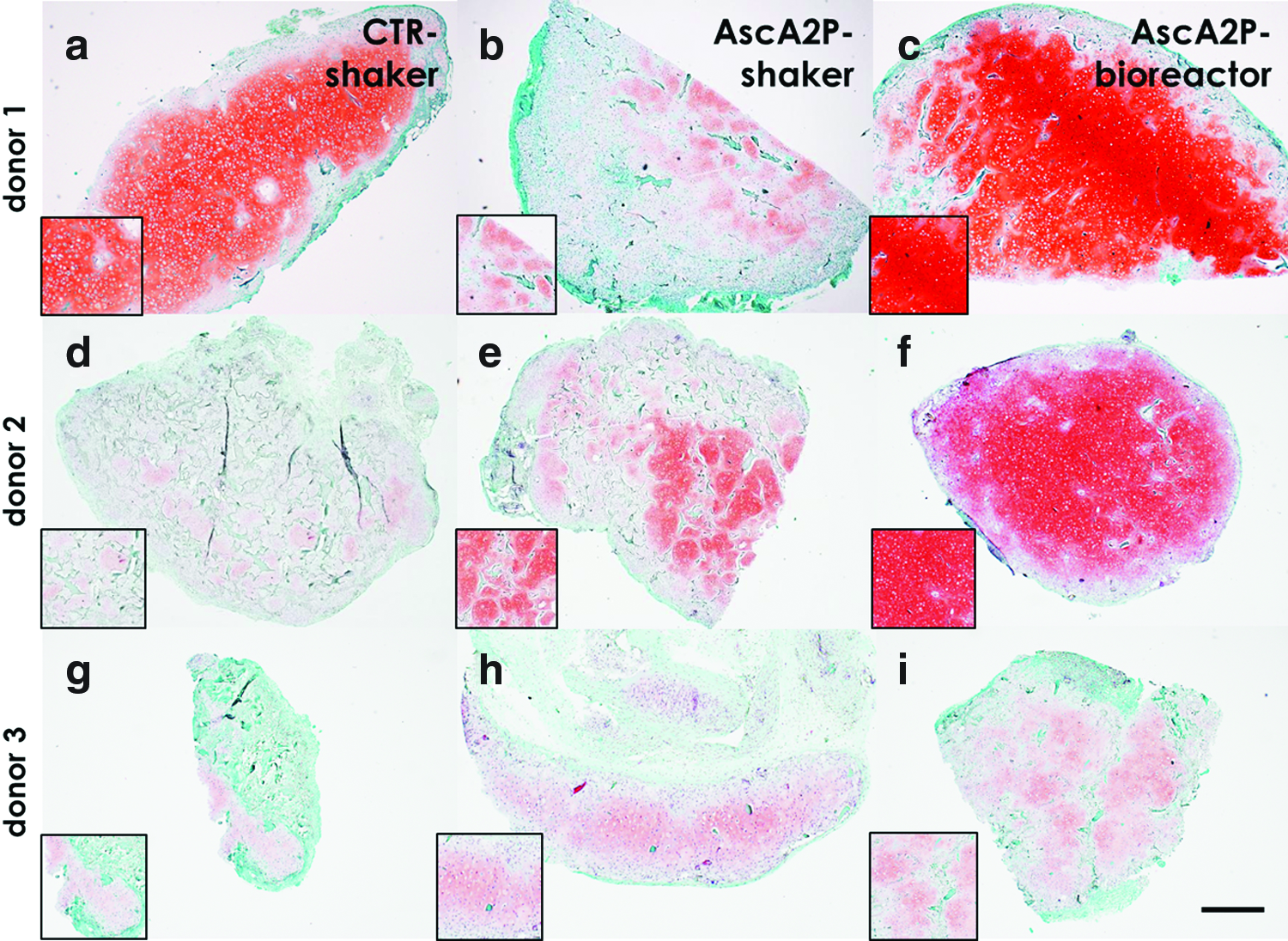
Bioreactor-based digestion improved the in vivo chondrogenic potential of hAC at multiclonal level (four constructs per donor, three donors). Safranin-O pictures of the three donors tested
p<0.01.
p<0.001.
AscA2P, ascorbic acid 2-phosphate; GAG, glycosaminoglycan.
Discussion
We have shown that the addition of an antioxidative factor (AscA2P) to the digestion medium, combined with the use of a perfusion-based bioreactor device increased the cell yield per gram of cartilage tissue biopsy up to 41.5%. Remarkably, the isolated hAC displayed an enhanced clonogenicity and propensity to proliferate. The tested parameter of osmolality, found to positively modify the digestion yield in bovine cartilage 32 and the proliferation rate of hAC,16,33 did not modulate the yield of isolated cells.
The beneficial effects of using AscA2P on the properties of the recovered chondrocytes could be due to the reduction of oxidative stress, through the inhibition of reactive oxidative species (ROS). Yudoh et al. 14 have indeed shown that by using antioxidative agents (i.e., AscA2P 100 μM) during AC cultures it is possible to prevent ROS-induced detrimental effects, such as reduction in GAG production, telomere length, and replicative capacity. The statistically significant and more prominent beneficial effect introduced by the AscA2P in the bioreactor-based system prompted us to speculate that this might be due to the fact that ROS production may be increased by the direct perfusion. Investigating this hypothesis will be a goal of future studies. Since the diffusion of the factors through the tissues is enhanced under perfusion, it can also be possible that the higher cell yield in bioreactor is mediated by a more efficient cellular intake of ascorbic acid.
The use of a perfusion bioreactor systems, usually employed for dynamic cell seeding and culture and hitherto never used for cell isolation from native tissue during cartilage digestion, allowed not only to further enhance the performance and reproducibility of the isolation process but also to improve the intrinsic cartilage forming capacity of the retrieved cells at clonal and multiclonal level. The cartilage pieces could be easily transferred in the bioreactor chamber and subjected to enzymatic digestion, making the whole procedure less user-dependent and thereby reducing inter-donor and inter-sample variability.
In the perspective of ex vivo generation of chondrocyte-based cartilage substitutes, our developed protocol could have a beneficial impact on manufacturing process and related costs by decreasing the time of the required in vitro cell expansion phase due to the higher cell isolation yield coupled with enhanced proliferation capacity. As an example, we may assume (1) to receive a biopsy weighing 75.5 mg 34 and (2) to have a synergistic improvement introduced by the AscA2P both in the isolation yield and in the proliferation rate. From our data it can be calculated that by adding 200 μM of AscA2P in the digestion solution and by performing the digestion procedure with perfusion bioreactors, the minimal cell density required to perform scaffold-based chondrocyte transplantation—that is, 2×10⁶ cells/cm2—could be reached 8 days earlier than using the CTR-shaker condition (25 vs. 17 days). 35 This would be a relevant advantage to reduce the labor intensive and thus costly serial cell passaging in monolayers.
The observed increase in the number of cells expressing markers associated with the mesenchymal progenitor phenotype and/or the intrinsic chondrogenic potential of hAC—such as CD90, 30 s100, 29 CD49e, 27 CD44, and CD49f 28 —along with the trend versus a higher clonogenicity in the AscA2P group, suggest that the improved cell yield and performance might be linked to the isolation and/or the preservation of specific subpopulations of progenitor cells during the digestion. This hypothesis is also supported by the increased frequency of chondrogenic clones found in the AscA2P-containing experimental groups. However, the actual existence of such populations is highly debated and the association of the expression of the modulated markers with these chondrogenic subpopulations was beyond the scope of this study. To address this pivotal open question, sorting specific subpopulations immediately after digestion will have to be combined with the establishment of quality markers able to predict the intrinsic chondrogenic potential of isolated cells. 19
Perfusion-based bioreactor systems could be also employed to solve another issue that may still impair the standardization and automation of the process. The first step of cutting the cartilage biopsy, necessary to increase the surface area available for enzymatic matrix digestion, may be indeed minimized by tuning the flow rate of the digestion medium perfused through the tissue, as much as needed to mechanically help/accelerate matrix dissociation. This may also have a positive impact on the cell yield, since cell death is described along the edges of each slash. 36
The use of a perfusion-based bioreactor opens the perspective of a streamlined tissue engineering approach where all the phases of graft manufacturing, from cell isolation to expansion and if necessary matrix production could be performed in single closed automated system.31,37 Furthermore, in a tissue engineering approach, freshly isolated hAC could be dynamically seeded in 3D scaffolds within the same closed bioreactor system, and induced to first expand and then differentiate directly within the biomaterial and under optimized perfusion rates. 38
Finally, we envision that described method may be extended to other tissue types (e.g., bone marrow isolation from mouse compact bone 39 ) to improve and standardize the isolation of the initial cellular material with an immediate effect in research and clinical applications.
Footnotes
Acknowledgments
We wish to thank Francine Wolf for her technical assistance, Dr. Christoph Hilker for performing osmolality measurements, and Dr. Atanas Todorov for the ectopic implantation in nude mice. This work was partially supported by the Eurostars project E!6065 IsoCart.
Disclosure Statement
The authors have a potential conflict of interest related to their specified industry affiliations, as CellCoTec BV and Cellec Biotek AG operate respectively in the areas of cell-based cartilage repair and of perfusion-based bioreactor systems.