




Abstract
Objective:
To construct an automatic decellularization platform (ADP) for preparing xenogenic extracellular matrices (ECMs), and to demonstrate that automatic decellularization for preparing xenogenic ECMs reduces processing time, requires fewer attendee hours, and is as effective as the manual gold standard preparation protocols.
Materials and Methods:
A soft tissue ADP was constructed and ovine aorta was harvested (n=9). Manual and automatic decellularization was performed on aortic tissue specimens and both groups were compared. The presence of acellularity was assessed with viability/cytotoxicity assays, and the presence of residual ovine DNA was determined with gel electrophoresis and spectrophotometry. Scaffold integrity was characterized with scanning electron microscopy (SEM) and uniaxial tensile testing.
Results:
Acellularity was confirmed with both preparation techniques and DNA concentrations measuring 540±130 and 590±270 ng/mg wet weight and the control measuring 6690±1210 ng/mg wet weight (p<0.05). SEM demonstrated no differences in the surface architecture of ECMs prepared by both techniques. Uniaxial testing demonstrated no significant differences in the incremental elastic moduli E below a stretch ratio of 2.70λ in both groups and a large reduction in E was recorded when both groups were compared with control samples above a stretch ratio of 1.7.
Conclusion:
Automatic decellularization of ovine aorta is as effective as gold standard manual decellularization protocols. Future research will focus on optimizing the automated decellularization technique and on upscaling protocols.
Introduction
A
Materials and Methods
Overview of experimental design
An ADP was constructed and comprised a biological tissue compartment (tissue digestion chamber), a multiwell solute facility with fully automated control (Fig. 1). Ovine aorta was decellularized manually and compared with the ADP. Feasibility of both decellularization processes was assessed by comparing residual DNA levels, acellularity, residual DNA fragment sizing, and the incremental elastic modulus.
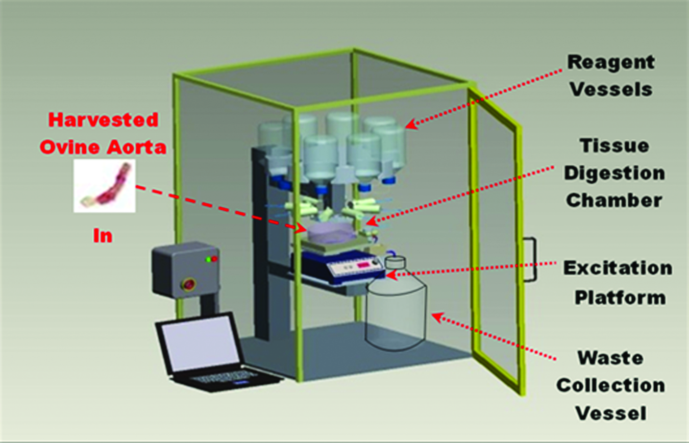
Automated decellularization platform; ovine aorta tissue specimens are loaded into the tissue digestion chamber, which is attached to the waste collection vessel. Reagent vessels are filled and the excitation platform speed selected. The decellularization protocol sequencing program is downloaded to the programmable logic controller (PLC), setting dispensing sequences and temperature parameters. Reagents are pumped into the tissue digestion chamber under PLC control, with the chamber evacuated of depleted reagents and residues to the waste collection vessel under instructions of the PLC program. On completion, the resulting extracellular matrices (ECMs) are removed to cold storage. Color images available online at
Ovine aorta sample preparation
Ovine aorta was harvested directly following euthanasia from 7-month-old females at the abattoir (n=9) (Gaelic Meats Ltd., Limerick, Ireland). Tissue specimens were transported on ice to the laboratory storage facility in a solution of phosphate-buffered saline (PBS) after euthanasia and maintained at −20°C for 2 weeks. Strips of ovine aorta were prepared by performing a circumferential incision, producing tubular segments, which were then incised into rectangular strips, facilitating fiber orientation postdecellularization for manual (n=9) and automated decellularization (n=9).
Decellularization protocol
Manual decellularization was performed according to the protocols described by Teebken et al. 6 and Steinhoff et al. 9 In brief, sterilization was achieved by immersing the tissue samples in a glass beaker with 250 mL of PBS and 0.02% sodium azide (NaN3), placing them in an incubator at 37°C for 30 min, and storing at −20°C for 2 weeks (Sigma-Aldrich, St. Louis, MO). Freezing was also used to aid in cell lysis by means of physical disruption by ice crystal formation in the cell and ECM ultrastructure, as it has little impact on the mechanical properties of the resulting ECM.4,11,12
Manual decellularization
Decellularization reagents were obtained from Sigma-Aldrich, Dorset, United Kingdom, unless indicated. PBS was added to the vessel containing the specimens until they were completely immersed and placed on a continuous excitation platform for 15 min at 45 oscillations per minute (opm) (Fisher Scientific UK Ltd., Loughborough, United Kingdom). Remaining residues were drained from the vessel on completion, and the samples were sterilized using 0.02% v/v NaN3 for 30 min at 37°C. A solution of 0.1% v/v Trypsin with 0.01% v/v EDTA and 0.3% sodium azide in PBS was prepared and the tissue samples were immersed in this solution and agitated at 45 opm for 24 h at 37°C. The solution was then drained from the vessel and any debris removed. A second solution of Trypsin/EDTA/EGTA sodium azide 0.1%/0.05%/0.2%/0.3% v/v PBS solution was added to the vessel until the samples were completely covered and agitated at 45 opm for 24 h at 37°C. Samples were then drained of the Trypsin/EDTA/EGTA solution, rinsed twice for 15 min in PBS, and then stored in 50 mL of deionized water/50 mL of PBS with 2 mL of anfirm/antibiotic solution and 200 mg of NaN3 at 4°C.
Automatic decellularization platform
In brief, the primary constituents of the ADP are illustrated in Figure 1(Centre for Applied Biomedical Engineering Research [CABER], University of Limerick, Limerick, Ireland). It comprised a biological tissue digestion chamber, a multiwell solute facility, and a waste disposal partition with fully automated control using a programmable logic controller (PLC). The tissue digestion chamber was located on a continuous excitation platform in the center of the ADP directly underneath the reagent dispensing nozzles and included temperature control. The tissue digestion chamber was connected to the waste collection vessel.
Automatic decellularization methods
Tissue specimens were loaded into the tissue digestion chamber. Gentle agitation was set at 45 opm. Reagents were loaded into reagent storage vessels and placed on the ADP. Specific reagent quantities utilized for the automatic decellularization protocol were as follows: Reagent vessel 1, 300 mL PBS, 0.1% v/v Trypsin, 0.01% v/v EDTA, and 300 mg NaN3; Reagent vessel 2, 300 mL PBS, 0.1% v/v Trypsin, 0.05% v/v EDTA, 0.2% v/v EGTA, and 300 mg NaN3; Reagent vessel 3, 1200 mL PBS; Reagent vessel 4, 100 mL deionized water, 100 mL PBS, 2 mL mixture of anfirm/antibiotic solution, and 400 mg NaN3; The control sequence used to automatically decellularize the ovine aorta is listed in Table 1 showing the sequencing for each step of the protocol. On completion, the tissue digestion chamber along with the decellularized contents was removed from the ADP and the resulting ECMs stored at 4°C until subsequent analysis took place.
Reagent vessel 1 is emptied into the tissue digestion chamber set at 37°C with 24 H of agitation, and then used reagents are evacuated. PBS solution is pumped from reagent vessel 3 into the tissue digestion chamber for 0.5 h (×2 cycles) at room temperature under gentle agitation. Spent PBS is then evacuated to waste. Reagent vessel 2 is emptied into the tissue digestion chamber at 37°C with 24 h of agitation, and then used reagents are evacuated. PBS solution is pumped from reagent vessel 3 into the tissue digestion chamber for 0.5 h (×2 cycles) at room temperature under gentle agitation. Spent PBS is then evacuated. Antibiotic solution is pumped from reagent vessel 4 to the tissue digestion chamber.
PBS, phosphate-buffered saline.
Scanning electron microscopy
To assess and characterize the structural integrity of the ovine aorta ECM prepared by the manual and automatic decellularization protocols, scanning electron microscopy (SEM) was performed on the ovine aortic ECMs pre- and postdecellularization. Control specimens were examined immediately on arrival at the laboratory. The scaffolds were fixed in 2% glutaraldehyde, 1% p-paraformaldehyde, and 0.1 M PBS (pH 7.4) at room temperature for 1 h and thereafter were dehydrated in a series of graded ethanol concentrations (50–100% ethanol; Bio Sciences Ltd., Dun Laoghaire, Ireland.). The samples were then mounted onto metallic stubs with double-sided carbon tape. Thin layers of gold and palladium were applied to each sample with an automated sputter coater. The samples were then examined under a Jeol CarryScope scanning electron microscope (Hertfordshire, United Kingdom) at 10 kV. Specimens were mounted with half the studs exposing the luminal surface and the other half exposing the adventitial surface.
Verification of cell removal
An analysis of the ovine aortic tissue both before and after the application of the decellularization protocols was performed to establish the acellularity of the remaining ECM.
Viability/cytotoxicity assay
Viability/cytotoxicity fluorescence assays were performed using a fluorescent light microscope (transmission) (Nikon Eclipse TE200, Nikon, Alton, United Kingdom) on both experimental groups to compare and illustrate the cell presence or absence following the application of the decellularization protocols (Bio Sciences Ltd.). 13 For the viability/cytotoxicity assays, control specimens and postdecellularization specimens were stained using calcein and ethidium homodimer-1 (Molecular Probes, Eugene, OR). Calcein is actively converted to calcein-AM in viable cells, which appear green under a fluorescence microscope. Ethidium homodimer-1 only accumulates in dead cells, which appear red (Bio Sciences Ltd.).
Tissue DNA extraction
DNeasy assays were performed for purification of total genomic DNA from both control and decellularized specimens (Qiagen, Germantown, MD). DNeasy tissue extraction kits contain a buffer system, which is used to induce cell lysis, followed by selective binding of the DNA to the DNeasy membrane. Purified DNA is eluted in a low-salt buffer for extraction. A specially formulated storage buffer, including 20 μL of proteinase K, was used for total specimen tissue lysis with an activity of 600 mAU/mL. A tissue lysis buffer labeled ATL is also part of the kit and was used to break cell membranes. The resulting DNA-laced solution was stored at −20°C.
DNA quantification (spectrophotometer)
Control and decellularized specimens had their DNA levels quantified pre- and post-decellularization using a NanoDrop spectrophotometer (Thermo Fisher Scientific, Inc., Waltham, MA). Residual DNA concentrations were measured using the NanoDrop's UV light at a wavelength of 400 nm. Three microliters of DNA was aliquoted into a mini tube with 3 μL of molecular grade H2O and proceeded by 1 min of vortexing. Three measurements of DNA concentrations (n=3) were taken per specimen (n=9) and an average result calculated. NanoDrop software calculated the DNA/RNA levels for each specimen. Results from the spectrophotometer are presented in ng/μL.
DNA electrophoresis–DNA fragment sizing
DNA Electrophoresis was used as a means for detecting minute traces of DNA before and after performing the manual and automatic decellularization protocol for both control and processed specimens (n=9). 14 A 1.2% agarose v/v gel was prepared containing 1× TAE buffer (0.04 M Tris–acetate, 1 mM EDTA) and 0.25% bromphenol blue (Thermo Fisher Scientific, Inc.). Prepared buffers comprised the specimen buffer 50 mM Tris–HCl, pH 7.6, 0.25% bromphenol blue, 60% glycerol, and TAE running buffer (0.04 M Tris–acetate, 1 mM EDTA; Thermo Fisher Scientific, Inc.). Electrophoresis was then performed at a voltage gradient of 5 V/cm. DNA was prepared for analysis by mixing the stock DNA solution with deionized formamide, at a concentration of 60% (v/v) formamide, with 1/10 sample volume of 10×loading dye (50 mM Tris–HCl, pH 7.6, 0.25% bromphenol blue, 60% glycerol; Thermo Fisher Scientific, Inc.). The UV illuminator and GeneSnap software were used to visualize the resulting RNA bands on the gel (Syngene Europe, Cambridge, United Kingdom). To quantify the different DNA fragment base pair (bp) sizes, an ovine DNA ladder was inserted for scaling purposes. 14
Mechanical characterization
Strips of aorta tissue were resected along the aorta's circumferential axis. The dog-bone dumbbell configuration (British standard BS EN 10002-1:1990) cutter (4×25 mm) was used to produce identical samples cut from each specimen. Uniaxial tensile testing was performed by clamping each prepared specimen into a custom-fabricated tensile tester with a 50 N load cell (Mecmesin, West Sussex, United Kingdom). Load displacement information was recorded and processed to develop stress–strain relationships for the ovine tissue using the uniaxial tensile test setup. There are several approaches that can be used to calculate strain. In this study, we used digital displacement measurements similar to Holzapfel and Sommer 15 and Khanafer et al. 16 Uniaxial data collected during this study were converted to the true stress (Cauchy stress) (σT) (MPa) and stretch ratio (λ) relationship.16,17 Strain measurements (n=9) were taken with the initial gauge length (GL) set at 20 mm throughout. The incremental elastic modulus (E) was measured in (MPa). Tissues were maintained in the tissue transport medium at 20°C throughout (Dulbecco's phosphate-buffered saline; Innoprot, Bizkaia, Spain). Strain was applied with an incremental extension rate of 1 mm/s and the magnitude of the load was measured in Newtons (N).16,17
Statistical analysis
Data are expressed as mean±standard deviation. A one-way analysis of variance (ANOVA) with Dunnett's comparison was used to determine whether (a) the residual DNA content remaining in the ECM was comparable between the manually and automatically decellularized ovine aorta and (b) the reduction in DNA was significant when decellularized specimens were compared with the control. Differences were considered significant at p<0.05 (SPSS 21.0 for Windows).
Results
Characterization of ovine aorta following manual and automatic decellularization
Characterization of the ovine aorta tissues and resulting scaffolds with SEM, before and after manual and automatic decellularization, demonstrated the ultrastructure of the luminal and abluminal surfaces. Findings showed that the luminal surface of the specimens prepared by manual (Fig. 2a) and automatic decellularization (Fig. 2c) was porous and that the collagen elastin layers were still intact. The abluminal surfaces were more fibrous with diminished integrity of the surface layer as illustrated in Figure 2a and b. By comparison, no appreciable surface structural changes resulted as a consequence of automating the decellularization process.

Ovine aorta following the application of the manual decellularization protocol; scanning electron microscopy (SEM) image of the luminal surface
Assessment of acellularity
Results for the viability/cytotoxicity assay are illustrated in Figure 3. The presence of nonviable cells was indicated by red fluorescence in control samples for both the manual (Fig. 3a) and automatic (Fig. 3c) protocols. The absence of red fluorescence in both protocols was indicative of acellularity (Fig. 3b, d).
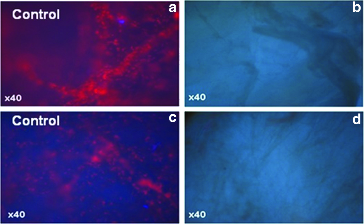
Results of viability/cytotoxicity assay for ovine aortic tissue following the application of the manual and automatic decellularization protocols; red fluorescence indicating cell presence in the control (manual)
Molecular analysis
Although both decellularization protocols were effective at eliminating all intact cells, the presence of residual DNA was confirmed on molecular analysis (Fig. 4). Both manual and automatic decellularized ovine aorta specimens contained residual DNA fragments remaining within their resulting ECMs. These fragments contained large quantities of highly shared base pairs ranging from 500 to 10,000 bp.

Results of molecular analysis; manually decellularized ovine aorta following the application of the manual decellularization protocol
DNA quantification (NanoDrop)
Both manual and automatically decellularized ovine aorta specimens displayed a significant reduction in residual DNA after the preparation processes. Control tissue contained 6690±1210 ng/mg wet weight of DNA, manually decellularized ovine aorta contained 540±130 ng/mg wet weight of DNA, and automatically decellularized ovine aorta contained 590±270 ng/mg wet weight of DNA. Manual decellularization demonstrated a reduction in DNA by 91.93% (p<0.05) and automatically decellularized ovine aorta saw a reduction of 91.18% (p<0.05). A summary of residual DNA levels remaining in the resulting ECMs relative to the ovine aortic control is illustrated in Figure 5.
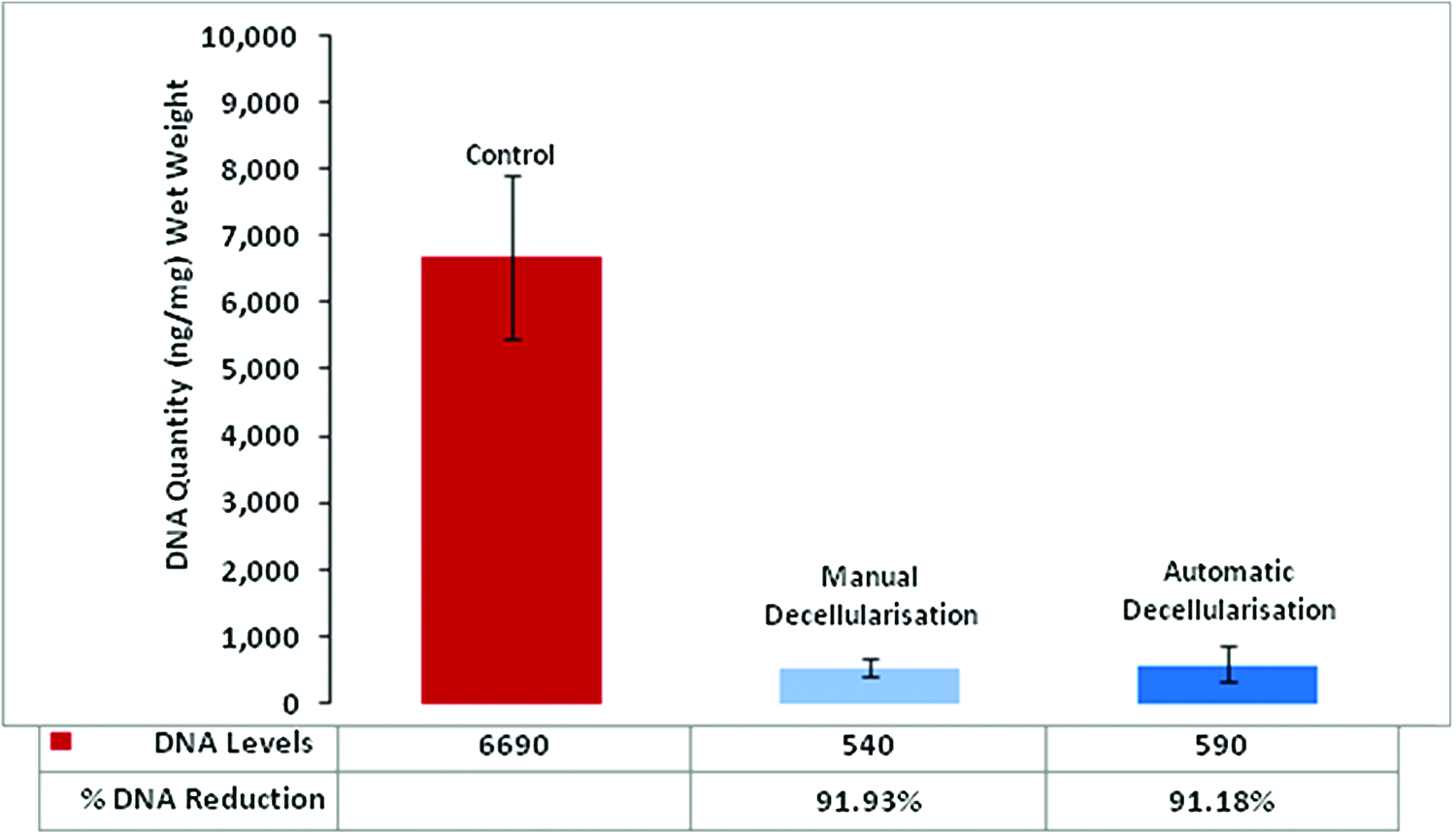
Results of DNA quantification (ng/mg wet weight) of residual DNA levels (ng/mg wet weight); control, manually decellularized, and automatically decellularized ovine aorta (average thickness; manual decellularization specimens t=0.79±0.28 mm and automatic decellularization specimens t=0.75±0.31 mm). Color images available online at
Mechanical properties
Comparative analysis of uniaxial tensile characteristics for specimens prepared by manual and automatic decellularization protocols is illustrated in Figure 6 and summarized in Table 2. Cauchy stress–stretch ratio curves between the control, manual, and automatic decellularized specimens were constructed based on average data. Average stretch ratio to failure occurred at 1.334±0.369 MPa, 2.14λ±0.13λ for the control; 0.637±0.128 MPa, 2.69λ±0.38λ for manually decellularized; and 0.728±0.228 MPa, 2.92λ±0.16λ for automatically decellularized ovine aorta (Fig. 6b). Incremental elastic modulus E at 1.30λ measured 0.070 MPa for the control, 0.137 MPa for manually decellularized, and 0.130 MPa for automatically decellularized ovine aorta. Incremental elastic modulus E at 2.00λ measured 0.635 MPa for the control, 0.291 MPa for manually decellularized, and 0.245 MPa for automatically decellularized ovine aorta (Fig. 6a). Statistical analysis demonstrated no significant differences for E or the tensile strength of ovine aorta following the application of the manual and automatic decellularization protocols below 2.70λ. By comparison, both manual and automatically prepared ECMs changed significantly from the control above a stretch ratio of 1.7 with a very large increase in ultimate failure strain and also a large decrease in the ultimate tensile strength.

Comparison of incremental elastic modulus E following the application of the manual and automatic decellularization protocols to ovine aortic (OA) control; both manually (black) and automatically (blue) decellularized specimens displayed similar incremental elastic moduli with a large decrease in stiffness in comparison with the control (red) when the stretch ratio is over 1.7
GL, gauge length; ↑, increasing; ↓, decreasing.
Resulting ECM
Images of the resulting ECM derived from ovine arterial tissue, postmanual and automatic decellularization, are illustrated in Figure 7. Structural integrity of both tissues remained intact with no notable differences. The total manual decellularization protocol processing time was 77 h versus 50.7 h for automatic decellularization (p<0.05), representing a 34.2% reduction in processing time for the automatic decellularization protocol with no attendee hours required.
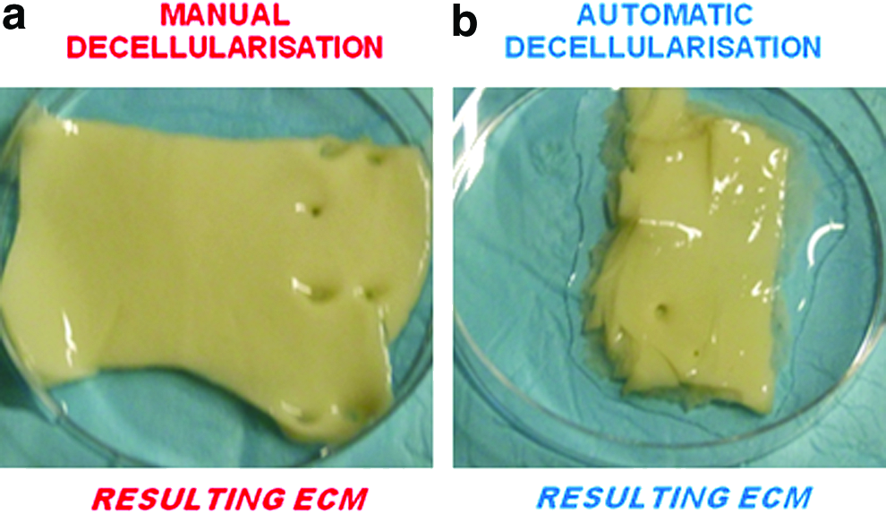
Ovine aorta resulting ECM; manually decellularized tissue specimen
Discussion
The demand for a reliable tissue-engineered vascular substitute has increased due to the increasing global prevalence of cardiovascular disease. 3 Decellularized ECMs provide an attractive option for vascular surgeons as they are inert, biodegradable, and readily available. Automatic decellularization is a necessary evolutionary processing step in ensuring that emerging tissue engineering markets can grow rapidly, meeting the world's growing demands for xenogenic-derived ECM vascular substitutes. Although manual decellularization has been in existence for decades, little research effort has been applied to the upscaled production of decellularized soft tissues. In this present ex vivo study, a comparative analysis was performed on manually decellularized ovine aortic tissue and automatically decellularized tissue. Important findings were that no significant differences were found between the manual and automatically prepared ECMs. Furthermore, automation also reduced the ECM processing time by 34.2% and eliminated the need for technical attendees during the processing steps.
To meet clinical requirements for an effective vascular substitute, an ideal graft material should facilitate cell adhesion to promote endothelization of the graft.12,18 As the application of a decellularization protocol can impact significantly on the surface characteristics of the resulting ECM, surface character structural changes were examined.4,5 To negate surface character changes as a result of automation, this aspect of processing needed to be understood. The surface structure alteration phenomenon is usually a multifactorial issue with the reagents used being the primary cause of disruption. The selection of agitation and its duration is an important factor when evaluating a decellularization protocol as it can amplify the effects produced by the reagents. 12 As automation was applied to the original protocols adapted from the literature, it was imperative that these effects were investigated. Upon comparison of the manually and automatically decellularized ovine aorta (Fig. 2), there was no significant difference between resulting surface features on either group of ECMs and these findings suggest that automatic decellularization protocol is capable of decellularization with minimal disruption to ovine arterial tissue's luminal and abluminal surfaces.
It is a well-established fact that the mechanical integrity of the resulting ECM is critical as an ideal vascular substitute should be compliant with the recipient vessel postimplantation.6,18–20 To verify that automation does not have a critical effect on the mechanical properties of decellularized ovine arterial tissue, uniaxial tensile testing was performed on control specimens and on manually and automatically decellularized tissue. Characterization focused on the Cauchy stress–stretch ratio of the resulting ECMs and also their ultimate tensile strength. Stretch ratio at failure was higher for both the manual and automatically decellularized tissues compared with the control. In this present study, the incremental elastic moduli were almost identical when comparisons were performed between the manual and automatic decellularization protocols with only minor differences in the ultimate tensile strength and the average strain to failure for both groups.
Significant changes occurred in the mechanical properties of both manually and automatically decellularized specimens upon comparison with the native control beyond a stretch ratio of 1.7. It has been widely reported that the use of trypsin/EDTA/EGTA has been attributed to the disruption of the ECM ultrastructure and collagen fiber kinematics, thus affecting the load-bearing capacity of a scaffold.4,11,12 In addition, the elastin content is greatly reduced by this treatment and therefore the initial high-stretch characteristics of the native control are no longer exhibited by either manually or automatically decellularized specimens (Table 2). Similar behaviors were reported by Gilbert et al., 4 Badylak et al., 11 and Crapo et al. 12 Since a combination of trypsin/EDTA/EGTA was used as the enzymatic decellularization agent in the preparation process, it is expected that the treatment will adversely disrupt the resulting ECM's ultrastructure, therefore affecting the mechanical properties.
It has been shown previously that freezing does not adversely affect the mechanical behavior of soft tissue.4,11,12,21 Freeze–thaw cycles produce minor disruptions of the ECM ultrastructure due to ice crystal formation within the collagen structures. However, in load-bearing robust tissues the influence of freeze–thaw processing on mechanical properties is minimal.4,11,12,21 It has also been shown that freezing does affect the cellular properties of tissue. Intracellular crystals disrupt the cell phospholipid bilayer outer membrane and also some of the inner structures of the cells.4,11,12 Therefore, the effects of freezing on the tissue are minimal regarding mechanical properties and beneficial as the first step in the decellularization process.
As the ADP is an unattended process, a complete reduction in attendee input was achieved once initiated. A 34.2% reduction in the overall process time was also achieved compared with manual decellularization techniques. This finding may have major implications for the commercialization of ECM-derived graft substitutes as it clearly demonstrates the cost advantages of automation, and as cost is considered to be one of the main barriers to market for ECM-derived graft substitutes, the ADP has demonstrated its ability to reduce cost inputs for the current manual decellularization protocols and provides an effective platform to produce quality ECM products for a target audience. 3
Although the preliminary findings of this study are promising, there are important limitations. Our results demonstrate that manual and automatic decellularization protocols failed to produce an ECM that was entirely free of DNA. Levels of residual DNA were identical in both groups; however, the reduction was not sufficient to produce a viable arterial graft for in vivo applications compared with the thresholds proposed by Gilbert et al. and Zheng et al. (<50 ng dsDNA per mg ECM dry weight), at <200 bp DNA fragment length.10,22 Preliminary results suggest that by following the current decellularization protocols that are available, decellularized biological material could have the potential to elicit an immune response in vivo due to the presence of remaining DNA fragments in the ECM. As the ADP process is controlled by a program running on a PLC, it will allow modification of the current protocol and further parameter optimization is necessary to reduce DNA levels below clinically relevant thresholds.10,21
Conclusions
Results from the current study demonstrate that automated decellularization of xenogenic ECMs is feasible and less labor intensive than the manual process. Future research will focus on optimizing the automated decellularization protocol and on upscaling techniques.
Footnotes
Acknowledgment
This study was supported by the Irish Research Council for Science Engineering and Technology (IRCSET), Embark Initiative, 2009.
Disclosure Statement
There are no conflicts of interest for any of the authors.