
Abstract
The present article investigates the use of a novel electrospun fibrous blend of poly(glycerol sebacate) (PGS) and poly(butylene succinate-butylene dilinoleate) (PBS-DLA) as a candidate for cardiac tissue engineering. Random electrospun fibers with various PGS/PBS-DLA compositions (70/30, 60/40, 50/50, and 0/100) were fabricated. To examine the suitability of these fiber blends for heart patches, their morphology, as well as their physical, chemical, and mechanical properties were measured before examining their biocompatibility through cell adhesion. The fabricated fibers were bead-free and exhibited a relatively narrow diameter distribution. The addition of PBS-DLA to PGS resulted in an increase of the average fiber diameter, whereas increasing the amount of PBS-DLA decreased the hydrophilicity and the water uptake of the nanofibrous scaffolds to values that approached those of neat PBS-DLA nanofibers. Moreover, the addition of PBS-DLA significantly increased the elastic modulus. Initial toxicity studies with C2C12 myoblast cells up to 72 h confirmed nontoxic behavior of the blends. Immunofluorescence analyses and scanning electron microscopy analyses confirmed that C2C12 cells showed better cell attachment and proliferation on electrospun mats with higher PBS-DLA content. However, immunofluorescence analyses of the 3-day-old rat cardiomyocytes cultured for 2 and 5 days demonstrated better attachment on the 70/30 fibers containing well-aligned sarcomeres and expressing high amounts of connexin 43 in cellular junctions indicating efficient cell-to-cell communication. It can be concluded, therefore, that fibrous PGS/PBS-DLA scaffolds exhibit promising characteristics as a biomaterial for cardiac patch applications.
Introduction
M
Electrospinning is one of the simplest and most cost effective methods available for the preparation of fibrous mats mimicking the ECM structure. Electrospun fibers possess very high surface area to volume or mass ratios and suitable interconnected pores, resulting in their characteristic architecture, which is suitable to enhance cell adhesion, proliferation, and differentiation. Such fibrous matrices also exhibit suitable mechanical strength which is tunable 11 and can mimic that of the native tissue.
As a result, many natural 12 and synthetic polymers 13 have been used to produce fiber mats through electrospinning. 14 Synthetic polyester poly(glycerol sebacate) (PGS) has attracted significant interest for cardiac patch applications, also in combination with other polymers,15–18 due to the relevant properties of PGS in terms of biocompatibility and bioresorbability. PGS is prepared by polycondensing glycerol and sebacic acid. Glycerol, particularly, has been approved to be used as humectant in foods by the US Food and Drug Administration, 17 whereas sebacic acid is the natural metabolic intermediate in ω-oxidation of medium to long-chain fatty acids.19,20 PGS has been shown to be safe in vivo18,21 and is an inexpensive polymer with tailorable mechanical properties and degradation kinetics. 16 However, further investigations about PGS-based materials are required to enhance their biological and mechanical performance for cardiac tissue engineering applications.
In this study, the potential of PGS prepolymer blended with poly(butylene succinate-butylene dilinoleate) (PGS-DLA) was evaluated, as a new biomaterial for cardiac tissue engineering, using C2C12 cells and 3-day-old rat CMs and analyzing adhesion, cellular metabolic activity, and cell-to-cell communication. Due to the low viscosity of the PGS prepolymer it cannot be electrospun on its own, therefore, a new system is presented here, according to which PGS prepolymer was blended with poly(butylene succinate-butylene dilinoleate) (PBS-DLA), which is a new random copolymer possessing high biocompatibility and biodegradability.22,23 The resulting blend was then electrospun, minimizing the limitations associated with each individual polymer. The advantage of this method was that it allowed the creation of new electrospun components without the need to synthesize a new copolymer.
Materials and Methods
PBS-DLA preparation
PBS-DLA (60-40) with a molecular weight ∼50×103 g/mol−1 was prepared at the Polymer Institute, West Pomeranian University of Technology, Szczecin, Poland. PBS-DLA was synthesized to produce a random copolymer containing 60 wt% of PBS segments and 40 wt% of DLA segments, respectively. The polymer synthesis was carried out as detailed previously in Kozlowska et al. 22 In summary, the esterification reaction between DLA, succinic acid, and 1,4-butanediol (1,4-BD) was carried out in the presence of magnesium–titanate organometallic complex (Mg–Ti) under intensive stirring and upon programmed temperature raising from 100°C to 200°C with a heating rate of 1.5°C/min. The polycondensation reaction was carried out at 245–250°C, ∼0.4 kPa, and in the presence of Mg–Ti catalyst. The process was considered complete when the observed power consumption of the stirrer motor signaled that the polymer of highest melt viscosity was obtained. The copolyester was purified before further processing.
PGS preparation
PGS was synthesized according to the process reported by Wang et al., 17 which involves two steps, that is, a prepolycondensation step obtaining a transparent viscous like gel followed by a crosslinking step. For the prepolycondensation step an equimolar mixture (0.1 M) of glycerol and sebacic acid was heated at 120°C under inert nitrogen atmosphere to form the prepolycondensed prepolymer. The PGS in this study was used in the prepolymer condition without further crosslinking.
Fabrication of two-dimensional PGS/PBS-DLA films
The fabrication of the desired nonporous two-dimensional (2D), PGS/PBS-DLA matrices were carried out by solvent casting as mentioned in the previous work. 24 In summary, the two polymers (PGS and PBS-DLA) were dissolved in dichloromethane (DCM) for 2 h, then casted in a covered glass Petri dish.
Fabrication of three-dimensional PGS/PBS-DLA fibrous mats through electrospinning
Fiber mats of PGS/PBS-DLA were fabricated in different compositions, that is, 50/50, 60/40, and 70/30. Polymer solutions (20 wt%) were prepared by dissolving the different polymer blends in DCM:methanol (7:3). The solutions were prepared and dissolved overnight under constant stirring. The PGS/PBS-DLA blend solutions were placed in a glass syringe (lure-lock type, 10 mL) and connected to a metal syringe needle (20 gauge, inner diameter 0.8 mm) and placed on the pump (R99-E syringe pump; Razel). The solution was electrospun with the following process parameters: 15 cm distance between needle tip and collector (aluminum foil), 1.6 mL/h flow rate and a voltage of 20 V, which was supplied directly from a high DC voltage power supply for 4 h. The obtained fiber mats had an average nominal thickness of 100±0.01 μm.
Preparation of the samples for in vitro cytocompatibility studies
The electrospun membranes were cut into circular discs (15 mm in diameter) for in vitro cell culturing studies. All samples were pretreated with 70% ethanol for 24 h before seeding, where the ethanol treatment has a dual effect as a sterilization method and leached out any unreacted monomers or toxic reagents. Later the samples were washed three times in phosphate buffered saline. CellCrown inserts (Scaffdex) were used to help to push down the thin flexible polymer fiber mats from floating, thus the cells could attach on the surface of the samples.
In vitro studies with C2C12
Mouse skeletal myoblast cell line C2C12 (DSMZ) were cultured with RPMI on the indicated matrices, supplemented with 10% fetal bovine serum (FBS) and 5% penicillin/streptomycin at 37°C in a humidified atmosphere containing 5% CO. Cells were passaged every third day using a solution containing 0.05% trypsin. C2C12 cells were cultured with a concentration of 105 cells/well in supplemented RPMI for 72 h and the medium was changed every day throughout the entire experiment. Given that these cells proliferate at a rapid rate it is likely that beyond a short period of time cells will start detaching from the substrate. It was, therefore, decided to limit the study to 3 days.
In vitro studies with postnatal CMs
Postnatal CMs were cultured on the different PGS/PBS-DLA fiber mats and gelatin- and fibronectin-coated glass coverslips were used as controls. CM cells were isolated from 3-day-old Sprague Dawley rats as previously described.
25
The isolated CMs were preplated for 2 h in 2 mM
Cytotoxicity assay
WST-8 was used to quantify cell metabolic activity; three separate experiments (n=3), three replicates each, were performed. Fluorescence values (mean±standard deviation of the mean) were calculated. The assay was performed after 8, 24, and 72 h of culture of C2C12 cells and after 2 and 5 days of culture of the CMs. Thus, the assay was used to evaluate cytotoxicity, cell adhesion, cell viability, and cell growth on the developed matrices as an indirect measurement of cell mitochondrial activity.
Cell morphology and attachment
Cell morphology and adhesion were studied through scanning electron microscopy (SEM) and immunocytochemistry studies to visualize cytoskeleton organization and cell nuclei. Cells seeded on PGS/PBS-DLA matrices were fixed with 4% PFA in phosphate buffered saline for 15 min at room temperature and were permeabilized with 0.1% Triton X-100 in phosphate buffered saline for 15 min at room temperature. C2C12 cell morphology, cytoskeleton organization and protein expression were detected through immunofluorescence analysis after incubation with Alexa Fluor®555 Phalloidin (1:60; Invitrogen), which binds to F-actin, for 1 h at room temperature. Nuclei were counterstained with 4–6-diamidino-2-phenylindole (DAPI) after 8, 24, and 72 h in culture. CMs were stained for comparative evaluation of their protein expressions, thus evaluating the functional capability of the cells after seeding on the electrospun fibrous mats. The expression of two different proteins, namely alpha-actinin (α-Act) and connexin 43 (Cx43), were used for this study. In brief, after 5 days of cell culture, the cellular constructs were washed with phosphate buffered saline and fixed with 4% PFA for 10 min. Then, samples were blocked with 5% bovine serum/0.2% Tween 20/phosphate buffered saline for 20 min. Samples were stained overnight at 4°C with monoclonal mouse primary anti-α-Act antibodies and rabbit primary anti Cx43 antibodies at a dilution of 1:100 and 1:50, respectively. The scaffolds were then washed thrice with phosphate buffered saline, each time for 5 min, and incubated with the appropriate secondary antibodies for 45 min at room temperature. The secondary antibodies used in this study were Alexa 488 donkey anti-mouse IgG for anti-α-Act or Alexa 594 donkey anti-rabbit IgG antibody for anti-Cx43, at a dilution of 1:200. The nucleus of the cells was counterstained with DAPI for 10 min. Then, the samples were washed thrice with phosphate buffered saline for 5 min, mounted onto glass slides and viewed under a Keyence BZ9000 microscope. For the proliferation and attachment assessment the number of DAPI-stained nuclei on the surface of the scaffold for both the C2C12 culture and CMs were counted using the image editing software ImageJ.
Chemical structure assessment of PGS/PBS-DLA fibers
Chemical analyses of the polymeric films were carried out in attenuated total reflectance mode (ATR) using FTIR spectroscopy (Nicolet 6700; Thermo Scientific). The penetration depth was 2–3 μm. The analyses were performed under the following conditions: Spectral range between, 4000 and 530 cm−1; Window material, CsI: 16 scans at a resolution of 4 cm−1.
Surface studies of PGS/PBS-DLA fibers
To study the surface morphology of different PGS/PBS-DLA matrices and to demonstrate the effect of PBS-DLA on the surface morphology, a LEO 435 VP SEM was used. Samples were prepared by placing them on 8 mm diameter aluminium stubs, which were sputtered with gold–palladium for 1 min. The SEM images were captured at an acceleration voltage of 10 kV. The apparent porosity of the fiber mats, as well as fiber diameter, and fiber diameter distribution were analyzed from SEM micrographs using an ImageJ. Furthermore, the theoretical porosity was calculated through the following formulae:
Where, W is weight and V is volume of the sample, δBulk is the bulk density and δTrue is the true density of the polymer blend system. The true density is calculated for each blend composition (x:y) where x is the PGS content and y the PBS-DLA content. The densities of PGS and PBS-DLA were measured. The wettability of the scaffolds was evaluated using static contact angle measurements. The reference liquid was deionized water (3 μL), and was placed on every sample by means of a gas tight microsyringe forming a drop. Photos (frame interval: 1 s, number of frames: 100) were taken to record the shape of the drops. The water contact angles on the specimens were measured by analyzing the recorded drop images using the Windows-based KSV CAM software. Five repeats of each reference liquid for each sample were carried out. The experiments were done on a KSV CAM 200 optical contact angle meter (KSV Instruments Ltd.).
Mechanical properties of PGS/PBS-DLA fibers
Tensile tests were carried out using a Zwick Universal Testing Machine Z050 at room temperature. The samples were cut into rectangular strips that were 5 mm wide, 3 cm long, and 100 μm thick. The measurements were carried out at a crosshead speed of 10 mm/min and a pretension of 0.2 N was applied. A 50 N load cell was used for all experiments.
Statistical analysis
The data sets (n=3) have been expressed along with their mean standard deviation. The data, where appropriate, were compared using the one-way ANOVA and differences were considered to be significant when *p<0.05 and **p<0.01.
Results
Morphology characterization of electrospun scaffolds
Randomly oriented, uniform, and bead-free fibers of different polymer compositions were successfully fabricated with PGS/PBS-DLA compositions 50/50, 60/40, and 70/30. SEM micrographs and the diameter distribution of the different fiber mats (Fig. 1) show the uniform and bead-free fibers over a large area in all three different blends, which were the result of the optimized parameters covered throughout this study. The average fiber diameters were 1.22±0.20, 1.20±0.16, and 1.18±0.23 μm, for PGS/PBS-DLA 50/50, 60/40, and 70/30 fibers, respectively. The parameter optimization was carried out with regard to the applied voltage, flow rate, distance between the needle and the collector, as well as the polymer concentration of the electrospinning solution.
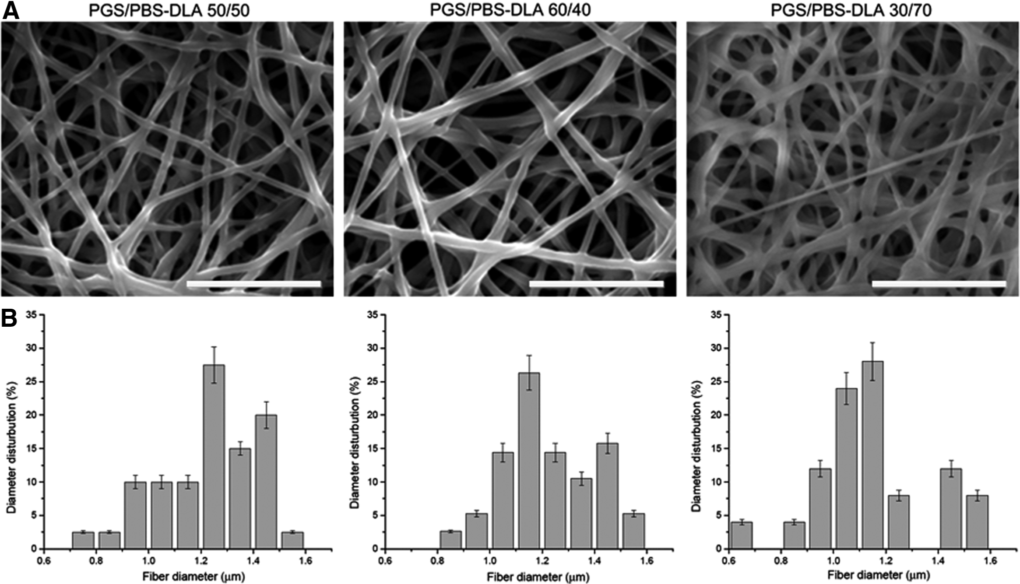
Morphology characterization of electrospun scaffolds.
The surface properties of the electrospun fiber mats were investigated by water contact angle measurements. The water contact angle for the electrospun PGS/PBS-DLA fibers was found to be 49.1±6.3, 44.5±6.5, and 38.5±10.0 for 50/50, 60/40, and 70/30 PGS/PBS-DLA fibers, respectively, showing a clear reduction in water contact angle with increasing content of PBS-DLA. The measurements showed no significant difference in water contact angle compared with the corresponding dense film blends of the same concentration. This shows that the hydrophilicity of PGS/PBS-DLA blend was not influenced by the fabrication process.
To evaluate the porosity of the fiber mats the density of both the neat PGS and PBS-DLA was measured to be 1.20±0.10 g/cm3 and 1.23±0.15 g/cm3, respectively. Following Equations (1) to (3), the theoretical porosity of the fiber mats was calculated and summarized in Table 1.
PGS, poly(glycerol sebacate); PBS/DLA, poly(butylene succinate-butylene dilinoleate).
Chemical assessment through FTIR
To investigate the successful fabrication of the electrospun PGS/PBS-DLA fibrous blends ATR-FTIR signature spectra of the neat polymers and electrospun fibers of different PGS/PBS-DLA concentrations were performed as a preliminary structural characterization (Fig. 2). The different PGS/PBS-DLA fibrous mat FTIR spectra possessed all peaks corresponding to both neat PGS and neat PBS-DLA, but it can be seen that there is a great deal of overlap as both polymers are polyesters. The only two exceptions are the peak at 980 cm−1, which corresponds to PBS-DLA and the peak at 3400 cm−1, for the hydroxyl group, which can be assigned to PGS. The intensity of this peak increased with increasing amounts of PGS in the blends. The FTIR demonstrates that the fibers are a simple blend where no reaction has taken place between the two polymers.

FTIR spectra of neat PGS and PGS/PBS-DLA of 50/50, 60/40, and 70/30 electrospun fibrous mats. Color images available online at
Mechanical behavior
Since the fiber mats were very delicate, the electrospun fibers were cut into the specific dimensions required (as mentioned previously) for the tensile testing before removing the fibers from the aluminum foil. Then the aluminum foil was carefully removed by soaking the samples in water, after which the samples were allowed to dry before mechanical testing. The stress–strain curves of the electrospun PGS/PBS-DLA fiber mats of different PGS and PBS-DLA concentrations were obtained using a universal testing machine. The cast films with equal composition of PGS and PBS-DLA were also tested for comparison.
The stress–strain curves of the neat PGS, neat PBS-DLA, 60/40 PGS/PBS-DLA blend film and fibers of different PBS-DLA contents are depicted in Fig. 3. It was observed that in general the fiber mats exhibited higher strains (similar to native cardiac tissue—120%) compared with both the neat films and the blended films. Furthermore, the Young's modulus of the electrospun fibers increased with increasing the PBS-DLA content. Table 2 summarizes the mechanical properties of all films and electrospun fibers.
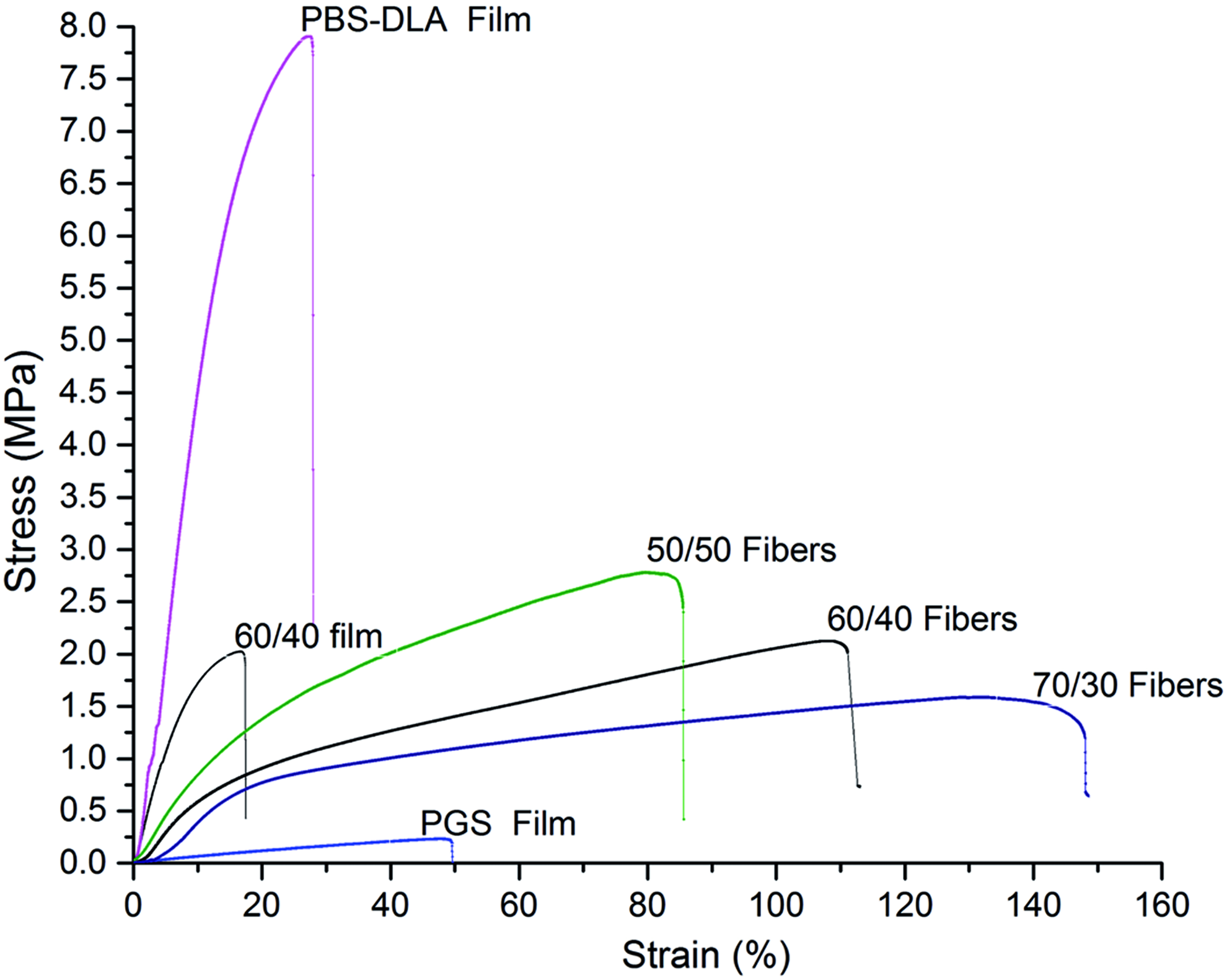
Stress–strain curves of the different fiber mats in comparison to neat PGS film, PBS-DLA film, and 60/40 blend. Color images available online at
Biocompatibility assessment—in vitro studies with C2C12
Initial cell viability studies performed with C2C12 revealed no significant differences between the 50/50 and 60/40 fiber blends for the myoblast cell viability after 8, 24, and 72 h of seeding (Fig. 4A). However, the 70/30 fibers showed significantly lower cell viability only at 8 h of culture. In addition, neither cell death nor morphological disorder was observed (Fig. 4B). Thus, it was concluded that PGS/PBS-DLA fibrous mats produced from this process did not display a cytotoxic effect, due to the presence of remnant solvents or from the polymer release.
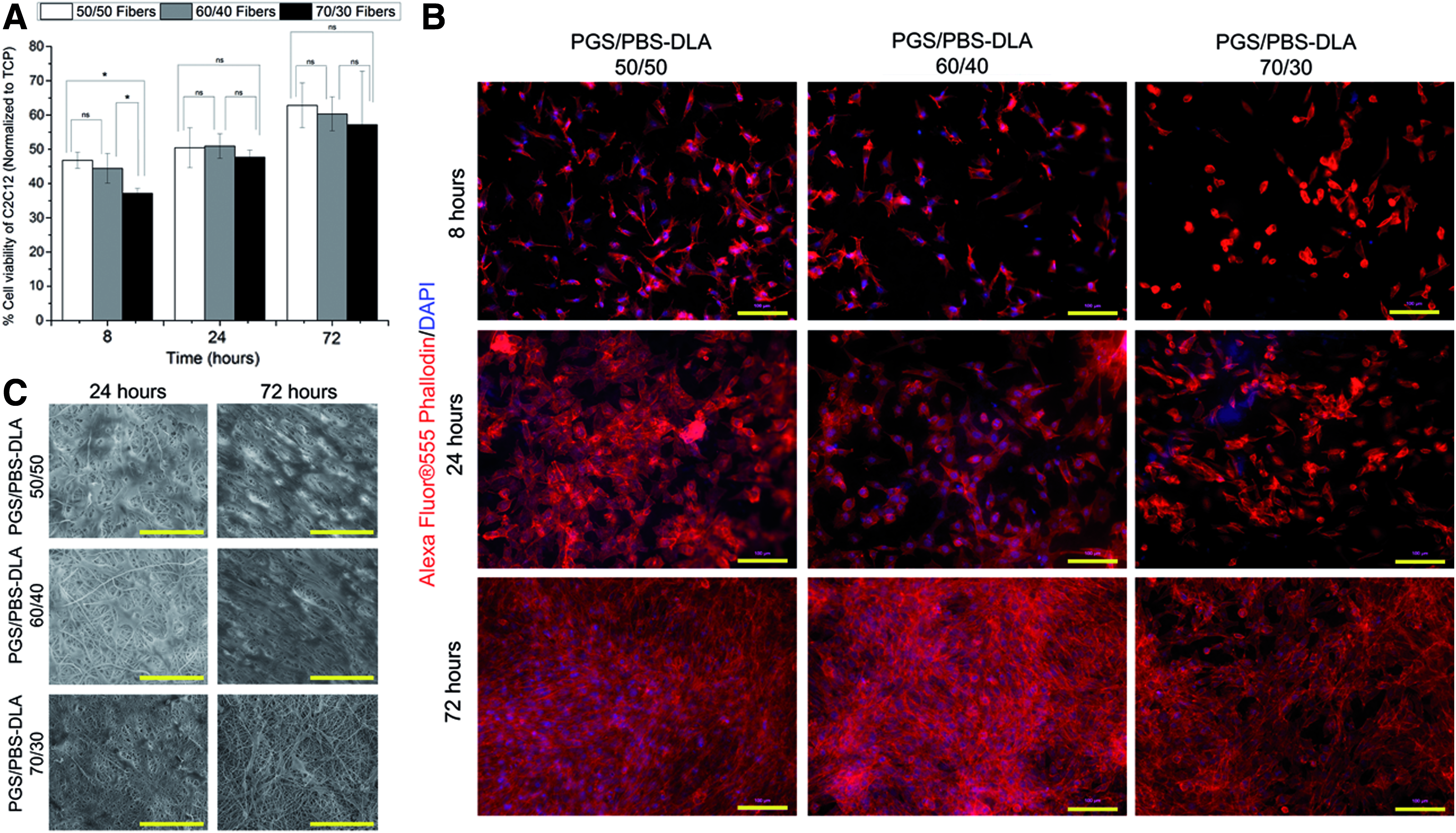
C2C12 attachment and proliferation on the different PGS/PBS-DLA fibers.
Cell morphology and attachment
The cell morphology was examined to continually check the biocompatibility of all PGS/PBS-DLA fiber mats. The cell morphology of the C2C12 after seeding the cell for 8, 24, and 72 h on the different fiber mats was revealed by immunostaining (Fig. 4B). SEM micrographs illustrated the cell attachment on the electrospun PGS/PBS-DLA 50/50, 60/40, and 70/30 fiber mats (Fig. 4C). By comparing the filopodium and lamellipodium (Fig. 4B) in each sample condition, it was noted that filopodium and lamellipodium growth developed well, except for the 70/30 fibers, for which rounded unhealthy cells were observed after 8 h of culture. Interestingly, 72 h after seeding, the seeded cells exhibited a typical myoblast-like morphology on all substrates, showing a correct cytoskeleton organization highlighted by F-actin immunostaining, irrespective of the different PBS-DLA content in the fibers, which was also confirmed by SEM analysis (Fig. 4C). In addition, by comparing, it was seen that filopodium and lamellipodium growth increased as the content of PBS-DLA increased. However, the C2C12 cell density was lower on the 70/30 fiber samples. The PGS/PBS-DLA fibrous mats demonstrated, thus, to be of high biocompatibility, which makes them suitable candidate for the intended application as cardiac patches.
Biocompatibility assessment—in vitro studies with postnatal rat CMs
Three-day-old postnatal CMs were cultured on the different PGS/PBS-DLA fiber mats and on gelatin- and fibronectin-coated glass coverslips as controls and cell viability was monitored after 2 and 5 days of culture, using WST-8 assay. The biocompatibility experiments were performed with all five scaffolds.
Adhesion and morphology
The adhesion of the cells is a crucial aspect in cardiac tissue engineering and tissue engineering in general. For these experiments an intermediate composition, namely 60/40 PGS/PBS-DLA polymer, was considered. Fibers were cultured with 3-day-old postnatal rat CMs with different horse serum concentration (0%, 0.5%, 5%) to investigate the adhesive properties of the blend fibers and the effect of the horse serum on the cell attachment.
CMs isolations are highly enriched in CMs (>90%), but contain also nonmyocytes such as endothelial cells, fibroblasts, and smooth muscle cells. Thus, specific CM staining was used to quantitatively analyze CM attachment. Our data demonstrated that the use of horse serum enhanced the CM attachment and morphology (Fig. 5A). At a concentration of 0.5% serum fiber mats were densely covered with CMs that exhibited well-developed sarcomeric structures. At a concentration of 5% horse serum, the mats were densely covered with cells, but the percentage of CMs appeared to have decreased. Quantitative analyses confirmed that there was an increase in nonmyocytes (Fig. 5B), which might be due to horse-serum-induced nonmyocyte proliferation.

Cardiomyocyte attachment on PGS/PBS-DLA 60/40 fibers depends on the serum (horse) concentration.
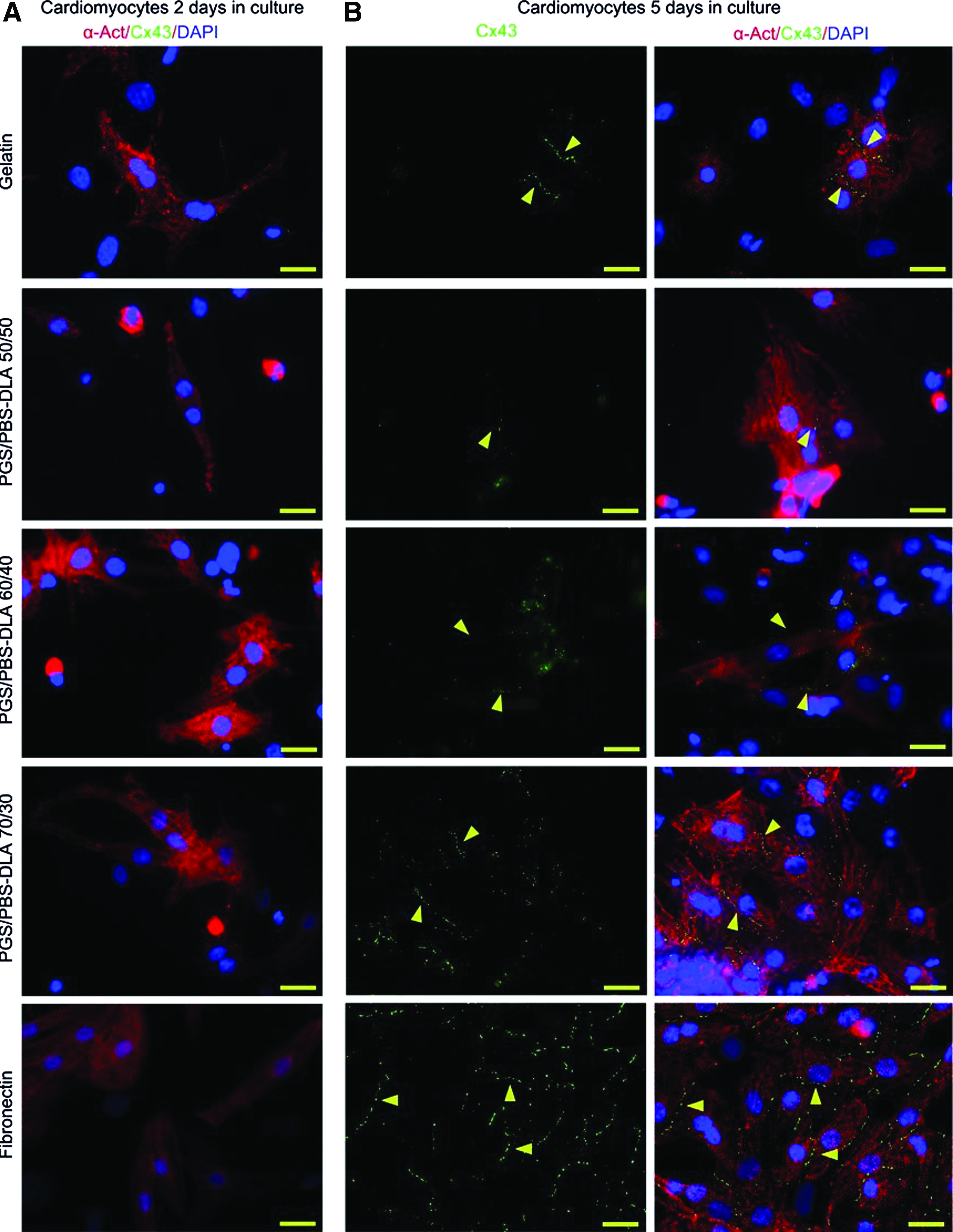
Cx43 expression
In a cardiac tissue engineering construct, contractility is a great indication of its success. Contractility of CMs depends on well differentiated sarcomeres (aligned and in parallel) and synchronized contractions through the electrical coupling between cells. Only cultures with 0.5% serum developed well differentiated sarcomeric structures (Fig. 5A). To further determine whether the 60/40 PGS/PBS-DLA fibers are suitable to generate cardiac patches, the expression of the gap junction protein Cx43 26 was analyzed. CMs attached to 60/40 PGS/PBS-DLA fibers exhibited well-organized sarcomeres and expressed along cellular junctions high amounts of Cx43 (Fig. 6). In summary, 60/40 PGS/PBS-DLA fibers promote sarcomere maturation and alignment, as well as cell-to-cell communication.

PGS/PBS-DLA 60/40 fibers stimulated with 0.5% horse serum promote cell-to-cell communication. Attached cardiomyocytes were stained with antisarcomeric α-Act (cardiomyocytes, red), anti-Cx43 antibodies (intercellular gap junctions, green), and DAPI (nuclei, blue). Scale bars: 20 μm. Color images available online at
Behavior of the postnatal rat CMs on the different fiber blends
To determine the cell viability of CMs on different materials, mitochondrial viability studies were performed 2 and 5 days after seeding (Fig. 7A). Quantitative scores indicated that samples with higher PGS content showed better mitochondrial activity. Furthermore, the measured overall mitochondrial activity of CMs on fibers was higher than on gelatin and showed comparable results for CMs on fibronectin. This suggests that PGS/PBS-DLA fibers are not toxic for CMs and that the CMs behave on this material similar as on their natural substrate fibronectin. Quantitative analyses confirmed that there was an increase in nonmyocyte (Fig. 7C, D) attachment, independent of the matrix. In contrast, CM in (Fig. 7B, D) the attachment was dependent on the culture duration and matrix. After 2 days significantly more CMs attached on all indicated matrices compared with gelatin. After 5 days an increase in mitochondrial activity was indicated on PGS/PBS-DLA fibers and fibronectin compared with gelatin. Besides, there was no difference between the 70/30 fibers and the fibronectin. Morphological studies revealed well-developed sarcomere structures in CMs on all fiber mats after 2 days (Fig. 8A) and 5 days (Fig. 8B) of culture, respectively. However, the density of CMs on the 70/30 fibers was higher than for the other two compositions, which is in agreement with the higher mitochondrial activity measured (Fig. 7A). Thus, the increased mitochondrial activity probably does not only reflect an increased activity per cell, but an increase in the number of cells attached, as shown by the attachment study after day 2. In addition, CMs exhibited a more pronounced cell-to-cell communication based on Cx43 expression (Fig. 8B).

Cardiomyocytes exhibit high metabolic activity on PGS/PBS-DLA fiber mats.
Evaluation of cardiomyocyte cell morphology after
Discussion
Current treatments for MI like early revascularization (percutaneous or surgical) or thrombolysis can significantly improve the outcome of patients. However, the most effective treatment is indeed heart transplantation, which is, however, limited by the donor's availability, in addition, implantation of a ventricular assist device is only a transient solution. Thus alternative treatment options are needed, such as regeneration through stem cell therapy 27 or induction of CMs proliferation. 28 However, these approaches have not yet been proven to be effective as a human therapy for heart failure.
One alternative option to treat heart failure might be the engineering of three-dimensional (3D) cardiac tissues. Previously, it had been shown that implantation of CMs embedded in a mixture of type I collagen gel and Matrigel can improve left ventricular function after MI. 4 This study exemplifies the potential of cardiac tissue engineering; however, this collagen gel Matrigel mixture used is not applicable for clinical practice. The first clinical application of tissue engineering for ischemic heart disease showed feasibility and safety of grafting cell-seeded collagen type I matrix onto the epicardium. 29 The study showed that the cell-seeded collagen matrix helps to normalize cardiac wall stress and enhances the thickness of the infarcted area with viable tissues. 29
The heart is continuously under dynamic stretching making cardiac tissue regeneration challenging. Materials for cardiac tissue engineering must allow the attachment of CMs, exhibit specific mechanical properties and be tailorable to the specific needs of the patient. Therefore, it is important to identify materials suitable to generate tailorable 3D cardiac tissue patches that can be applied to patients. In this study, we introduce a promising new matrix (PGS/PBS-DLA fiber mats) for cardiac tissue regeneration. PGS has been extensively studied, especially in the field of cardiac tissue engineering supporting the attachment and growth of CMs, endothelial cells, and mesenchymal stem cells. PGS has been demonstrated to be a promising material for contractile tissue due to its tailorable mechanical properties and degradation kinetics, which can be designed to the required properties. 16 However, PGS fibers are very difficult to obtain owing to the low viscosity of the PGS prepolymer. This is a problem as surface topography is a very crucial aspect in tissue engineering. It is not only crucial for cell guidance, but most importantly the surface topography decides how the immune system reacts to the foreign material. 30 Thus, to mimic the fibrous ECM structure of the myocardium, in the present study, the novel polymer PBS-DLA was blended with PGS to achieve an electrospunable PGS-based material.
Based on the quantitative scores of the C2C12 cytotoxicity test, it was concluded that the extracts of the fiber mats displayed no cytotoxic reactivity. However, to develop a biomaterial for tissue engineering it is important to achieve an adequate interaction between the cells and the matrix. 31 CMs are characterized by relatively poor attachment to random surfaces and rather slow attachment and spreading even to components of its native matrix. Our data clearly demonstrate that CMs attach and spread much faster on PGS/PBS-DLA fiber blends than on gelatin. Especially CMs on 70/30 fibers responded to extracellular stimuli in the same way as CMs attached to fibronectin. The interaction between cells and 70/30 fiber mat appears to be satisfactory.
Furthermore, the sarcomeres of CMs loaded on 70/30 fibers mats were aligned. In addition, CMs on 70/30 fibers expressed higher levels of the gap junctional protein Cx43 than on the 50/50 and 60/40. This expression was similar to the expression in CMs on fibronectin. This study thus suggests that 70/30 fibers enable CMs maturation and together with their superior mechanical properties these fibers might support contractile strength and electrical coupling of the implant to the myocardial tissue.
The CM results contradicts those of the C2C12 cells, where the 70/30 fibers showed the lowest cell viability; on the contrary when seeded with CMs these fibers possess similar behavior to that of fibronectin. There are two factors which can influence the cell-to-substrate interaction, namely the stiffness of the fibers and the chemical composition of the different blends. In case of the C2C12 cells it is speculated that the chemistry of the fibers was the key factor influencing cell proliferation and viability. However, in the case of the CMs the cells seem to be more sensitive to the matrix stiffness, showing better attachment and maturation on the softer matrix. 32 The observed differences between the different fiber blends might be due to the fact that 70/30 fibers in contrast to 50/50 and 60/40 have relatively low Young's modulus due to the lower PBS-DLA content.
In the future it will be important to further optimize the loading of the 70/30 fibers mats with CMs. Moreover aligned fibers should be prepared to guide the cells morphology considering that native cardiac tissue is anisotropic.
Conclusions
Microstructured electrospun PGS/PBS-DLA fiber mats were confirmed to provide mechanical strength, flexibility, and guidance for both C2C12 and CMs. Immunostaining analyses and SEM micrographs identified these fiber mats as good adhesives for C2C12 cells and primary neonatal rat CMs. However, fiber blends with high PGS content (70/30) had a toxic effect on C2C12 cells. In contrast, CM attachment to 70/30 fibers was comparable to the attachment to fibronectin, the natural matrix protein of CMs. Furthermore, the behavior of CMs was dependent on the percentage of horse serum in the culture medium. At an optimal concentration of 0.5% serum, CMs exhibited well-developed sarcomeric structures and cell-to-cell contacts. This suggests that PGS/PBS-DLA fiber mats allow CMs to exhibit high contractile and electrical signal propagation capabilities. Collectively, this particular electrospun fiber blend shows promising structural and functional properties to develop suitable cardiac tissue constructs.
Footnotes
Acknowledgments
The authors acknowledge partial financial support from the ERC Starting Grant MINATRAN 211166. Aga Kozlowska is acknowledged for experimental assistance, Alina Grünewald for her technical support and Prof. Ben Fabry for providing access to his laboratory. This work was also supported by an ELAN-Programm Grant by the Friedrich-Alexander-Universität Erlangen-Nürnberg (to F.B.E.).
Disclosure Statement
No competing financial interests exist.