
Abstract
Over the past decade, silk fibroin (SF) has been emergently used in peripheral nerve tissue engineering. Current approaches aiming at producing SF-based nerve guidance conduits (SF-NGCs) used dissolved silk based on either aqueous solutions or organic solvents. In this study, we describe a novel procedure to produce SF-NGCs: A braided tubular structure of raw Bombyx mori silk is subsequently processed with the ternary solvent CaCl2/H2O/ethanol, formic acid, and methanol to improve its mechanical and topographical characteristics. Topographically, the combination of the treatments results in a fusion of the outer single silk fibers to a closed layer with a thickness ranging from about 40 to 75 μm. In contrast to the outer wall, the inner lumen (not treated with processing solvents) still represents the braided structure of single fibers. Mechanical stability, elasticity, and kink characteristics were evaluated with a custom-made test system. The modification procedure described here drastically improved the elastic properties of our tubular raw scaffold, favoring its use as a NGC. A cell migration assay with NIH/3T3-fibroblasts revealed the impermeability of the SF-NGC wall for possible invading and scar-forming cells. Moreover, the potential of the SF-NGC to serve as a substratum for Schwann cells has been demonstrated by cytotoxicity tests and live-dead stainings of Schwann cells grown on the inner surface of the SF-NGC. In vivo, the SF-NGC was tested in a rat sciatic nerve injury model. In short-term in vivo studies, it was proved that SF-NGCs are not triggering host inflammatory reactions. After 12 weeks, we could demonstrate morphological and functional reinnervation of the distal targets. Filled with collagen, a higher number of axons could be found in the distal to the graft (1678±303), compared with the empty SF-NGC (1274±146). The novel SF-NGC presented here shows promising results for the treatment of peripheral nerve injuries. The modification of braided structures to adapt their mechanical and topographical characteristics may support the translation of SF-based scaffolds into the clinical setting. However, further improvements and the use of extracellular matrix molecules and Schwann cells are suggested to enable silk tube based conduits to bridge long-distance nerve gaps.
Introduction
T
However, autografting carries several disadvantages such as limited number of donor sites for graft harvesting and associated donor site morbidity, including loss of nerve function, painful neuroma formation, and hyperaesthesia.6,7 These negative aspects have led to the search for alternative approaches. Besides nervous tissue, other autologous materials, such as vein grafts8,9 or muscles,10,11 have been used to bridge nerve gaps. However, the use of these substances is both preclinically and clinically unsatisfactory.
Recent advances in tissue engineering have opened new opportunities in peripheral nerve repair. Artificial nerve guidance conduits (NGCs), composed of synthetic or natural polymers, are currently being investigated for bridging nerve defects.5,12,13 The rationale behind using an NGC is to entubulate the nerve stumps to provide a protective micro-environment for the regenerating peripheral nerve. While numerous synthetic and natural biomaterials have been evaluated, both preclinically and clinically, for the bridging of nerve defects, their therapeutic benefits14,15 still remain unsatisfactory.
In the past few years, silk fibroin (SF) has attracted considerable interest as a biomaterial that is suitable for applications toward peripheral nerve regeneration. SF has been shown to possess characteristics that favor its use as an NGC, such as mechanical stability, slow degradation rate, biocompatibility, and its ability to support nerve regeneration.2,16 Apart from biocompatibility, an NGC should act as a barrier for infiltrating fibroblasts and provide mechanical resistance against compression and kinking by the surrounding tissue. Most of the current approaches to create tubular structures use electrospinning, as this process can be well controlled.2,17–20 Other techniques include dipping,21,22 gel spinning,23,24 or molding. 25 All the preparation techniques mentioned earlier are based on dissolved SF, which is then used to create a tubular construct. For the first time, to our knowledge, our study attempts to use textile-engineered raw silk constructs as the starting material for bridging a nerve defect. To improve the mechanical properties favoring its use as an NGC, these braided tubular structures are further processed by treatment in a ternary solvent system of a CaCl2/H2O/ethanol solution and then re-stabilized with formic acid (FA).
The aim of this study was to develop a novel method to generate an SF-NGC with distinct mechanical and anisotropic properties, and to prove its biocompatibility and functionality both in vitro and in vivo in a rat sciatic nerve injury model.
Materials and Methods
Unless otherwise noted, all reagents were obtained from Sigma and are of analytical grade.
Design and preparation of silk conduits
White raw Bombyx mori silkworm fibers of 20/22 den, 250 T/m, were purchased from Testex AG. The tubular silk conduit was fabricated in cooperation with a commercial braiding company (Edelrid GmbH). Six single silk fibers form a twisted yarn, representing the raw material for the commercial braiding machines. Figure 1 shows the tubular structure designed from six intertwined twisted yarns. The resulting raw silk conduit was degummed by boiling in 0.2 M boric acid in a 0.05 M sodium borate buffer (pH=9.0). 26 Batches of 2 g of silk conduits were boiled twice in 500 mL of degumming solution for 45 min, with an intermittent exchange of the degumming solution. After degumming, scaffolds were thoroughly washed in double-distilled water (ddH2O) and air-dried before further processing (Fig. 1).
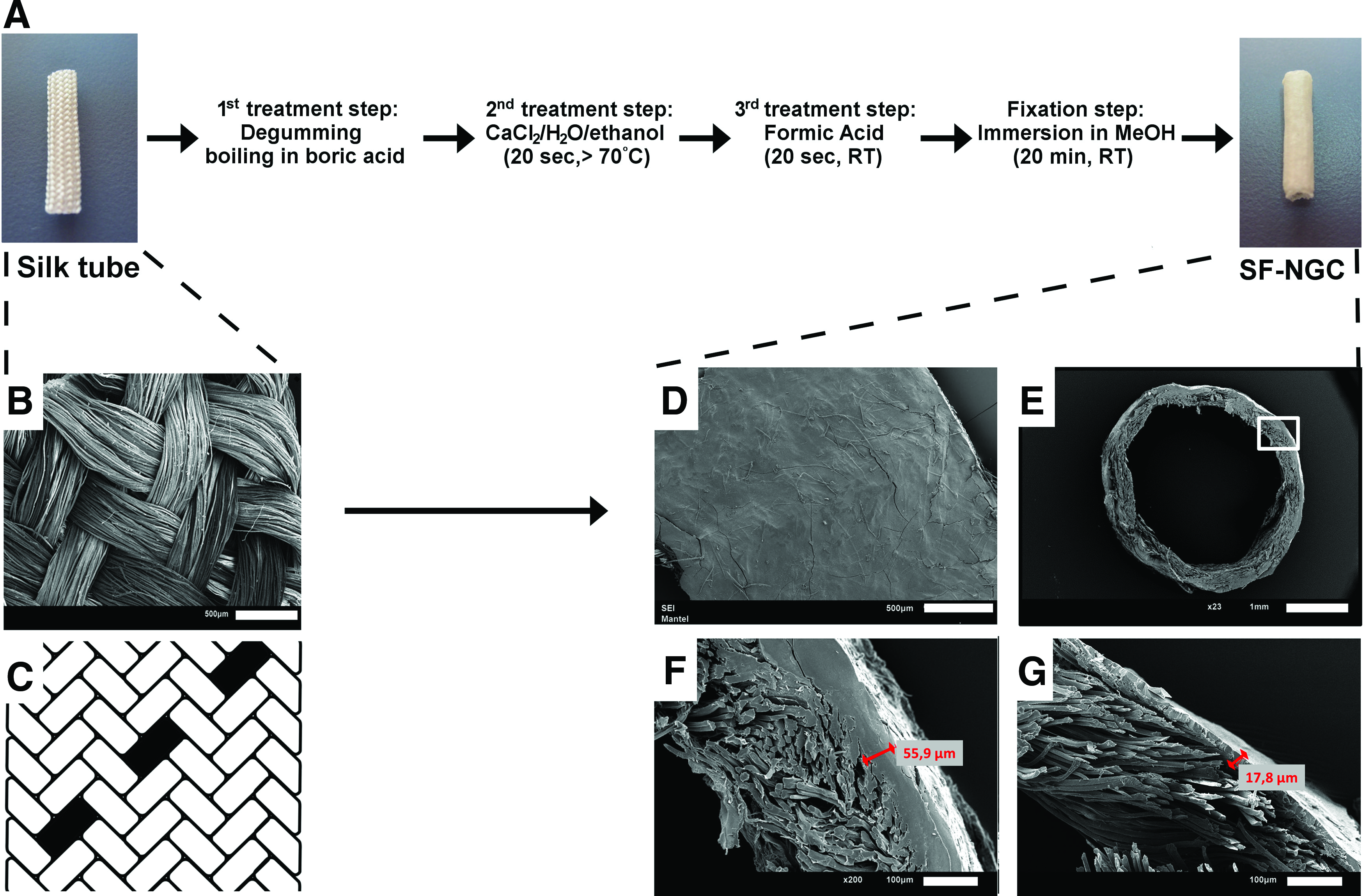
Overview of the preparation method of the silk fibroin (SF) nerve guidance conduit (NGC).
The degummed SF tubes were placed on an acrylonitrile butadiene styrene rod, having a diameter of 2 mm, and dipped into a boiling solution of the ternary solvent calcium chloride/distilled water/ethanol (CaCl2/H2O/ethanol) at a molar ratio of 1:2:8 for 20 s. Immediately after etching the outer surface, the tubes were dipped in 100% of FA at room temperature for 20 s. The tubes were then fixed in methanol for 20 min and subsequently washed thoroughly with ddH2O (Fig. 1). The tubes were dried under laminar airflow and sterilized by autoclaving before use.
Endurance and fatigue tests
To test the elasticity of the SF-NGC in comparison to the unprocessed initial tubular SF-scaffold, a custom-made compression test machine was built (Supplementary Fig. S1; Supplementary Data are available online at
To evaluate the effect of the various treatment components, four types of tubes have been evaluated (Fig. 2). The first type was produced by degumming of the raw tube followed by methanol treatment. The second and third types were created by either CaCl2/H2O/ethanol- or FA treatment, both of which were fixed with methanol. The fourth type of tube was produced as described earlier, combining all sequential treatments (Fig. 1).

Effects of the treatment steps on the surface of the SF fiber-based NGC. Panels show scanning electron micrographs of the degummed samples treated with the single treatments of calcium chloride, ethanol, and water in a molar ratio of 1:2:8 (CaCl2/H2O/ethanol) or FA and the the effect of the combined treatment (CaCl2/H2O/ethanol+FA), respectively. In the case of the combined treatment, SF constructs were treated with CaCl2/H2O/ethanol and FA, subsequently for 20 s each, resulting in a total treatment time of 40 s. For comparison, the single-step treatments were carried out for 40 s.
Before testing, respective samples were hydrated in phosphate-buffered saline (PBS) overnight. For testing, the conduits were fixed with cannulas (B. Braun) in a Sylgard-plated Petri dish (Sylgard® 184; Dow Corning Europe S.A.) and covered with PBS. The mechanical test regimen consisted of 1000 cycles of compression (300 ms duration and 58.8 MPa load) and release. After testing, the tubes were air-dried overnight at room temperature and an ∼1 mm thick slice was cut out from them at the impression site for morphological analysis. The deformity after the compressions was assessed by scanning electron microscope analysis.
Scanning electron microscope analysis
Samples were fixed in 2.5% glutaraldehyde in cacodylate buffer overnight at room temperature, then washed, dehydrated through graded ethanol changes followed by treatment with hexamethyldisilazane, and allowed to air-dry in a fume hood. Coating with Pd-Au was performed through the use of a Polaron SC7620 sputter coater (Quorum Technologies Ltd.), and the samples were examined using a JEOL JSM-6510 scanning electron microscope (JEOL Ltd.) operated at 15 kV.
Cell culture experiments
NIH/3T3
NIH/3T3 cell line was purchased from European Collection of Cell Cultures. NIH/3T3 cells were cultured in Dulbecco's modified Eagle medium (DMEM) containing 10% fetal bovine serum (FBS; Lonza Ltd.) supplemented with 2 mM L-glutamine, 100 U/mL penicillin, and 0.1 mg/mL streptomycin in plates coated with 0.2% gelatin solution.
Schwann cells
Schwann cells were isolated from rat sciatic nerves as described by Kaewkhaw et al. 27 All animals were housed in the facilities of the Ludwig Boltzmann Institute for Experimental and Clinical Traumatology in a temperature-controlled environment. Animals were provided with food and water ad libitum. All experimental protocols were approved by the City Government of Vienna, in accordance with the Austrian law and Guide for the Care and Use of Laboratory Animals as defined by the National Institute of Health.
The sciatic nerves were dissected free of connective tissue and minced after removing the epineurium. Nerve fragments were incubated with 0.05% collagenase for 1 h at 37°C, subsequently filtered through a 40 μm cell strainer, and centrifuged at 400 g for 6 min. After washing the cell pellet in DMEM containing 10% fetal calf serum, the pellet was resuspended in Schwann cell culture medium consisting of DMEM-
Schwann cells were seeded on the inner wall of the SF-NGC at a concentration of 105 cells/mL. Three groups were set: In group 1, only degummed (boric acid treatment) silk tubes were used; in group 2, the silk tubes were further treated with CaCl2 (degumming+CaCl2); and in group 3, tubes received a full treatment completed with FA (degumming+CaCl2+FA). As a final step, all tubes were fixed with methanol. After 2 h, cells were supplied with Schwann cell culture medium. Schwann cell attachment to the inner wall structure of the SF-NGC and viability were evaluated after 48 h with Calcein AM/propidium iodide staining (Invitrogen).
Cell permeability
A cell migration assay was designed to verify the cell impermeability of the SF-NGC. A 100 μL fibrin clot (Tisseel; Baxter) containing platelet-derived growth factor-AA (PDGF-AA) was used to induce cell migration. Before the addition of 250 U/mL of thrombin to induce polymerization, 10 ng of PDGF-AA (Peprotech Austria) was thoroughly mixed in fibrinogen. The resulting dense fibrin structure provides a slow release of PDGF-AA. This fibrin clot was then placed inside the investigated tubes, and the assembled constructs were pinned in silicone-coated (Sylgard 184; Dow Corning Europe S.A.) 12-well plates. Besides enabling the fixing of the constructs to a Petri dish, Sylgard 184 is known to discourage cell adhesion as a result of its hydrophobic character 28 and it, therefore, prevents cell migration from one clot to the other over the surface of the cell culture plate. As a result, the only way for the cell to move from one clot to the other is to pass through the SF-NGC. This thereby permits the assessment, if any, of the NGC's cell permeability. A second 100 μL fibrin clot containing 2.5×10⁵ NIH/3T3 fibroblasts was placed on top of the tube. For this clot, cells were suspended in fibrinogen and then the polymerization was initiated with 2 U/mL thrombin. The generated loose fibrin structure allows fibroblasts to migrate from the clot toward the chemotactic stimulus. As a positive control, the fibrin clot with cells was separated from the clot containing PDGF-AA using the nylon mesh of a cell strainer with a 100 μm pore size (Becton Dickinson Ltd.). The constructs were completely covered with cell culture medium. On day 6, cell migration was evaluated by staining the PDGF-AA-containing fibrin clot with Calcein AM (Invitrogen).
Cytotoxicity assay
To test cytotoxicity of the prepared SF-NGC, 1 g of the material was immersed in 5 mL cell culture medium for at least 24 h. In parallel, 0.2×10⁵ Schwann cells per well were seeded onto 24-well plates. Then, the medium containing leach-out products from the material was filtered (0.22 μm; Rotilabo) and used to change media in the cell cultures. Standard culture medium was used as a negative control. After 72 h, cell culture medium was aspirated and the respective cell culture medium containing 650 μg/mL [3-(4,5-dimethylthiazol-2-yl)-2,5-diphenyltetrazolium] (MTT) bromide was added to each well. After 1 h of incubation at 37°C and in 5% CO2, the medium was aspirated and MTT formazan precipitate was dissolved in dimethyl sulfoxide by shaking mechanically in the dark for 20 min. Aliquots of 100 μL of each sample were transferred to 96-well plates. The absorbance at 540 nm was read immediately on an automatic microplate reader (Spectra Thermo; TECAN Austria GmbH). Optical density values were corrected for an unspecific background.
Animals and surgery
All animals were housed in the facilities of the Ludwig Boltzmann Institute for Experimental and Clinical Traumatology in a temperature-controlled environment. Animals were provided with food and water ad libitum. All experimental protocols were approved by the City Government of Vienna in accordance with the Austrian law and Guide for the Care and Use of Laboratory Animals as defined by the National Institute of Health.
A total of 28 female Sprague–Dawley rats (Animal Research Laboratories), weighing between 250 and 350 g, were used in the experiments. Possible adverse effects against the implanted SF-NGCs and the initial axon outgrowth into these tubes were evaluated on day 7, 10, and 21 day (n=2, 6, 2, respectively). Eighteen animals were randomly assigned into three different treatment groups: autologous grafting (n=6), SF-NGC (n=6), and collagen-filled SF-NGC (n=6) for 12 weeks of observation. The animals were weighed and anesthetized in a fume box with 3.5% isoflurane (Forane®; Abbott) at a flow rate of 800 mL/min. Subsequent anesthesia throughout the surgical procedure was maintained using 2.5% isoflurane via a nosepiece. The right lower limb in each animal was shaved and disinfected with povidone-iodine (Betaisodona®; Mundipharma) at the mid-thigh level. All the following surgical procedures were carried out under an operating microscope (Leica M651; Leica Microsystems). The sciatic nerve was exposed and an 8 mm segment of the sciatic nerve was excised, resulting in a 10 mm gap due to elastic retraction. In the autologous grafting group, the excised 8 mm segment of the sciatic nerve was rotated 180° and then sutured to the proximal and distal stumps using Ethilon 8/0 epineurial sutures (Ethicon-Johnson and Johnson). In both SF-NGC groups, the conduit was implanted by insertion of the proximal and distal nerve stumps into the 12 mm tube and coaptated to the conduit by two epineurial sutures. In the SF-NGC-collagen group, the lumen of the SF-NGCs was filled with 8 μL of collagen solution (Type I, 2.5 mg/mL; Millipore). Afterward, the wound was closed in anatomical layers. The analgesic treatment was administered in the form of 0.75 mg/kg bodyweight (BW) meloxicam (Metacam®; Boehringer Ingelheim) and 1.25 mg/kg BW butorphanol (Butamidor®; Richter Pharma AG) immediately before the surgical procedure and for 2 days thereafter.
Tissue sampling, perfusion, and immunohistochemistry
Twelve weeks after surgery, the animals were deeply anesthetized by inhalation of 3.5% isoflurane and euthanized with 110 mg/kg BW ketamine hydrochloride (Ketasol®; Dr. E. Graeub AG) and 12 mg/kg BW xylazine (Rompun® 2%; Bayer AG) intraperitoneally. The animals were perfused transcardially with 4% paraformaldehyde in a 0.1 M phosphate buffer (pH 7.4). The autologous transplants or the implanted SF-NGC were harvested under the operating microscope along with the proximal and distal nerve stumps.
The nerve grafts or conduits (n=6 per group), removed from perfusion-fixed animals, were immersion fixed for 6 h in 4% paraformaldehyde and then cryoprotected in 30% sucrose in PBS. Parallel cryostat sections were cut on a Leica 1850 cryostat (Leica Microsystems), and sections were either stained with cresyl violet or processed for 200 kDa neurofilament immunohistochemistry. Sections were treated with a 1% milk powder solution and then incubated either with an anti-200 kDa rabbit neurofilament antibody (Abcam Ltd.; Lot. No.: ab8135, rabbit, 1:1000) or with biotinylated Griffonia (Bandeira) simplicifolia lectin B4 (GSA-B4, 1:200; Vector Labs) overnight at 4°C. The sections were then processed for incubation with an anti-rabbit Alexa 546 or secondary antibody (1:400) or with streptavidin Alexa 488 (to visualize 200 kDa neurofilament or Griffonia simplicifolia isolectin, respectively) for 2 h at room temperature, protected with a coverslip, and investigated under an epifluorescence microscope (Olympus FX-50; Olympus Ltd.).
The sciatic nerve segment distal to the graft was transferred into a 2.5% phosphate-buffered glutaraldehyde solution after perfusion and immersion fixed for 24 h. Remnants of fixative were carefully washed out from the nerve, and the tissue was treated in 1% OsO4 in PBS (Agar Scientific) for 1 h, dehydrated in a graded ethanol series and propylene oxide, and embedded in Durcupan (Fluka). Semithin sections (0.4 μm thick) were cut 2 mm distal to the graft on a Leica Ultracut-R ultramicrotome and stained according to Rüdeberg. 29
Quantification of the Schwann cell densities
To quantify the cellular area on the luminal surface of tubes treated to various extents, we randomly selected three (500×500 μm) areas and photographed Schwann cells at a 10-fold primary magnification, using an Olympus BX50 epifluorescence microscope. Using ImageJ Software (NIH), we measured the relative density of Calcein AM-stained Schwann cells. The background/autofluorescence of unstained samples as a reference intensity was then subtracted from the intensity of Calcein AM-stained samples. The cellular area occupied on the total surface was divided by the total area and multiplied by 100. Furthermore, automatic thresholding was performed for each image by using the NIH ImageJ software to determine the threshold for the specific signal.
Locomotor tests—Gait analysis (CatWalk™)
To evaluate the functional recovery of the animals, the Catwalk gait analysis system (version 7.1; Noldus) was used. This method allows an objective quantification of multiple static and dynamic gait parameters.30,31 The animals were trained to use the runway before the surgery for a period of 3 weeks. Postoperatively, animals were evaluated once a week from week 4 to week 12. Various parameters for locomotor functional recovery, including print area, maximum intensity, stance time, and duty cycle, were determined. The intensity of the right hind paw was expressed as a percentage of the contra-lateral left hind paw. The Catwalk experiments were performed in a blinded fashion.
Electrophysiology
At the end of the defined regeneration period (12 weeks), electrophysiological analysis (NeuroMax-XLTEK) was carried out before sacrificing the animals. Stimulation electrodes were placed 2 mm proximal and 2 mm distal to the graft for calculation of the nerve conduction velocity (NCV). A needle electrode was placed as a recording electrode into the tibialis anterior muscle, and the sciatic nerve was stimulated for 0.05 ms first proximally and then distally to the graft, so as to achieve the supramaximal stimulation amplitude. The compound action potential, the amplitude, and the NCV were determined. All measurements were carried out at a temperature between 38°C and 39°C.
Statistical analysis
Statistical analysis was performed using Graph Pad Prism software (Graph Pad Software, Inc.). Normal distribution of data was tested with the Kolmogorov–Smirnov test. One-way analysis of variance followed by Tukey's post hoc test was used to assess statistical significance, and p-values below 0.05 were considered statistically significant. All graphs in this study are shown as mean±standard deviation.
Results
Structural changes during processing of silk tubes
Figure 1A–C shows the raw SF-NGC consisting of braided single silk fibers. After degumming, the SF-NGC was subsequently treated with CaCl2/H2O/ethanol and FA for 20 s each, followed by fixation with methanol (Fig. 1A). This treatment resulted in a fusion of the outer single silk fibers to a closed layer with a varying thickness ranging from 40 to 75 μm (Fig. 1D–F). Treatment with CaCl2/H2O/ethanol for <20 s resulted in a thinner outer layer (Fig. 1G). In contrast, the luminal wall of the tube, which was not treated with the various solvents, preserved its original braided structure (Fig. 1F). Figure 2 shows the time-dependent effects of CaCl2/H2O/ethanol and FA on the SF fibers. SF fibers solely treated with CaCl2/H2O/ethanol dissolve and precipitate, especially after 40 s. In contrast, FA-treatment alone disorganizes the original braided structure in a time-dependent manner.
Cytocompatibility assays
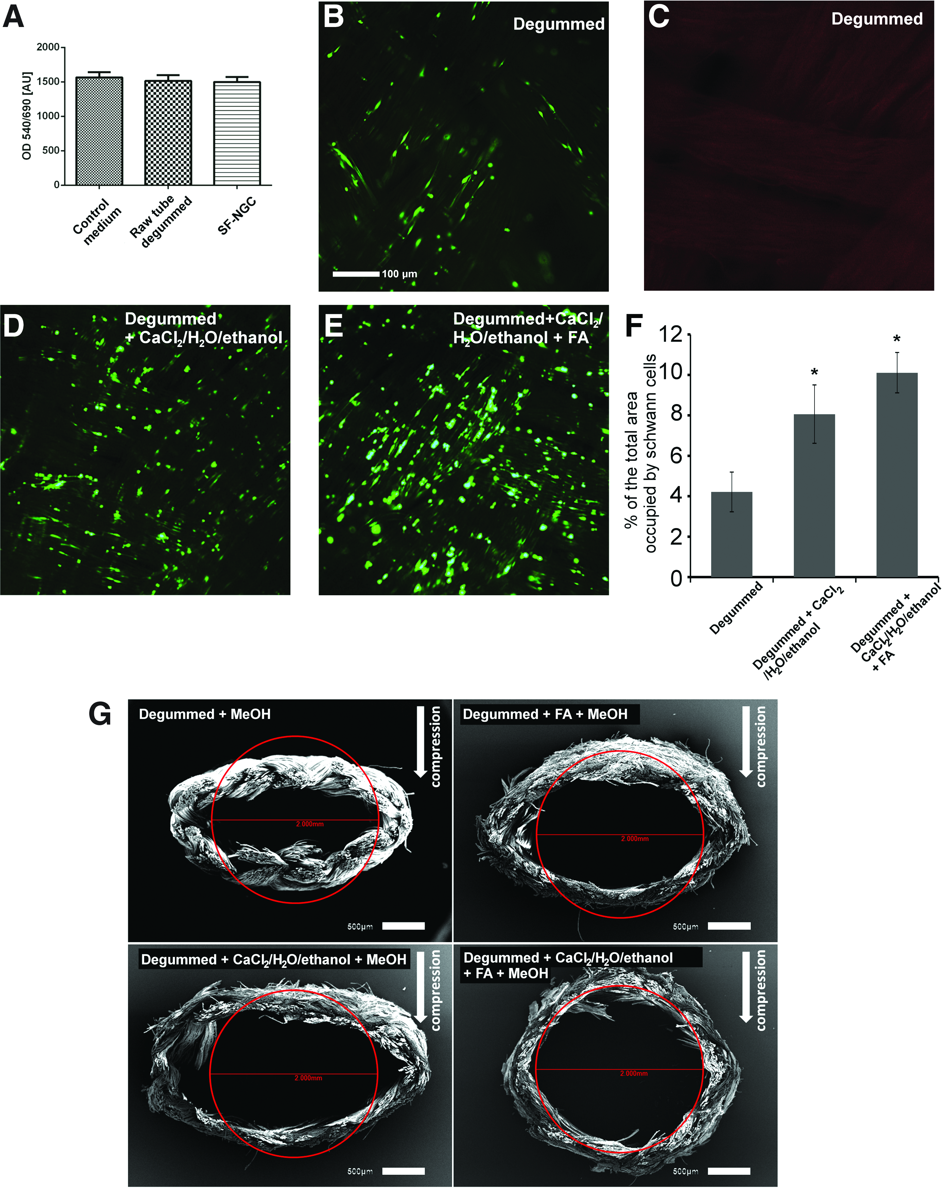
To investigate whether cytotoxic residuals were left in the SF-NGCs during the preparation procedure, an MTT assay was performed. Dissected material from the SF-NGC and the unprocessed raw silk scaffold were incubated in cell culture media, to collect substances that may leach out of the constructs. Treatment of cultured Schwann cells with these leach-out media resulted in no significant difference in the cell viability of Schwann cells in any treatment group (Fig. 3A). Viability and adhesion pattern of Schwann cells cultured on the luminal surface of the silk tubes processed to a various extent during the SF-NGC preparation procedure were tested with Calcein AM/propidium iodide staining. Treatment with CaCl2/H2O/ethanol and the full treatment procedure, including incubation with FA, induced significantly more Schwann cells to adhere to the luminal surface of the silk tubes than degumming only (Fig. 3B, D–F). On the other hand, propidium iodide staining did not reveal any nonviable Schwann cells on the luminal surface of these cultured silk tubes (Fig. 3C).
Results of the cytotoxicity, Schwann cell viability, and compression tests of the various conduits.
Endurance test
To verify the required elasticity of the SF-NGC, mechanical endurance tests were performed with a custom-made system (Supplementary Fig. S1). Degummed tubes remained compressed and flat after 1000 cycles of compression (Fig. 3G). FA or CaCl2/H2O/ethanol improved the elastic properties of the SF-NGCs, resulting in partial preservation of the lumen; however, substantial deformation was still present. On their own, none of these treatments were able to provide the SF-NGC with elastic properties to withstand external pressure whereas full treatment (degumming, CaCl2/H2O/ethanol followed by FA treatment) helped in improving the elastic properties of these SF-NGCs to become resistant to external forces (Fig. 3G).
Cell permeability
A cell migration assay was applied to test the impermeability of the SF-NGC wall to invading cells. The test was based on the chemotactic properties of PDGF-AA embedded in a fibrin clot (Fig. 4A). The efficacy of NIH/3T3 fibroblasts to penetrate and pass through the wall of the silk tube was tested. Degummed silk tube walls were suitable structures for the fibroblasts to migrate through their braided structure similar to positive controls, where the cells were able to pass through the mesh of a cell strainer (100 μm pore size) (Fig. 4B, C). In contrast, SF-NGC with a completely closed outer surface did not support the penetration of fibroblast into the wall of the conduit (Fig. 4C).
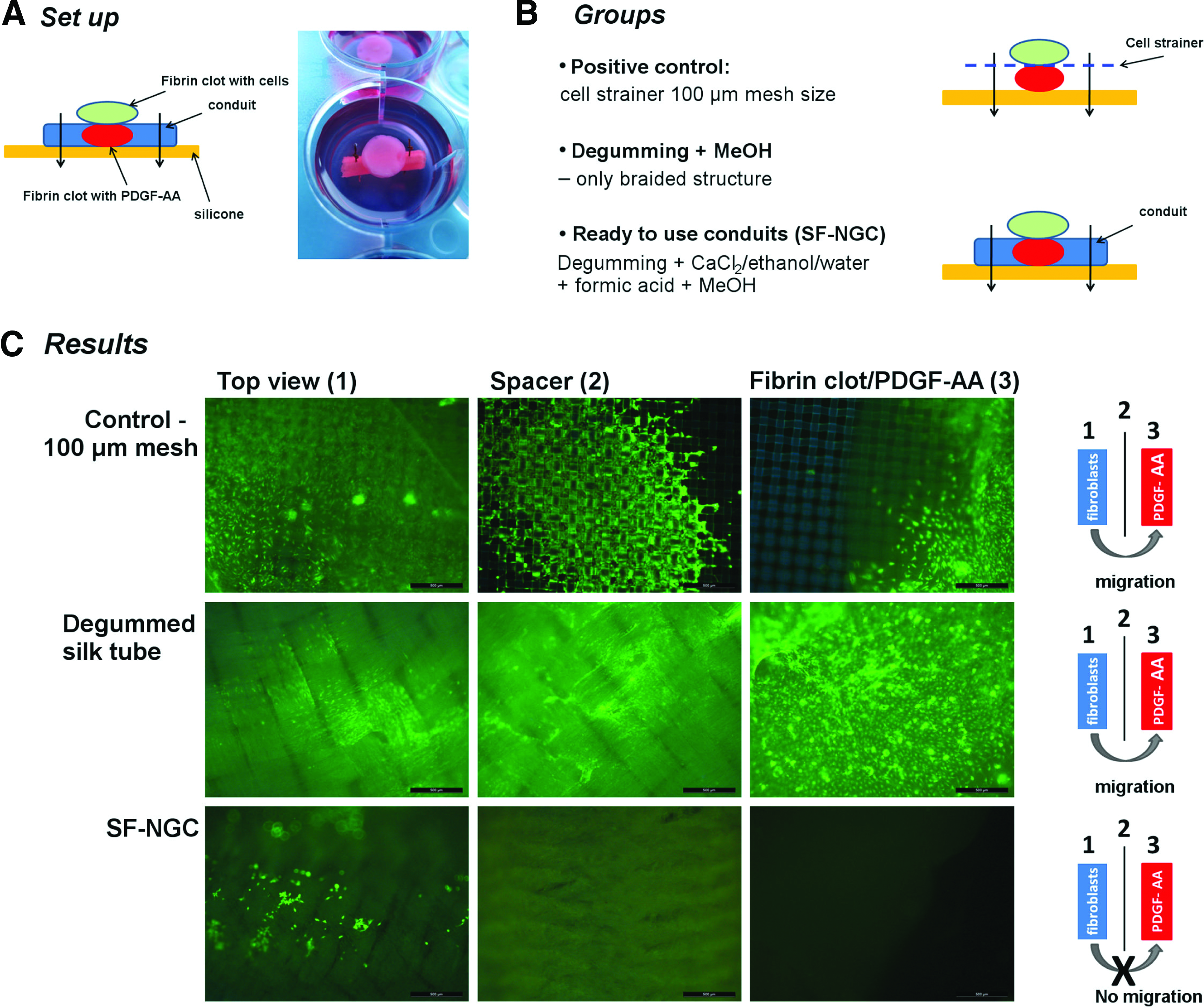
Results of the cell permeability assay using a fibrin clot containing NIH/3T3 fibroblasts and a second clot loaded with platelet-derived growth factor-AA (PDGF-AA) as a chemoattractant
Short-term in vivo studies—general observations
The implanted SF-NGCs were explored and thoroughly checked under the operating microscope at 1 and 3 weeks after surgery. Figure 5A shows the proximal and distal nerve stumps coaptated to the SF-NGC by two epineurial sutures at the time of surgery. One week after implantation, visual inspection revealed that the SF-NGC did not exhibit substantial degradation (Fig. 5B). Furthermore, no signs of inflammatory reaction or neuroma formation at the coaptation sites could be detected. The entire outer surface of the implanted graft was covered by a thin layer of connective tissue. Interestingly, the proximal as well as the distal end of the implanted SF-NGC shows a partial integration of the nervous tissue with the SF-NGC (Fig. 5B). Moreover, the thin layer of connective tissue on the surface of the SF-NGC contained small blood vessels (Fig. 5C, D). At 3 weeks after implantation, the lumen of the SF-NGC was completely filled with regenerated tissue (Fig. 5E). Careful dissection of the SF-NGC (Fig. 5F) revealed a complete reconnection of the proximal and distal nerve stumps.

Integration of the silk-fibroin conduit in the defect site.
Axonal regeneration
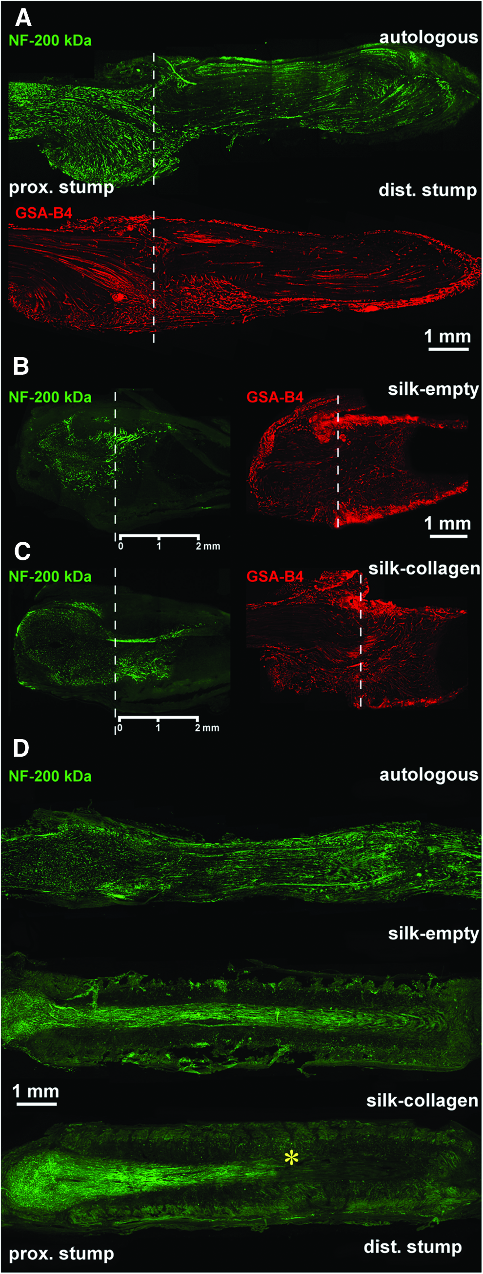
Our results showed that an 8 mm gap in the rat sciatic nerve could be bridged by implanting an SF-NGC in the gap. To compare the axon growth-promoting capacity of the three conduits used in the study, we looked at the axon outgrowth from the proximal nerve stump into the conduits 10 days after grafting by using neurofilament staining. Autologous nerve grafts were already populated with regenerated axons along their whole length at this time-point, and the axons approached the distal coaptation site (Fig. 6A). In contrast, the silk tubes were able to promote only limited outgrowth of the axons at this stage. The regenerating axons had grown to a distance of∼2 mm in both conduits (1.7 and 1.8 mm in empty tubes and 2 and 2.1 mm in collagen-filled tubes, n=2 in each group) without considerable difference between them (Fig. 6B, C). The autologous nerve grafts 10 days postoperatively are well vascularized (Fig. 6A). A similar range of vascularization of the silk tubes could be observed on day 10 after surgery. No considerable number of macrophages were seen in the implanted silk tubes (Fig. 6B, C).
Axonal regeneration and vascular ingrowth in the various experimental groups.
Three months after transplantation, the course of regenerated axons throughout the lumen of the implanted SF-NGCs and autologous nerve grafts was clearly visible (Fig. 6A–D). Although the axon bundle was present in the empty silk tubes, the immunohistochemical analysis did not reveal a significant staining pattern for neurofilament 200 kDa in the distal portion of the tubes. On the other hand, myelinated axon counts showed significantly less myelinated axons in the empty SF-NGC as well as in the collagen-filled SF-NGC compared with the autologous nerve graft (empty SF-NGC: 1274±146, collagen-filled SF-NGC: 1678±303, autologous nerve graft: 6252±474; Fig. 7). Accordingly, filling the silk tubes with collagen did not influence the short- and long-term regeneration of axons.
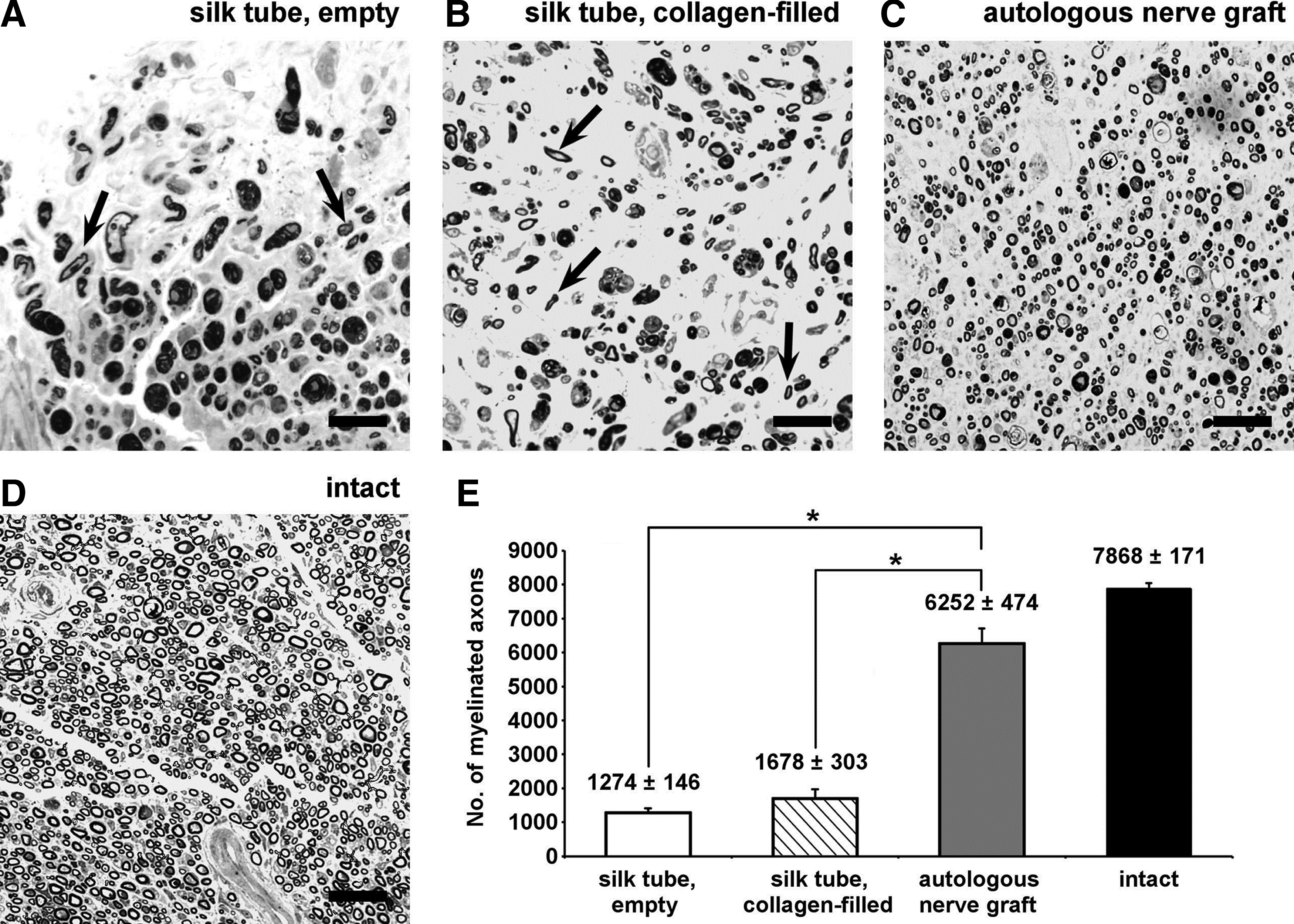
Numbers of myelinated axons in the various experimental groups 12 weeks after implantation. Arrows point to well-myelinated axons in the distal stumps
Functional recovery—Catwalk analysis
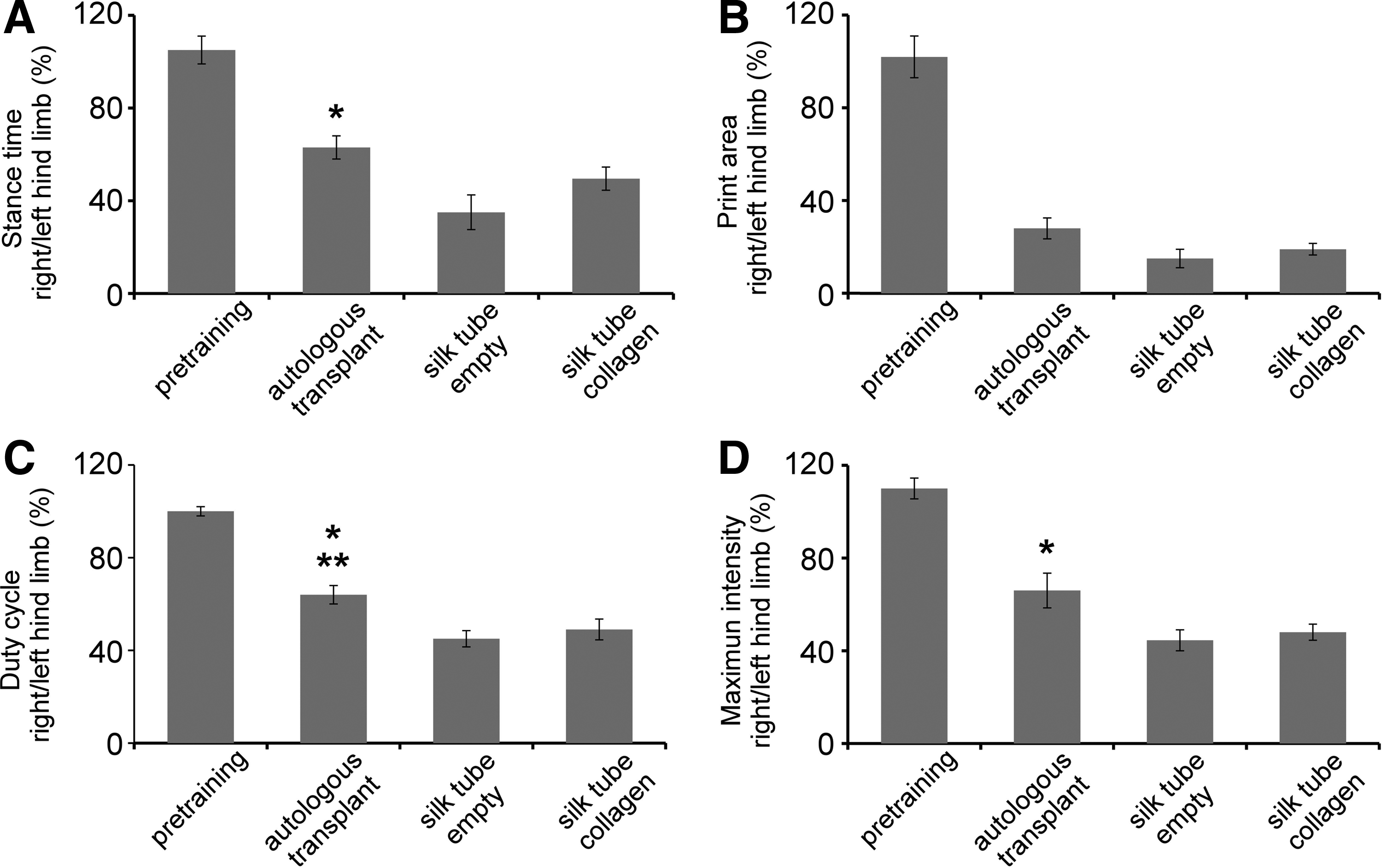
Twelve weeks after surgery, the functional recovery parameters (Fig. 8A–D), including mean stance time, mean print area, mean duty cycle, and the mean maximally exerted intensity of the right hind limb, were evaluated. In three out of four parameters (excluding limb print area), there was a significant difference between the extent of recovery of the autologous nerve grafts compared with the empty silk tube; whereas in the case of duty cycle, the animals receiving an autologous nerve graft performed significantly better in comparison to both silk tube groups. A minor, statistically not significant difference was found between the two silk tube groups in case of all parameters (Fig. 8A–D). It should be noted that animals treated with autologous nerve grafts displayed functional parameters approaching but never closely reaching the pretraining values.
Quantitative Cat-Walk gait analysis of locomotor functional recovery 12 weeks after implantation, including stance duration time
Electrophysiology
The results of the electrophysiological analysis strongly correlate with the functional data described earlier. Electrophysiological recordings were carried out 12 weeks after transplantation. Compound nerve action potential (CNAP) and NCV values were significantly improved in the autologous nerve grafting group (CNAP: 22.8±7.5; NCV: 49.2±14.2) compared with both silk tube groups (empty SF-NGC, CNAP: 6.5±3.1; NCV: 23.9±6.6. Collagen-filled SF-NGC, CNAP: 9.7±4.4; NCV 25.9±7.3; Fig. 9). No difference could be detected between the groups receiving the various silk tubes, although the animals with empty silk tubes displayed slightly impaired electrophysiological data.

Electrophysiological analysis of the effect of axonal regeneration through the various conduits. The compound nerve action potential area
Discussion
In this study, we have investigated the mechanical properties and biocompatibility of a novel NGC manufactured from a braided tubular structure of SF fibers. Moreover, the ability of this novel conduit to bridge a peripheral nerve gap and support the regeneration of injured rat sciatic nerve axons has been tested.
The nature of the chemical treatment to transform a braided structure into a mechanically resistant, nonpermeable, and flexible tube for externally invading cells made it necessary to investigate cytotoxicity and cellular viability before in vivo implantation. The mixture of CaCl2/H2O/ethanol dissolves native silk fibers,32,33 while methanol induces the formation of β-sheets, leading to a crystalline-like structure of the SF.34,35 FA functions as a solving and crystallizing agent. Um et al.36,37 reported that FA induces an ordered structure and molecular arrangement. As the end result, this combination treatment resulted in a homogenous crystalline-like outer layer of the nerve conduit wall. In addition, by controlling the incubation times, we were able to design the structure and thickness of the outer crystalline layer. All together, the treatment steps resulted in the generation of a mechanically stable tubular conduit.
Apart from the favorable mechanical properties, the question remained as to whether this construct maintained its biocompatibility, was able to prevent invasion of connective tissue cells from the environment, and provided a supportive luminal surface for proliferating Schwann cells. According to our findings, these conduits fulfilled all these requirements.
Our short-term in vivo studies have provided evidence that the implanted silk tube conduits were able to integrate into the host environment without generating significant inflammatory reactions and, on the other hand, could successfully bridge an eight-millimeter-long nerve defect. These features may enable this type of silk tube conduit to act as a strong candidate for nerve repair. From a practical point of view, the best available nerve conduit is an autologous nerve graft, frequently regarded as the gold standard for experimental and clinical use of nerve grafting. There is, however, an urging need for nerve conduits in clinical use when autologous nerve grafts are not available. These conduits should preferably fulfill a number of requirements: They should be biocompatible, long enough to bridge large defects, able to support Schwann cell proliferation followed by rapid axonal regeneration, and accept external vascular ingrowth, while they resist invasion of external cell populations, especially that of fibroblasts.
It is evident that a chemically inert silk tube bears several of the features mentioned earlier but it is still unlikely to guide a significant number of degenerating axons over long distances. The longest distances that can still be bridged by artificial or natural conduits are frequently called “critical gap,” and they are believed to range between 2 and 6 cm in humans.37–39 Gaps longer than 6 cm can only be reconnected by using autologous nerve grafts or nerve allografts.40–42 The intriguing question is raised as to how nerve conduits should be altered to make them suitable for grafting in long nerve defects. The silk tube conduit presented in this study is likely to undergo a number of further modifications to suit these requirements. It could be argued that by making the silk tubes permeable for growing vessels and modifying their luminal environment to foster axonal regeneration the silk tube conduits would be transformed into structures with features closely resembling peripheral nerve grafts. Such conduits should carry features usually present in an intact or freshly degenerated peripheral nerve, for example, the presence of axonal growth-promoting cells (such as Schwann cells or Schwann cell-like cells)43–45 and the extracellular matrix compounds produced by these cells. Recently, advances in experimental bridging of larger nerve defects have been made, including the strategies outlined earlier. We suggest that the next generation of biologically inert silk tube conduits could possibly include treatment with the peripheral nerve-specific extracellular matrix molecules fibronectin and laminin along with sequential transplantation of Schwann cells or Schwann cell-like cells into the conduit.
These novel methodological approaches may open new horizons in the field of peripheral nerve regeneration and repair and may contribute to better treatment opportunities of large human nerve defects.
Conclusion
In this study, we describe the production of a novel NGC based on raw silk textile tubular structures. The chemical treatment of the raw silk tube resulted in a biocompatible and mechanically stable conduit that was able to bridge relatively short gaps in the rat sciatic nerve. It can be concluded that these silk tube conduits are subject to further studies and modifications to produce cellularized bioartificial conduits that would support long-distance nerve regeneration.
Footnotes
Acknowledgments
The financial support by the NewTissue Project (FFG #818412 and City of Vienna MA27#06-06) and the City of Vienna Competence Team reacTissue Project (MA27#12-06) is gratefully acknowledged. The authors thank Mrs. Tricia Behling for the language review. For their support of the in vivo study, they thank the whole animal facility team, especially Karin Hahn and Claudia Keibl (DVM), from the Ludwig Boltzmann Institute for Experimental and Clinical Traumatology, Vienna. Moreover, the authors would like to thank Prof. Martijn van Griensven for fruitful discussions.
Disclosure Statement
No competing financial interests exist.
References
Supplementary Material
Please find the following supplemental material available below.
For Open Access articles published under a Creative Commons License, all supplemental material carries the same license as the article it is associated with.
For non-Open Access articles published, all supplemental material carries a non-exclusive license, and permission requests for re-use of supplemental material or any part of supplemental material shall be sent directly to the copyright owner as specified in the copyright notice associated with the article.