
Abstract
In vitro degradation rates of calcium phosphate bioceramics are investigated using a large variation of soaking protocols that do not all match the dynamic conditions of the perfused physiological environment. Therefore, we studied the effect of stirring and fluid perfusion on the in vitro degradation rate of apatitic calcium phosphate cements (CPC) containing poly(lactic-co-glycolic acid) (PLGA) microspheres. The composites were soaked in phosphate-buffered saline up to 6 weeks under unstirred, stirred, or perfused conditions followed by analysis of mass loss, compression strength, porosity, crystal phase composition, and morphology of the cement composites. The results showed that fluid perfusion reduced the decrease in pH and corresponding degradation rates, while nonperfused soaking conditions (i.e., stirred and unstirred conditions) resulted into more extensive acidification, the rate of which increased with stirring. After 2 weeks, the formation of a secondary brushite phase was observed for cement composites soaked under nonperfused (i.e., stirred and unstirred) conditions, whereas this phase was not detected in cements soaked under perfused conditions. The degradation rate of cement composites decreased in the order unstirred>stirred>perfused, as evidenced by quantification of mass loss, compression strength, and pore morphology. To summarize, we have demonstrated that soaking conditions strongly affected the in vitro degradation process of CPCs. As a consequence, it can be concluded that the experimental design of current in vitro degradation studies does not allow for correlation to (pre-)clinical studies.
Introduction
C
Numerous chemical and physical properties such as the density and the composition of the material affect the degradation rate of apatitic CPCs. Among all these factors, the nano- and microporosity of apatitic CPCs are considered to be of critical importance. Therefore, a wide variety of strategies have been explored to increase the porosity of apatitic CPCs. A recent approach focused on the inclusion of degradable polymeric microparticles into apatitic CPC.
Poly(lactic-co-glycolic acid) (PLGA) is a synthetic polymer, which has been extensively studied for applications in tissue engineering and regenerative medicine. PLGA is highly biocompatible and biodegradable, which renders this polymer suitable as drug delivery transport for sustained release of therapeutic biomolecules. The degradation of PLGA occurs by the hydrolysis of ester linkages, producing two acidic monomers, that is, lactic and glycolic acids.6,7 By introducing PLGA microparticles into CPC, the porosity of CPC is increased and the degradation is accelerated due to the formation of acidic by-products that are able to dissolve calcium phosphate phases. Recent studies have indicated that the degradation rate of CPC/PLGA cement composites can be tuned by controlling the chemical and/or morphological parameters of the PLGA microparticles, such as the molecular weight, lactide to glycolide ratio, particle morphology (hollow or dense), and end-group functionalization.8,9
Numerous CPC formulations have been developed and evaluated in vitro during the last decades. Nevertheless, the in vitro degradation rates of calcium phosphate bioceramics are generally investigated using soaking protocols that show a large variation in study design and do not match the dynamic conditions of the perfused physiological environment. For instance, the type of soaking medium, its volume, the pH, the rate, and the type of medium refreshment (static or dynamic) differed to a large extent between the studies. Generally, three main soaking situations can be discerned:
(1) unstirred, (2) stirred, and (3) perfused. In the first category, samples are incubated in a soaking medium, which can be refreshed or not at a regular frequency during the study period, without any mechanical stirring.10–12 Under stirred soaking conditions, the soaking medium is stirred at a fixed rotation speed,13,14 while samples are subjected to a continuous dynamic flow under perfused conditions. Previously, the degradation process of CPC has been mainly studied in vitro at unstirred conditions.15,16
However, a great variability was observed between the in vitro and in vivo results. 17 Based on this observation, we hypothesized that current in vitro testing methods cannot reliably predict in vivo degradation rates of CPCs. Therefore, the aim of this study was to investigate the influence of stirring and perfusion on the in vitro degradation behavior of CPCs containing PLGA microparticles. In more detail, we hypothesized that perfused conditions would lead to decreased degradation rates due to more effective removal of degradation by-products that are responsible for dissolution of the calcium phosphate matrix. To test this hypothesis, the composites were soaked in phosphate-buffered saline (PBS) up to 6 weeks under unstirred, stirred, or perfused conditions followed by analysis of mass loss, compression strength, porosity, crystal phase composition, and morphology of the cement composites.
Materials and Methods
Materials
PLGA microspheres were synthesized from PURASORB® PDLG 5002A (molecular weight=17 kDa, acid terminated, lactide/glycolide ratio 50:50; Corbion Purac). Polyvinyl alcohol (PVA, 88% hydrolysed, molecular weight 22 kDa; Acros), isopropanol (IPN, analytical grade), and dichloromethane (DCM, analytical grade; Biomet Merck) were used as solvent for the synthesis.
The cement powder mixture consisted of 95 wt% commercially available α-tricalcium phosphate (α-TCP; CAM Bioceramics BV) and 5% w/w precipitated hydroxyapatite (pHA; Biomet Merck). The α-TCP was ball milled at 500 rpm for 4.5 h using three laboratory balls, followed by the addition of pHA to the ground powder during another hour of milling. The ball milling was performed under dry conditions.
Synthesis of PLGA microspheres
The synthesis of the PLGA microspheres was described in detail in previous studies.9,18 Briefly, 0.2 g PLGA was dissolved in 2 mL DCM in a 20-mL glass tube. The solution was transferred into a stirred beaker containing 150 mL of a 0.3 w/v% PVA solution. Five minutes later, 100 mL of 2% IPN solution was added. After 1 h of stirring, the microspheres were allowed to settle for 1 h and the clear supernatant was decanted. The remaining suspension was centrifuged and the final supernatant decanted. Finally, the microspheres were frozen at −20°C, freeze dried for 24 h, and stored at −20°C.
The morphology of the PLGA microspheres was determined by light microscopy (Leica/Leitz DM RBE Microscope system; Leica Microsystems AG) and scanning electron microscopy (SEM, 6340F; JEOL). The average size of the microspheres was determined by digital image software (Leica Qwin W, Leica Microsystems AG) using a sample size of at least 300 microspheres.
Preparation of CPC/PLGA composites
PLGA microspheres (0.4 g) were added to 0.6 g of ground CPC powder in a 2-mL plastic syringe (yielding a PLGA content of 40 wt%), followed by the addition of 0.32 mL of a 3% NaH2PO4 solution (Biomet Merck) to the powder mixture and 25 s of shaking (Silamat® Mixing apparatus; Vivadent) to form the CPC/PLGA paste. Subsequently, the paste was injected into a polytetrafluorethylene (PTFE) mold (cylinder shaped, diameter=4.5 mm, height=9 mm) to obtain specimens of similar dimensions. Subsequently, the samples were left in a furnace at 37°C overnight to allow for hardening, after which the samples were stored at room temperature.
In vitro degradation studies
Cement composites were soaked at three different soaking conditions, that is, (1) unstirred, (2) stirred, and (3) perfused (Fig. 1). For the unstirred condition, sample holders were prepared from titanium mesh (height: 4.5 cm, diameter: 1.5 cm, and pore size: 1 mm) and vertically mounted on a metal stick. Each sample holder contained seven cement composites and was immersed in a transparent borosilicate glass flask filled with 3 L of PBS buffer (pH=7.4) at 37°C.
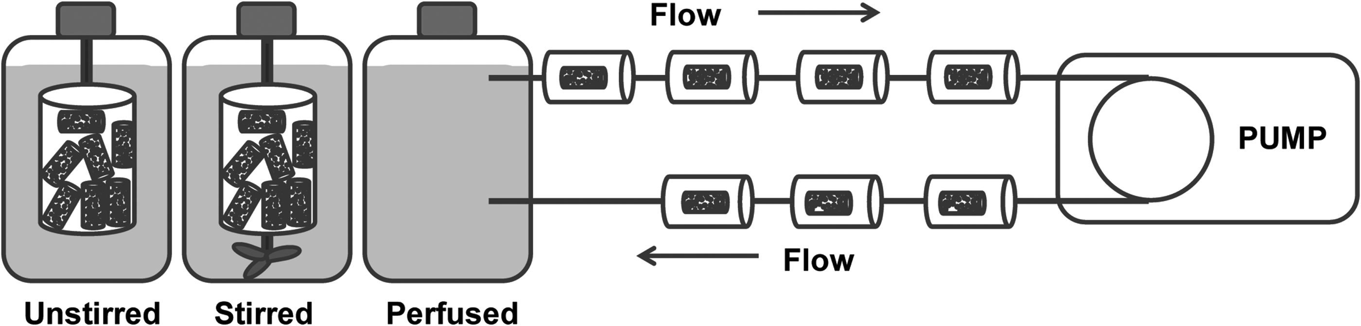
Schematic of three different soaking conditions.
A 3-paddle stirrer (P100; Applikon® 1010 Bio Controller) was used for stirring at 200 rpm for stirred conditions and 0 rpm for unstirred conditions. In addition, a perfused flow system was designed, which consisted of flow chambers with a rectangular-shaped exterior (length=60 mm, width=20 mm, and height=20 mm) and a cylinder-shaped interior (diameter=8 mm, height=60 mm) made of polymethylmethacrylate (PMMA). Seven flow chambers were loaded with one sample each and connected in series using standard silicon tubes (L/S® Precision Pump Tubing, Masterflex®; Metrohm Applikon B.V.). A borosilicate glass flask filled with 3 L of PBS (37°C, buffered at pH 7.4) was used as medium reservoir. A peristaltic pump (EV045, Verderflex; Verder Liquids B.V.) was connected to the flow chambers and the glass flask, which allowed PBS to flow through the samples at a rate of 1 mL/min (Fig. 1). For each of the three groups, 28 CPC/PLGA samples were fabricated and immersed in PBS. After an incubation time of 1, 2, 4, and 6 weeks, seven samples were taken out at each time point for further analysis (n=7).
Analysis of samples and suspensions
The pH of the PBS buffer was measured using a pH electrode (Orion; Sigma-Aldrich®) for all soaking conditions. To determine the mass loss as a function of time, the sample weight was measured before and after soaking. Microcomputed tomography (μ-CT; Skyscan 1172) was used to observe the distribution of PLGA microspheres and the porosity of the composites as a function of in vitro degradation. Tomographs were recorded at a voltage of 100 kV and a current of 98 μA. The scanning resolution was set to 7.08 μm at a rotation step of 0.9°.
To test the compressive strength, samples were placed in a testing bench (858 Mini Bionix II®; MTS Corp) and compressed at a cross-head speed of 0.5 mm/min. After testing the compressive strength, solid samples were ground to powder and analyzed by powder X-ray diffraction (XRD; PW 3710, Philips) to determine the crystal phase of the cement composites from 10° to 40° 2θ with a step size of 0.05° 2θ and a counting time of 20 s. The average crystallite size of the CaP depositions was calculated by using the Scherrer formula and the full width at half maximum of the (002) reflection of apatitic depositions. 19 Samples were cut using a razor blade (GEM® Scientific, American Safety Razor Co.) to obtain cross-sections for morphological analysis using SEM (JEOL6340F, Field Emission Scanning Electron microscope, operated at 10 kV).
Statistical analysis
The data were calculated as mean±standard deviation. Significant differences were determined by analysis of variance (two-way ANOVA). The results were considered significant if p<0.05. Calculations were performed using GraphPad Instat® (GraphPad Software, Inc.).
Results
pH test
The formation of acidic degradation by-products (lactic acid and glycolic acid) during degradation of the CPC matrix and PLGA microspheres (average size 94.8±29.8 μm) decreased the pH of the soaking medium for all experimental groups. After 6 weeks of degradation, the pH decreased from 7.4 to 6.5 and 6.33 under unstirred and stirred conditions, respectively. The pH was reduced only slightly to 7.2 after 6 weeks of perfused soaking (Fig. 2).

The change of pH of incubating phosphate-buffered saline (PBS) as a function of time during the in vitro degradation.
Mass loss
The mass loss of CPC/PLGA composites as a function of the soaking time is presented in Figure 3. After 1 week of soaking, no difference between the three experimental groups was observed. After 2 weeks, however, the mass loss under the unstirred conditions was significantly higher (27.4%±2.9%) than under stirred (12.8%±3.1%) and perfused (10.0%±1.0%) conditions. Accordingly, after 4 weeks of degradation, similar results were observed. Samples continued to lose mass under unstirred (47.8%±2.3%) and stirred conditions (35.2%±3%), whereas the amount of mass loss did not increase under perfused conditions (10.0%±1.7%). Finally, the mass loss after 6 weeks of degradation stabilized under unstirred conditions (45.7%±1.9%), while the mass loss kept increasing under stirred (44.4%±1.2%) and perfused conditions (19.7%±1.8%).

Percentage mass loss of calcium phosphate cements/poly(lactic-co-glycolic acid) (CPC/PLGA) composites during degradation time up to 6 weeks in PBS at 37°C. Data are presented as mean±standard deviation (n=7).
Compressive strength
A quantitative evaluation of the compressive strength of PLGA/CPC composites as a function of soaking time is presented in Figure 4. During the first week of incubation, the compressive strength increased for all groups (unstirred: 16.3±2.3 MPa, stirred: 17.1±2.3 MPa, and perfused: 16.3±4.0 MPa). With increasing soaking time, however, the mechanical strength of cement composites decreased upon soaking under unstirred and stirred conditions. On the contrary, under perfused conditions, samples displayed an increased compressive strength. After 2 weeks of soaking, perfused samples showed the highest strength (22.6±4.1 MPa), while the mechanical strength under stirred conditions (13.1±1.6 MPa) was considerably higher than under unstirred ones (4.9±1.0 MPa). After 4 weeks, the compressive strength of the degrading composites was reduced for all experimental groups.
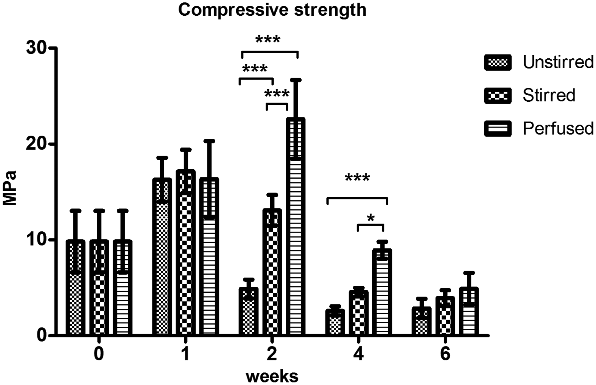
Compressive strength of CPC/PLGA composites as a function of incubating time. Data are presented as mean±standard deviation (n=5). *indicating p<0.05 and ***p<0.001.
However, compared to the other conditions (unstirred: 2.6±0.5, stirred: 4.6±0.4), the samples tested under perfused conditions still revealed the highest compressive strength (8.9±0.9 MPa). After 6 weeks of degradation, the strength under perfused conditions (4.9±1.6) was decreased further and no significant differences were observed anymore between the three experimental groups (unstirred: 2.8±1 MPa, stirred: 3.9±0.8 MPa).
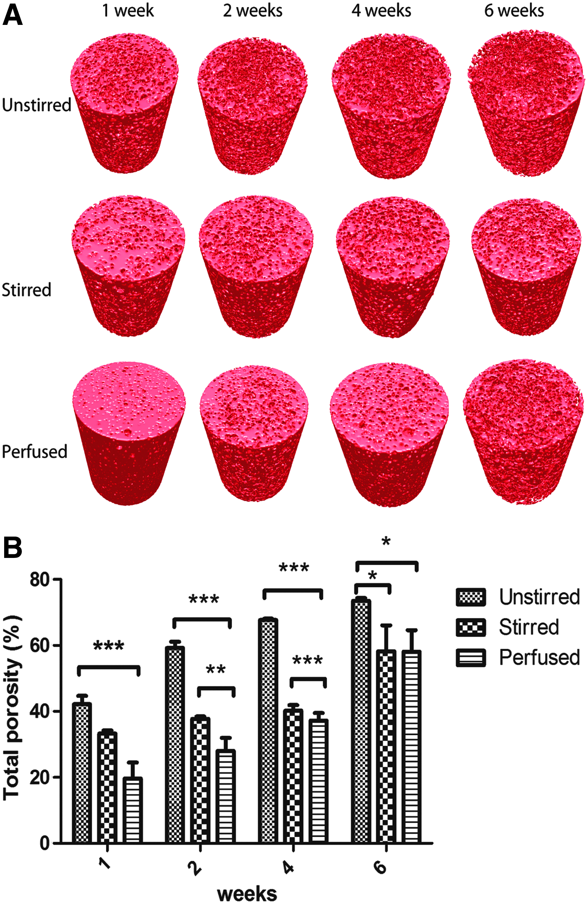
Microcomputed tomography
The microcomputed tomographs provided an overview of the three-dimensional morphological changes of the CPC/PLGA composites under different soaking conditions. Cement samples revealed a highly porous structure after soaking under unstirred and stirred conditions, whereas a more dense morphology and significantly smaller total porosity were observed after 1, 2, and 4 weeks of soaking under perfused conditions (Fig. 5B).
X-ray diffraction
The XRD diffractograms of cement samples are shown in Figure 6 as a function of soaking time. All groups revealed major reflection peaks for hydroxyapatite (HA) (2θ=25.9° and 2θ=31.7°). Interestingly, after 2 weeks of soaking, brushite peaks (2θ=11.8° and 2θ=21.9°) emerged at a low diffracted intensity under stirred conditions and at a high intensity under unstirred conditions. After 4 weeks of degradation, the intensity of brushite peaks increased under stirred conditions. However, after 6 weeks of immersion, brushite peaks disappeared and only HA peaks were detected for all experimental groups. In addition, crystal sizes at 2θ=25.9° were found as 56.2, 56.2, and 44.1 nm after 6 weeks soaking under unstirred, stirred, and perfused conditions, respectively.
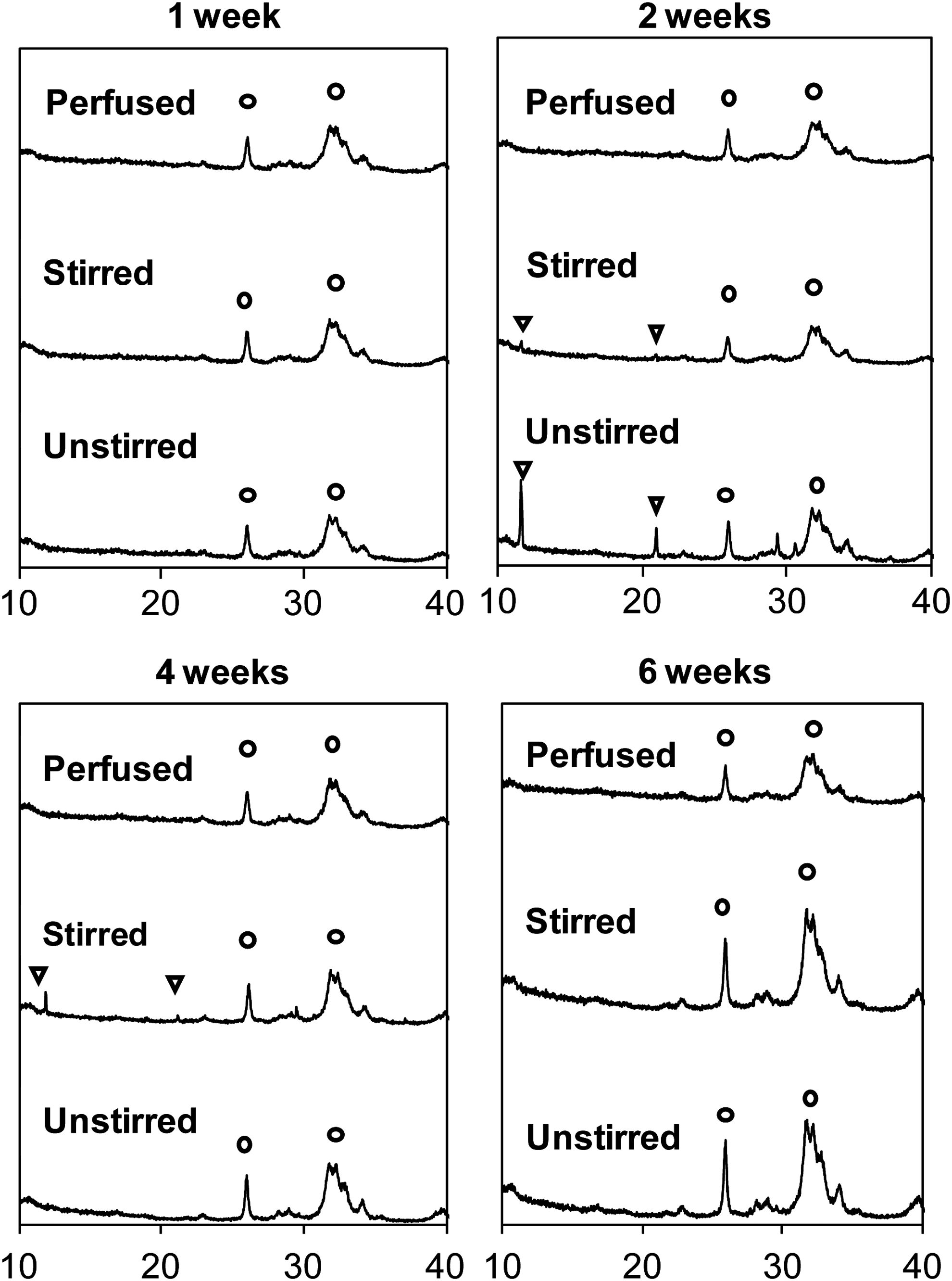
X-ray diffraction (XRD) analyses of the CPC/PLGA composites after incubation (◯=apatite peaks; ▽=brushite peaks).
Scanning electron microscopy
The degradation process of both the CPC matrix and the PLGA microspheres was monitored using SEM (Fig. 7). After 2 weeks of soaking under unstirred conditions, large pores of several tens of micrometers in diameter were formed resulting from the degradation of the PLGA porogens, which were occasionally filled with flake-like crystals and granular debris. These pores were also formed under stirred conditions, but the pores were still partially filled with organic remnants from the PLGA degradation process. In the cement samples soaked under perfused conditions, however, the pores did not form until 4 weeks of soaking, while flake-like crystals or granular debris were not observed. After 4 weeks of degradation, composites soaked under stirred or unstirred conditions revealed a highly porous structure, which were filled with flake-like crystals and granular debris under stirred conditions only. After 6 weeks of degradation, all tested samples exhibited a highly porous morphology without the presence of flake-like crystals, although organic remnants were still observed under perfused condition.
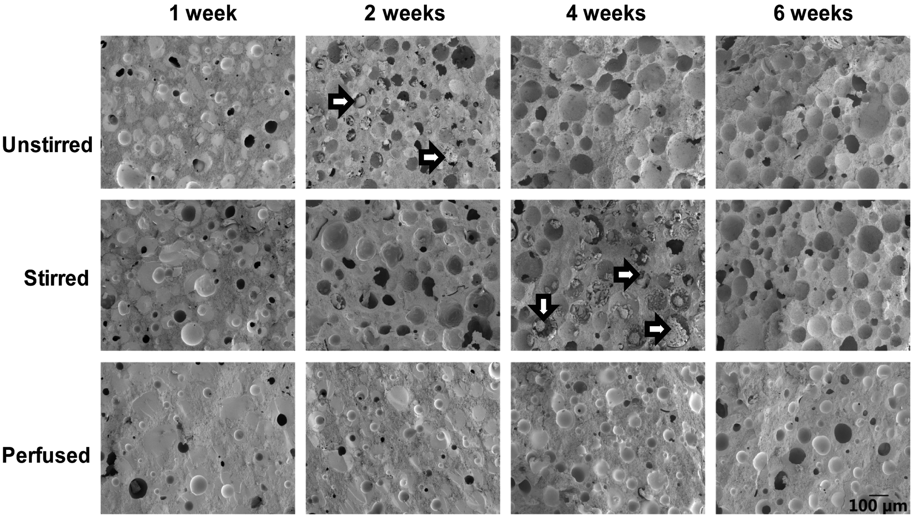
Scanning electron microscopy (SEM) images of CPC/PLGA composites after 1, 2, 4, and 6 weeks in PBS at 37°C (=brushite-like crystals).
Discussion
The aim of the present study was to characterize the effects of different soaking conditions on the degradation of CPC/PLGA composites in vitro. Three types of soaking conditions (unstirred, stirred, and perfused) were used to study the degradation process of CPCs containing 40 wt% of dense PLGA microspheres by monitoring pH, mass loss, compression strength, porosity, and morphology of the cements as a function of soaking time.
In the current study, we observed a slight mass loss and an increased compressive strength due to the transformation from α-TCP to HA for all experimental groups within the first week of soaking. For unstirred conditions after 2 weeks of degradation, the mass loss percentage was significantly higher than stirred and nonperfused conditions. Accordingly, the compressive strength of samples soaked under unstirred conditions was much lower. From then onward, the composite kept losing weight and strength until the end of the study period. For the stirred conditions, the main loss of mass and mechanical strength occurred after 4 weeks of degradation followed by a more gradual loss of remaining mechanical strength afterward until 6 weeks of soaking. In contrast, samples soaked under perfused conditions displayed a continuous increase in mechanical strength until 2 weeks of soaking. After this time point, the mass loss remained moderate (<20 wt%) for the entire study period.
The in vitro degradation process of PLGA scaffolds was divided into three stages by Pan et al.: (1) the initial quasi-stable stage; (2) the decrease-of-strength stage, and (3) the loss-of-weight and disruption-of-scaffold stages. 20 The first stage is characterized by decreased dimensions and increased mechanical strength of the porous scaffold. In the second stage, the weight and dimensions of porous scaffold are generally maintained, but the mechanical properties are considerably reduced. The most prominent feature of stage 3 is a significant weight loss and eventual disruption of the scaffolds. Although our system was a CPC/PLGA composite and not a purely polymeric PLGA scaffold, important aspects of the description of the degradation of PLGA scaffolds by Pan et al. are still applicable to out composite system.
Generally, the first week strongly resembles the first stage of in vitro degradation for all conditions. The decrease of strength occurred after 2 weeks of degradation under the unstirred condition, which indicates that the second stage occurred at an earlier time point compared to the other two conditions. We interpret the constant loss of mass and strength as a continuation of the second stage of degradation. In contrast to the unstirred and stirred conditions, the third stage of degradation was never reached for perfused samples. Previous studies on degradation of CPC/PLGA composites revealed that the PLGA-free cement phase, hardly dissolved in vitro after transformation to the apatite phase, whereas acidic degradation by-products of PLGA were mainly responsible for the degradation of the composites.9,17 Therefore, we assume that the decreased mass was mainly caused by the degradation of PLGA microspheres.
From the SEM micrographs, it can be concluded that the ceramic matrix of CPCs soaked under perfused conditions hardly dissolved for the entire study period. PLGA microspheres, on the other hand, degraded slower under perfused rather than stirred/unstirred conditions. As a consequence, the PLGA microspheres might have partially lost their integrity between week 2 and 4, resulting in flowable sphere remnants with a decreased load-bearing capacity.The combined evidence of XRD analysis revealed the presence of brushite-like crystals after 2 weeks of soaking under unstirred conditions, whereas samples soaked under stirred conditions contained brushite crystals after both 2 and 4 weeks of soaking. Under perfused conditions, the brushite phase was not detected throughout the entire duration of the study.
Summarizing, it was shown that brushite was formed at a faster rate under unstirred conditions vs. stirred conditions, whereas brushite was not formed at all under perfused conditions. Brushite (also known as dicalcium phosphate dihydrate [DCPD]) is generally formed at an acidic pH below 4.2.8,21 Even though the hydrolysis degradation by-products of PLGA are acidic, pH values of the soaking solutions remained considerably higher than a pH of 4.2. Therefore, it can be concluded that brushite crystals were formed due to local acidification yielding pH values in the vicinity of degrading PLGA microspheres below 4.2. This local acidification was attributed to autocatalytic PLGA degradation as well as a lack of perfusion under stirred and unstirred conditions.
Under perfused conditions, the cements were subjected to direct flow of the soaking medium through the nano- and micropores of the cements, thereby effectively removing acidic degradation by-products and reducing the rate of autocatalytic PLGA degradation.12,22 After 6 weeks, only apatite peaks were revealed as the main reflections for all experimental groups, which is in accordance with previous reports.23–25 Moreover, as determined by line broadening using XRD, the formed apatite crystals had an average size of 56 nm under stirred and unstirred conditions, whereas smaller crystallites were formed under perfused conditions (44 nm). We attribute this phenomenon to a higher degree of supersaturation caused by higher pH under perfused conditions. Generally, it is known that higher supersaturation ratios result into smaller crystals.
In agreement with our hypothesis, the present study clearly confirmed that the degradation rate of CPCs containing PLGA microspheres increased with decreasing fluid perfusion. Similar results were reported by Agrawal et al. who compared the effects of static and flow conditions on the in vitro degradation rate of polymer scaffolds and observed that fluid flow decreased the degradation rate significantly. 15 Similarly, it was observed that stirring delayed the in vitro degradation of PLGA-containing cements. These results raise the question whether the in vivo degradation rate of CPC/PLGA can be predicted by a properly designed in vitro study.
To answer this question, we have analyzed histological slides from a previous in vivo study 26 using thin-film X-ray diffraction analysis. In this particular study, similar CPCs containing PLGA microspheres were implanted into femoral condyles of rabbits and analyzed using histology (after embedding in polymethylmethacrylate) after 6 weeks of implantation. Only apatite peaks were observed at 26° and 32° 2θ, whereas brushite peaks were absent. Considering the fact that brushite peaks were also absent in the diffractograms of CPCs tested under perfused conditions, it seems reasonable to assume that perfused conditions mimic the highly perfused in vivo conditions more closely than stirred or unstirred conditions.
Nevertheless, some limitations of the present study need to be addressed as well. First, the study period was short compared to conventional time periods used for in vivo degradation studies. In addition, soaking media were not refreshed during the entire study period. These limitations complicate the correlation of in vitro to in vivo degradation studies even further.
Conclusions
To summarize, the degradation rate of CPC/PLGA cement composites decreased in the condition order unstirred>stirred>perfused, as evidenced by quantification of mass loss, compression strength, and morphology. We have demonstrated that soaking conditions strongly affected the in vitro degradation process of CPCs. As a consequence, it can be concluded that the experimental design of current in vitro degradation studies does not yet allow for correlation to (pre-)clinical studies.
Footnotes
Acknowledgment
J.A. acknowledges the China Scholarship Council (No. 2011704027).
Disclosure Statement
No competing financial interests exist.