
Abstract
A persistent challenge in tissue engineering is the fabrication of manipulatable scaffolds for implantation in clinical treatments and use in disease models for drug screening. Electrospinning of nanofibrous membranes is an emerging technology in artificial extracellular matrix (ECM) design that can offer precisely tunable microenvironments upon assembly into three-dimensional (3D) scaffolds that mimic the in vivo ECM structure. In this study, we report a facile and versatile strategy for preparing 3D multilayered constructs from Fe3O4/polycaprolactone (PCL)/gelatin nanofibrous membranes. This method combines membrane assembly with noncontact magnetic force to preserve the mechanical integrity and interconnectivity of the 3D scaffolds. An ordered layer structure can be achieved using a magnetic control technique through the addition of magnetic nanoparticles into the PCL/gelatin nanofibers. We first verified the magnetic properties and structures of magnetic nanofibers according to X-ray diffraction, hysteresis, scanning electron microscopy, and transmission electron microscopy. We tested the potential toxicity and osteogenic differentiation of mesenchymal stem cells seeded on the layered scaffolds. To add further functionality to the scaffolds, the membranes were coated with silver nanoparticles and shown to inhibit the growth of Escherichia coli and Staphylococcus aureus, which are responsible for most cases of infection-related implant failure. Finally, we tested the utility of magnetic membranes implanted in an animal model as a contrast agent for magnetic resonance imaging. Scaffolds formed using the presented magnetically guided fabrication strategy have the potential to mimic the structure and function of human tissues and also may be applied in disease models to study cell–cell interactions.
Introduction
T
The complex relationship between the extracellular environment and cell function further requires novel approaches for controlling the properties of tissue engineering scaffolds. Many studies have shown that cellular characteristics ranging from their functioning to morphogenetic expression differ when cells are grown on two-dimensional (2D) materials versus within a three-dimensional (3D) scaffold.5,6 A general consensus is that 3D scaffolds can more closely mimic the cells' native in vivo environment, but constructing an appropriate 3D scaffold and then guiding cellular behavior within the scaffold are major challenges, especially if multiple cell types are needed to reconstruct a tissue.7,8 Bottom-up approaches to designing scaffold materials have been the focus of extensive research in recent decades,9,10 and the reasons are two-fold. First, such approaches permit the directed construction of a tissue at different levels starting from the primitive basic structure at the molecular level (nanoscale or subnanoscale), to the cellular level (microscale), and up to the tissue level (macroscale). 10 Second, at each level, we can customize the features of the scaffold, such as including growth factors at the molecular level, seeding cells at the micro level, and regulating the physical environment at the macro level. 11
One type of 3D scaffold, 3D nanofibrous membranes, constructed through electrospinning have been shown to provide a better environment for tissue development than the traditional 2D cell culture platform. 12 This is because the 3D fibrous matrix provides a structure similar to that of the 3D fiber network of collagen and elastin in natural extracellular matrix (ECM). 13 To date, 3D nanofibrous structures have been fabricated using a number of different techniques. 4 One approach employs manual stacking of electrospun membranes to construct multilayered scaffolds with cells seeded on each layer.14,15 Magnetic nanoparticles have found particularly multiple applications in biomedicine,16–19 such as magnetic cell seeding, cell sheet construction, cancer hyperthermia treatment, and drug delivery vehicle. Because the directional movement of magnetic nanoparticles can be controlled in a magnetic field, their unique feature of orientational movement was reported by several pieces of research., For example, the methods about precisely controlled fabrication of magnetic fibrous scaffolds, such as the multilayered grids formed by the alignment of iron oxide (Fe3O4)/poly(vinyl alcohol) nanofibers 20 and the fibrous bundles composed of Fe3O4/poly(lactic-co-glycolic acid) nanofibers. 21 However, magnetically guided 3D microfabrication and its potential use in creating multifunctional scaffolds for engineered tissues as well as contrast agents and other applications have not been clearly demonstrated.
In the present study, we investigated a magnetically driven fabrication technique for preparing magnetically controllable layered membranes for various theranostic applications. We synthesized multilayered nanofibrous Fe3O4/polycaprolactone (PCL)/gelatin membranes loaded with superparamagnetic iron oxide nanoparticles (SPIONs). With the application of only a small and cheap magnetic bar, multilayered stacks of the membranes could be easily assembled and then disassembled with removal of the magnetic bar. To demonstrate the potential of the magnetically driven layers for use in fabricating multifunctional tissue engineering scaffolds, we first characterized the viability and morphology of mouse bone marrow-derived mesenchymal stem cells (mBMSCs) seeded on the membranes. Then we evaluated the osteogenic differentiation of mBMSCs seeded on the membranes of layered 3D scaffolds. We then coated the membranes with silver nanoparticles (Ag-NPs) and tested the antibacterial activity of the membranes. Finally, multilayered scaffolds were implanted in rats and observed through in vivo magnetic resonance imaging (MRI) to demonstrate their potential as a contrast agent.
Materials and Methods
Materials
PCL, gelatin, iron (II) sulfate heptahydrate, iron (III) chloride hexahydrate, and Pluronic® F-127 were purchased from Sigma-Aldrich. 2,2,2-Trifluoroethanol (TFE, 99%) was purchased from Alfa Aesar. mBMSCs were purchased from ATCC. All other chemicals were purchased from Sigma-Aldrich, unless otherwise indicated.
Preparation of magnetic nanoparticles
Fe3O4 SPIONs were prepared by the chemical coprecipitation technique reported previously. 22 In brief, 0.023 mol iron (II) sulfate heptahydrate and 0.046 mol iron (III) chloride hexahydrate were dissolved in deionized water (150 mL) at 45°C, and nitrogen gas was bubbled through the solution for 30 min. After mixing the iron salt solutions, 20 mL NH3•H2O (25%, v/v aqueous) was added immediately and the solution was stirred vigorously for 1 h. Then the SPIONs were collected by magnetic separation.
Electrospinning process
PCL and gelatin were dissolved separately in TFE to achieve 16% (w/v) solutions. The solutions were then combined at a ratio of 1:1 (v:v). SPIONs in 2% F-127 solution were sonicated for 1 h to obtain a uniform suspension. Then the required quantity of SPIONs was added to PCL/gelatin solution to achieve SPION concentrations fixed at 2.5, 5, and 10 wt%. The solutions were loaded in a 10-mL glass syringe equipped with a 0.8-mm needle, and the feed rate was held constant at 1 mL/h. The high voltage supply was 15 kV. Flat aluminum foil (10×10 cm) was grounded at a distance of 15 cm. Fibers collected on the aluminum foil were dried completely and crosslinked for 2 h in glutaraldehyde vapor.
Characterization of SPIONs and magnetic nanofibers
Transmission electron microscopy (TEM) observations were carried out on a Tecnai G2 F20 S-TWIN electron microscope (FEI Company). For observation by scanning electron microscopy (SEM; Hitachi E-1010), the membranes were air dried overnight and sputter coated with gold. Fourier Transform Infrared (FTIR) spectra of the SPIONs and nanofibers were recorded using a FTIR spectroscope (Thermo Scientific) over the wavenumber range of 4000 to 400 cm−1 at a resolution of 4 cm−1. The crystalline structure of the SPIONs was identified by X-ray diffraction (XRD) measurement (Bruker D8 Advance). The superparamagnetic properties of SPIONs and magnetic nanofibers were tested using the Superconducting Quantum Interference Device (SQUID, MPMS-XL-7; Quantum Design).
Evaluation of the biocompatibility of the magnetic nanofibrous membranes
Cell culture and seeding onto magnetic nanofibrous membranes
mBMSCs were maintained in 5% CO2 at 37°C and cultured in DMEM supplemented with 10% fetal bovine serum (FBS), and 1% penicillin–streptomycin, which was changed every 2–3 days. At 70–80% confluence, the cells were trypsinized and seeded onto both sides of membranes (3×10⁴ cell/cm2).
Cell viability on magnetic nanofibrous membranes
Cell metabolic activity and proliferation over time were assessed using the MTT assay. LIVE/DEAD assay (Molecular Probes) was performed after cells were cultured on membranes for 2 days.
mBMSC morphology on magnetic nanofibrous membranes
For cytoskeletal staining, samples of cells attached to membranes were fixed in 4% paraformaldehyde for 15 min on ice and washed three times with phosphate buffered saline (PBS). Samples were permeablized with 0.5% Triton-X100 in PBS and blocked with 5% bovine serum albumin. Then cells were stained with Alexa Fluor 488 fluorescein isothiocyanate (FITC)-phalloidin (Molecular Probes) for 20 min. Lastly, Hoechst 33258 nuclear stain (Molecular Probes) was applied, and samples were stored at 4°C before imaging with a microscope.
Magnetically guided fabrication of multilayered nanofibrous structures
Rapid assembly of 3D multilayered cell-laden structures
Using a simple layer-by-layer assembly technique, a magnet was applied to rapidly stack cell-laden membranes into multilayered 3D nanofibrous structures. mBMSCs were cultured for 3 days on 10 wt% magnetic membranes and then 3D constructs (n=3) were assembled by stacking five membranes with 50 μL fresh medium on each layer. A magnet placed under the culture dish kept the layers securely stacked. Finally, 2 mL of fresh culture medium was added. Constructs in the control group were cultured without a magnet under the culture dish.
Structural stability of 3D multilayered structures
After the assembled multilayered structures were kept in the culture for 3 days with a magnet under the culture dish, the magnet was released. The multilayer assembly in the cell culture medium was gently shaken, and the stability of 3D structures was observed visually.
Cell morphology within 3D layered structures
The samples were then embedded in optimum cutting temperature compound (VWR Corporate) and frozen. The samples were cut into 10-μm-thick longitudinal sections at −20°C and then stained with Alexa Fluor 488 FITC-phalloidin, which binds to the cytoskeleton filaments and Hoechst 33258 nuclear stain.
Differentiation of mBMSCs within magnetic 3D nanofibrous structures
Osteogenic induction of mBMSCs in 3D structures
mBMSCs in multilayered structures were cultured for 7 days in an osteogenic differentiation medium (DMEM containing 10% FBS, 100 nM dexamethasone, 50 μM ascorbate, 10 mM glycerophosphate, and 1% penicillin–streptomycin). The medium was changed every 3 days.
Real-time quantitative polymerase chain reaction analysis of osteogenic gene expression
The total RNA of mBMSCs grown on different substrates (n=3) was extracted according to the instructions of the TRIzol RNA extract kit, and 1 μg RNA samples were reversely transcribed for first strand cDNA synthesis. Real-time polymerase chain reaction was performed with an ABI 7500 qPCR system (Applied Biosystems). The following highly purified desalted gene-specific primers were used (5′–3′): alkaline phosphatase (ALP)-F: CTC CAA AAG CTC AAC ACC AAT G, ALP-R: ATT TGT CCA TCT CCA GCC G; bone sialoprotein (BSP)-F: CCA CAC TTT CCA CAC TCT CG, BSP-R: CGT CGC TTT CCT TCA CTT TTG; collagen 1α (COL1)-F: AAC AGT CGC TTC ACC TAC AG, COL1-R: AAT GTC CAA GGG AGC CAC; osteopontin (OPN)-F: CTA CGA CCA TGA GAT TGG CAG, OPN-R: CAT GTG GCT ATA GGA TCT GGG; and GAPDH-F: AGG TCG GTG TGA ACG GAT TTG, GAPDH-R: TGT AGA CCA TGT AGT TGA GGT CA. Quantification of gene expression was based on the CT value for each sample, and the average of three replicate measurements for each sample was calculated. The calibrator control group included samples of mBMSCs in the scaffolds collected on day 0. The targeted gene expression was normalized to that of GAPDH, which was used as a housekeeping gene. Relative expression levels of genes were determined by the 2−ΔΔCt method.
Nanosilver modification of magnetic nanofibrous membranes and test of antibacterial activity
To introduce antibacterial functionality, 10 wt% magnetic nanofibrous membranes were coated with Ag-NPs. Trisodium citrate was used to reduce the silver nitrate solution in an aqueous medium. Briefly, disc-shaped membranes 8 mm in diameter were immersed in 90 mM Trisodium citrate solution. For Ag-NP coating, silver nitrate solution was added to a final concentration of 30 mM. After 3 h, the membranes were removed from the solution and washed with PBS. An Agar plate method was used to evaluate the antibacterial activity of the Ag-NP-coated membranes against the Gram-negative bacterium Escherichia coli and the Gram-positive bacterium Staphylococcus aureus. Magnetic membranes without Ag-NP coating were tested as a control, and the Ag-NP on the membranes were chartered by SEM and EDX.
MRI of implanted magnetic nanofibrous scaffolds
For MRI, two 10 wt% magnetic nanofibrous scaffolds composed of three membrane layers each were implanted into the anterior abdominal subcutaneous fascia of Sprague-Dawley (SD) rats (male, 200 g weight, n=3). All SD rats were from the Laboratory Animal Center of Southern Medical University (Guangzhou, Guangdong, China), and all animal experiments were approved by the Laboratory Animal Center of Southern Medical University and carried out in accordance with the Regulations for the Administration of Affairs Concerning Experimental Animals. Nuclear MR relaxometry of the magnetic scaffolds in rats was performed using a clinical superconducting 3T whole-body MRI scanner (repitition time/echo time=3500/120 ms, field of view=170, 68, 17 mm, matrix=260×157, number of excitations=4, thickness/interval=2/0.6 mm) on the third day postimplantation.
Statistical analysis
Each experiment was run with at least three independent samples, and data are expressed as means±standard errors. Student–Newman–Keuls tests were used for multiple comparisons of different groups. Statistical significance was determined at a value of p<0.05.
Result and Discussion
Physical characteristics and biocompatibility of magnetic nanofibers
In the present study, we prepared magnetic nanofibrous PCL/gelatin membranes containing SPIONs for the purpose of assembling them into multilayered scaffolds with controllable complex architectures using only a simple magnet (Fig. 1). The properties of the SPIONs precipitated from Fe2+ and Fe3+ salt mixtures were characterized. TEM images showed that the diameters were 12.6±1.6 nm (Fig. 2A, B). PCL and gelatin were used for electrospinning because PCL provides physical stiffness, and gelatin promotes cell adhesion. Electrospun PCL/gelatin and magnetic nanofibers presented good uniformity with fiber diameters of 275±156 nm (Fig. 3A–D). The morphology and diameter of the nanofibers were not influenced too much because of the small diameter and narrow size distribution of the SPIONs. SPIONs concentration of 10 wt% or less had little effect on the smoothness of the fibers. TEM images of the nanofibers (Fig. 3E–H) showed that SPIONs were embedded in the nanofibers with small agglomerations of nanoparticles consistent with most electrospun magnetic nanofibers reported previously.23–25 The XRD results indicated diffractions of 220°, 311°, 400°, 422°, 511°, and 440° on crystal faces of the SPIONs spinel structure (Fig. 2C). XRD patterns of the Fe3O4/PCL/gelatin membranes (2.5, 5, and 10 wt% SPIONs) demonstrated incorporation of Fe3O4 nanoparticles (Fig. 2E). The saturation magnetization (Ms) values of the magnetic nanofibrous membranes containing different concentrations of SPIONs were 6.3 emu g−1 with 2.5 wt% SPIONs, 11.8 emu g−1 with 5 wt% SPIONs, and 19.2 emu g−1 with 10 wt% SPIONs (Fig. 2F), higher SPIONs content led to a higher Ms value of the scaffolds, but all of which were much lower than 65.2 emu g−1 of bare SPIONs (Fig. 2D).
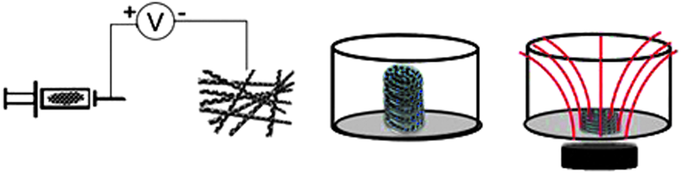
Schematic of magnetic nanofibrous membrane preparation through the addition of SPIONs into the PCL/gelatin solution. Layer-by-Layer assembly of 3D microfibrous structures (five-layer structures were stacked loosely and instably for cell culture without placement of magnet below the dish), but the five-layer structures became tightly stacked when a magnet was placed under the dish. 3D, three-dimensional; PCL, polycaprolactone; SPIONs, superparamagnetic iron oxide nanoparticles.

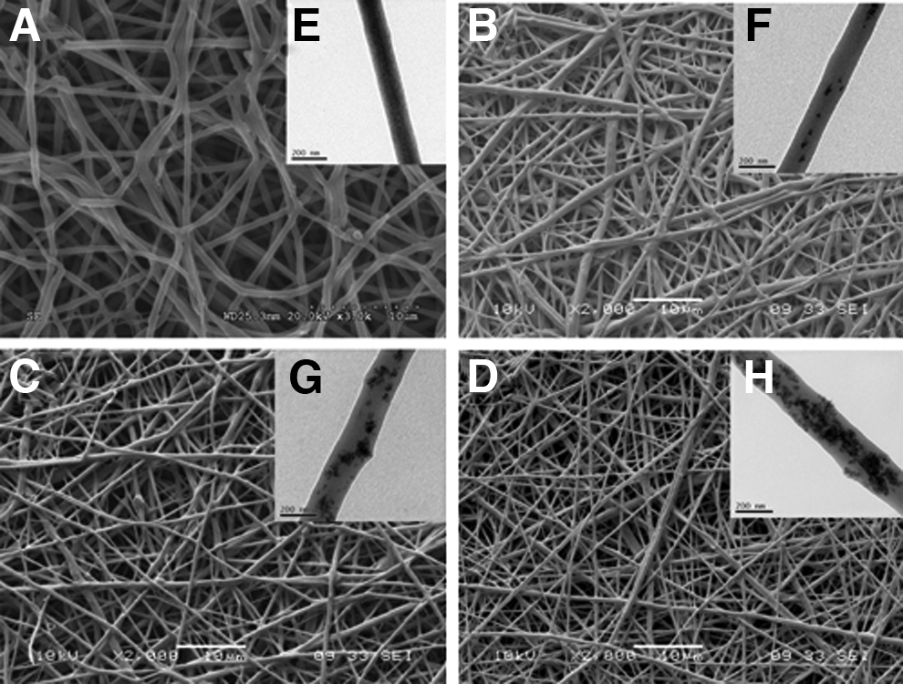
SEM images showing the microstructure of
The metabolic activity of mBMSCs seeded on magnetic nanofibrous membranes according to MTT assay was compared to that on control surfaces of cell culture plates and PCL/gelatin membranes (Fig. 4). No significant differences in mBMSC metabolic activity were observed for cells cultured on the magnetic membranes and control surfaces after 24 h or 3 days culture (p>0.05). The metabolic activity of mBMSCs after 3 days was much higher than that at 24 h on all magnetic membranes and control surfaces (p<0.05). These results suggest that the incorporation of SPIONs at 10 wt% or less does not compromise the cytocompatibility of PCL/gelatin nanofibers. The LIVE/DEAD assay (Fig. 5A–D) revealed that cells grew well on both control and magnetic PCL/gelatin membranes containing different concentrations of SPIONs. Observations of cell morphology showed that SPIONs at these concentrations did not inhibit mBMSC growth and maintenance of a spindle-like morphology (Fig. 5E–H).
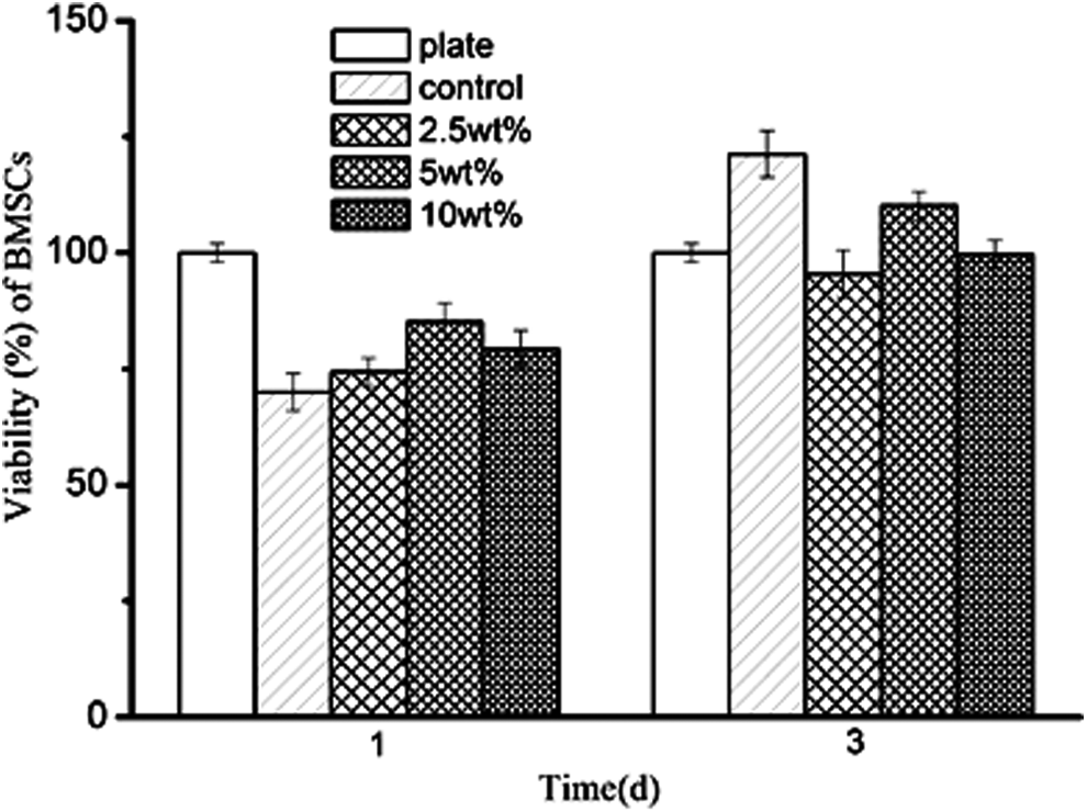
mBMSC viability on PCL/gelatin nanofibrous membranes and magnetic membranes containing different concentrations of SPIONs according to cell metabolic activity. mBMSC, mouse bone marrow-derived mesenchymal stem cell.
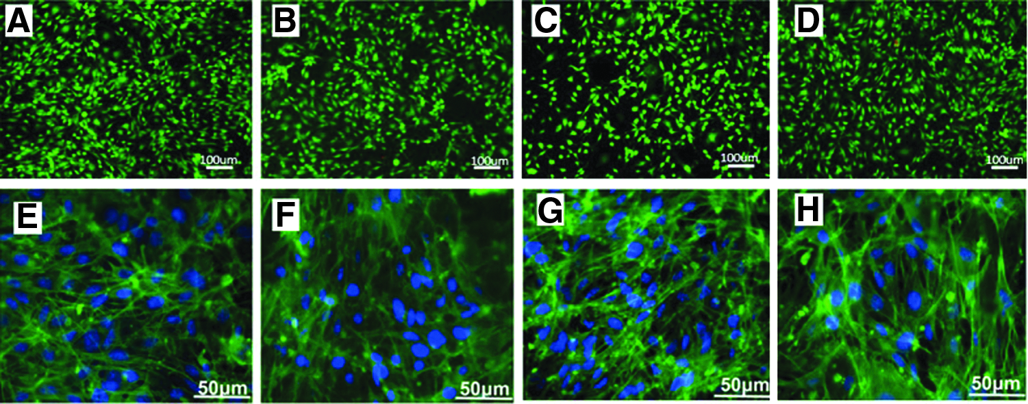
On the upper line LIVE/DEAD staining to distinguish live and dead cells after 2 days cultured on
Magnetic 3D multilayered nanofibrous scaffolds support cell growth and keep good stability
Many studies have focused on the design and intrinsic properties of nanofibers prepared through magnetic electrospinning,20,21,26 and the resulting nanofibers have been applied as a membrane for bone tissue engineering.27,28 However, to the best of our knowledge, there have been no reports on the fabrication of 3D multilayered structures using magnetic force to assemble nanofibrous membranes. Considering that cells are present in high density and intricately organized in a 3D architecture within tissues and organs, the single-layer 2D electrospun membranes are not suitable as a 3D template for cell growth and neotissue formation. In this study, we present a rapid and easy postprocessing strategy to create complex multilayered 3D structures using an externally applied magnetic field. Our magnetic PCL/gelatin membranes were easily transferred and stacked through placement of a magnet under the tissue culture dish (Fig. 6A, B and Supplementary Video S1; Supplementary Data are available online at
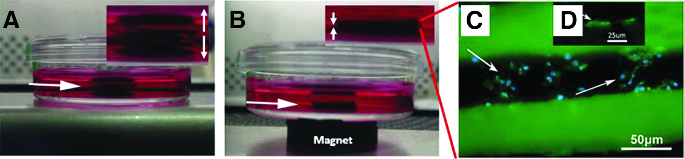
Rapid and facile strategy to create complex 3D structures from 10 wt% magnetic nanofibrous membranes using only a simple magnet.
When we tested the stability of the five-layer scaffolds constructed from the different membranes after 3 days of cell culture, we observed that all layers easily separated for the nonmagnetic control PCL/gelatin membranes (Supplementary Video S2). However, 3D multilayered structures fabricated with membranes containing 10 wt% SPIONs remained intact (Supplementary Video S3). We propose that the stability of the scaffolds even after removal of the magnet is due to the tight packing of the layers that occurs while the magnet is applied. Cells first attached to the surface of the magnetic nanofibrous membranes, and when the membranes were stacked to form the 3D scaffolds, a certain distance remained between adjacent surfaces (Fig. 6A) that decreased upon application of the magnetic force (Fig. 6B). With this reduction in the distance between membrane surfaces, the cells could form bridges between layers through the production of ECM that effectively glued the membranes together. After 7 days, cytoskeletal staining showed that a monolayer of mBMSCs had coated the single-layer magnetic membranes (Fig. 6D), whereas mBMSCs seeded within multilayered scaffolds showed a 3D ingrowth in the inner spaces (Fig. 6C). Compared with previous layer-by-layer nanofiber assembly methods, such as direct spinning of fibers onto a grounded medium surface and thermally induced phase separation, our new method enabled the directed assembly of nanofibrous cell sheets into 3D configurations. Therefore, the stability of our multilayered scaffolds not only demonstrated the significant potential for use of these scaffolds in tissue engineering applications, but also indicates that our system is scalable and could be used for high-throughput applications. Moreover, these nanofiber-based units could be used to form a complex 3D microarchitecture loading different types of cells on each layer.
Osteogenic differentiation of mBMSCs in 3D multilayered scaffolds
As a representative tissue engineering application, we investigated the osteogenic differentiation of mBMSCs within 3D multilayered scaffolds. The relative mRNA expression levels of osteogenesis-related genes in mBMSCs cultured in the multilayered scaffolds are shown in Figure 7. ALP expression was significantly greater on day 7 than on day 2, showing gradual upregulation with a marked increase at day 7 compared to that on day 0. ALP is an early marker for osteogenic differentiation and is expressed during the postproliferative period of ECM maturation. 30 BSP gene expression was higher on days 2 and 7 than on day 0, and that on day 7 was higher than that on day 2. BSP gene expression indicates copious formation of noncollagenous proteins. 31 COL1 was expressed during the initial period of proliferation and ECM biosynthesis, and it was higher on both day 2 and 7 than on day 0, but there was no significant difference between COL1 expression levels on day 2 and 7. OPN, a noncollagenous protein, showed remarkable upregulation during the first 2 days of osteogenic induction and low expression at day 7. Overall, these results indicate that mBMSCs attached within the fabricated 3D nanofibrous scaffolds retained their osteogenic differentiation potential and deposited ECM, in support of their potential as scaffold material in bone tissue engineering. Our 3D nanofibrous scaffolds not only offer facile control of microarchitectural features, but also the added magnetic NPs may provide more potential for guided bone regeneration.
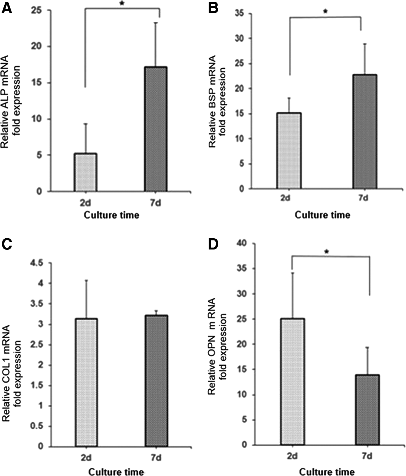
Real-time quantitative polymerase chain reaction analysis of osteogenic gene expression.
Coating of magnetic membranes with Ag-NPs to achieve antibacterial functionality
With nanofibers serving as the basic scaffold material, many functions can be incorporated, including antibacterial activity, which is especially relevant because bacterial infection is a significant cause of biomaterial implant failure. Some studies have already reported the modification of nanofibers for antibacterial application.32,33 In the present study, we applied a coating of Ag-NPs as an antibacterial agent to the surface of our magnetic nanofibrous membranes. The antibacterial effect of the Ag-NP-coated membranes against E. coli and S. aureus was demonstrated by the greater zones of inhibition observed for these membranes compared to noncoated membranes, which showed no antibacterial activity (Fig. 8A, B). The SEM and EDX result showed the Ag-NPs were coated uniformly (Fig. 8C–E), the Ag-NP size was 8.3±1.6 nm, and the MTT test (Supplementary Fig. S2) showed only little cytotoxicity in the coated membranes. These results demonstrate the potential application of Ag-NP-coated magnetic membranes as an effective antibacterial agent as well as the potential for generating multifunctional multilayered scaffolds.
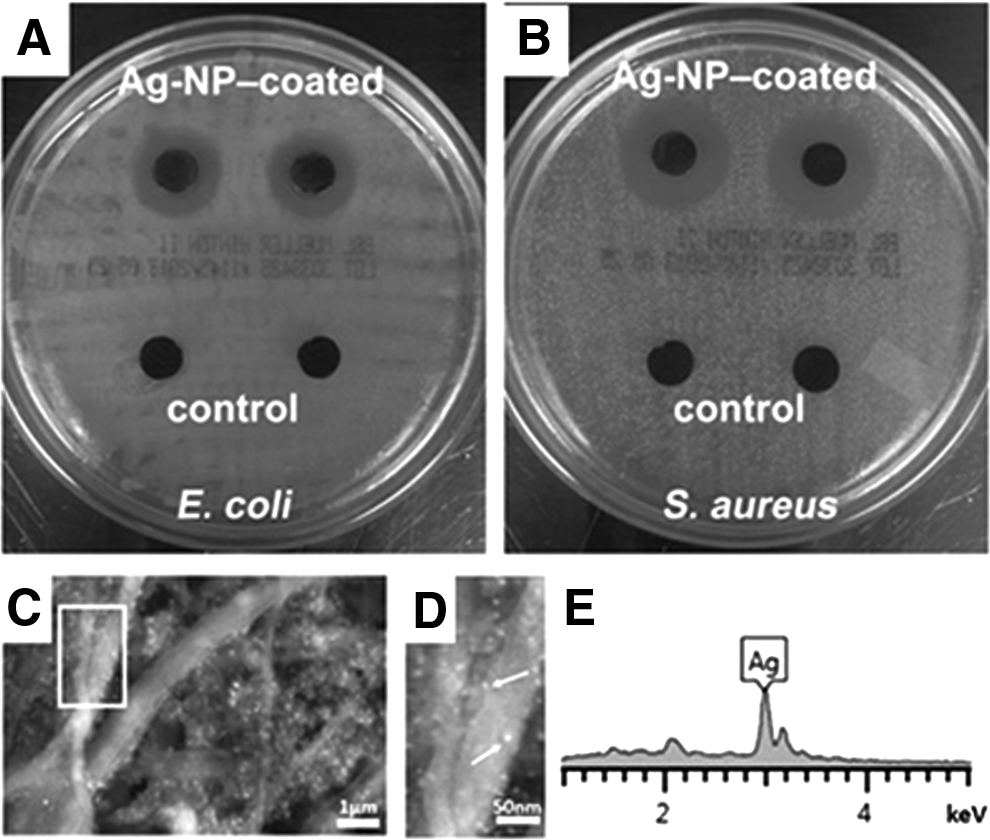
Photographs showing zones of
In vivo MRI of 3D multilayered scaffolds composed of magnetic nanofibrous membranes
The superior magnetic properties of SPIONs as well as their inherent biocompatibility have made SPIONs a candidate in many theranostic applications, such as contrast probes for MRI. 34 SPION loading in polymer scaffolds has been shown to effectively reduce T2 relaxation time, achieving signal attenuation on a T2 or T2*-weighted map for indication of abnormal biological activity. 35 In this study, we implanted 3D multilayered scaffolds composed of nanofibrous membranes, and MR images of the coronal and axial sections of T2-weighted images of rats taken 3 days postimplantation as shown in Figure 9. Only dark images were obtained in the anterior abdominal subcutaneous fascia due to the inherent high magnetic susceptibility of SPIONs. MRI has a very high sensitivity for visualization of iron oxides, which is beneficial to the spin-echo T2-weighted pulse sequence and demonstrates another potential application for the magnetic nanofibrous membranes as a theranostic implant for tissue engineering and controlled drug release.
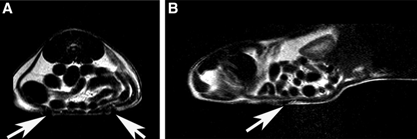
T2-weighted MR images of
Discussion
In this article, we present magnetic fabrication and packaging technologies that may offer rapid and easy layer-by-layer assembly of a biomimetic system for bone tissue engineering applications as well as multifunctional structures with theranostic functionality. In previous studies, the fabrication of complex and large functional bone tissues still faces challenges because of poor implant survival and integration. Multilayered biomimetic assembly approach is one of the most potent ways to mimic the structure of natural bone. However, most multilayered tissue scaffold systems are based on cell-laden gels such as Matrigel or collagen gel coated onto a surface first. 36 Here we use the biomimetic electrospun nanofibrous scaffolds that could promote initial cell adhesion, spreading, and proliferation, as well as mimic the natural ECM and promote osteogenic differentiation of MSCs. Also magnetic nanoparticles have been proven to promote osteoinduction37,38 and magnetic scaffolds can be assembled to form complex 3D tissue constructs in a controlled manner through magnetic field. 39
According to previous studies, during the first 4 days culture provides a static magnetic field that can accelerate the cell proliferation. 40 In our study, the cells proliferated also accelerated on the composite scaffolds composed of SIONPs, and a magnet under the cell culture dish also accelerated the cell contact between the two layers. The reason of osteogenic differentiation of MSC in these multilayered structure is also probably that each magnetic nanoparticle could be regarded as a single magnetic domain in static magnetic fields. In these composite scaffolds with 3D microenvironment which would express osteoinductive effect, the osteogenic gene expression proved the effect. So as previously reported that the novel strategy for osteoconductivity materials is engineering physical microenvironments,41,42 here we combined the topographical features of nanofibers, magnetic assembly, and multilayered 3D structure together to biomimic the ECM regulating the osteogenic differentiation of MSCs.
The magnetic multilayered structure provides a highly efficient tracking in real-time changes in the location of the scaffolds by using the noninvasive MRI technique. The SIONPs embedded in the polymer matrix could not be cleaned by macrophages through phagocytosis before the biodegradation of the nanofiber. As an MRI contrast agent, SIONPs enhance proton relaxation of scaffolds compared with surrounding tissues after implantation. 43 As seen in the in vivo MRI, the scaffold was observed to be significantly darker. In the future, these scaffolds can be used as better potential therapeutic nanocomposite implants to guide and monitor bone tissue development.
Moreover, various other parameters can be manipulated to further increase the functionality of the prepared magnetic membranes and scaffolds. For example, bioactive molecules have been incorporated into nanofibers for multiple biomedical applications. 44 In addition, the thickness of each membrane layer is tunable over a range of tens to hundreds of micrometers. The multilayered structure can meet the requirement for sufficient nutrient exchange within the scaffold, and different types of cells can be placed on the different membrane layers. Because the scaffolds can be easily and repeatedly assembled and disassembled, they will likely also be useful for studying cell–cell interactions in a controllable 3D environment. 45
Conclusion
In conclusion, the composite scaffolds composed of PCL/gelatin and different contents of SPIONs (2.5, 5, and 10 wt%) were synthesized and their morphology and magnetism property were investigated. Furthermore, the rapid construction of multilayered scaffolds was successfully achieved using a magnetic assembly technique. Scaffolds prepared using this strategy have the potential to mimic human tissues with respect to structure and function, as mBMSCs attached within the fabricated 3D nanofibrous scaffolds retained their osteogenic differentiation potential. Also these scaffolds may be used in disease models because of the antibacterial effect or as a theranostic implant to be the contrast probes for MRI. The future of the scaffolds involves the creation of multifunctional 3D materials in regenerative therapies.
Footnotes
Acknowledgments
This work was supported by the National Basic Research Program of China (Grant No. 2012CB619100), NSERC Discovery Grant and NSERC RTI Grant, Manitoba Health Research Council Establishment Grant, Dr. Moore House Fellowship, Manitoba Diabetes Foundation, Manitoba Institute of Child Health, China 863 Project (Grant No. 2012AA020504), and National Natural Science Foundation of China (No. 21304098).
Disclosure Statement
No competing financial interests exist.
References
Supplementary Material
Please find the following supplemental material available below.
For Open Access articles published under a Creative Commons License, all supplemental material carries the same license as the article it is associated with.
For non-Open Access articles published, all supplemental material carries a non-exclusive license, and permission requests for re-use of supplemental material or any part of supplemental material shall be sent directly to the copyright owner as specified in the copyright notice associated with the article.