
Abstract
By definition, osseointegration means close contact between bone and implant. Bone response is related to implant surface properties. Various surfaces have been studied and applied to improve the biological properties of the implant and thereby favor the mechanism of osseointegration. This strategy aims to promote osseointegration by means of a faster and stronger bone formation, improving stability during the healing process, and thus allowing for earlier loading of the implant. Dental implant osseointegration has so far been studied in various animal models. The development of a method based on tissue engineering for assessing the osseointegration process in vitro could prove a valid biomimetic alternative to sacrificing animals. In this study, flat cylindrical dental implants with moderately rough surfaces and machined implants were set in bovine bone blocks. Then, adipose-derived stem cells (ADSCs) were three dimensionally cultured onto these blocks in osteo-endothelial medium for up to 30 days to mimic the osseointegration process in vitro. Scanning electron microscopy (SEM) and gene expression were used to examine stem cell commitment. Mechanical pull-out tests were also performed. SEM analysis identified cells with an osteoblast morphology adhering to the surface of the implants after their removal. Gene expression analysis showed that ADSCs seeded onto the bone blocks were able to express osteoblast and endothelial markers. The implants with the moderately rough surface generated higher pull-out strengths when compared with the machined implants. Nevertheless, the pull-out test values were higher for implants placed in bone blocks with ADSCs than for those set in scaffolds without stem cells. Our results demonstrate the validity of the method adopted and its potential for use in the in vitro assessment of the biological behavior of dental implant surfaces.
Introduction
P
Research on the effects of different surface properties on osseointegration should ideally follow a hierarchical approach before clinical trials. 13 New biomaterials should be tested first in vitro, then in vivo in animal models to ascertain their safety and efficacy.13,14
Two-dimensional in vitro cell culture models are routinely used in preclinical research to examine cell morphology, adhesion, migration, proliferation, and differentiation at the biomaterial interface.13–15 Nevertheless, such results do not always correlate with in vivo performance, because the in vitro conditions cannot reproduce the dynamics of the in vivo environment and bone/implant interactions.13–16
In vitro results consequently need to be confirmed and validated in carefully designed animal experiments, before human clinical trials can begin. Dogs, goats, and sheep (long bones), and primates are preferably used for this purpose because their bone is similar to human bone in terms of structure and healing processes, but ethical issues currently restrict the use of large animal models in medicine.14,17
The most common tests used in the in vivo setting include histological, histomorphometric, biomechanical, and radiological analyses. Histological and histomorphometric analyses consist of qualitative and quantitative methods for measuring static parameters, such as bone density, amount and type of cellular content, or bone-to-implant contact (BIC).14,18,19 Dynamic histomorphometric parameters, such as the mineral apposition rate and fluorescence analysis, are used to characterize bone remodeling kinetics around different implant surfaces.13,14 Biomechanical assessments include the removal torque test and push-out and pull-out tests, which record the amount of force or torque needed to loosen the implant.13,14 Resonance frequency analysis is commonly performed as well. 18 Radiological assessments include conventional radiography or microcomputed tomography.18,19 Finally, the molecular assessment of osseointegration may provide new insight on the fundamental molecular events that support osseointegration in vivo. 20
The use of three-dimensional (3D) scaffolds loaded with human adipose-derived stem cells (ADSCs) has been investigated in the field of bone tissue engineering.21,22 ADSCs possess many advantages, including the easy harvesting, security, abundance, and higher yield if compared to bone marrow mesenchymal stem cells. The potential applications of ADSCs for a wide range of clinical disorders have also been reported.23,24 Blocks of hydroxyapatite were used as scaffolds and provided an excellent porous substrate for the diffusion, adhesion, growth, and proliferation of ADSCs. In vitro ADSCs osteo-endothelial commitment, which is essential for native bone to be mimicked, has also been described. 25
The aim of the present work was to reproduce the osseointegration process in vitro for the purpose of studying the dynamics of bone–implant interactions. A tissue engineering approach was used that involved placing dental implants in 3D bone-derived scaffolds seeded with stem cells. The ultimate goal was to overcome the limitations of in vitro methods, and to complement (and possibly replace) animal studies in this field.
Materials and Methods
Biomaterials
Dental implants
Customized cylindrical implants 6.6 mm long and 3.4 mm in diameter were used in this study (supplied by Sweden & Martina, Padova, Italy). The implant design was characterized by a titanium extension on the coronal side 2.5 mm in diameter and ∼10 mm long with a screw threaded end, a polished surface close to the body of the implant, and a polished hexagon-shaped intermediate portion. Two different types of titanium implants were tested: ZirTi®, zirconium sand-blasted acid-etched titanium implant (Ra 0.542 μm; Sa 0.545 μm) 26 and machined implant (Ra 0.404 μm; Sa 0.317 μm). 26 The two implants will be hereafter referred to as sand-blasted acid-etched implant and machined implant, respectively (Fig. 1a and 1b).
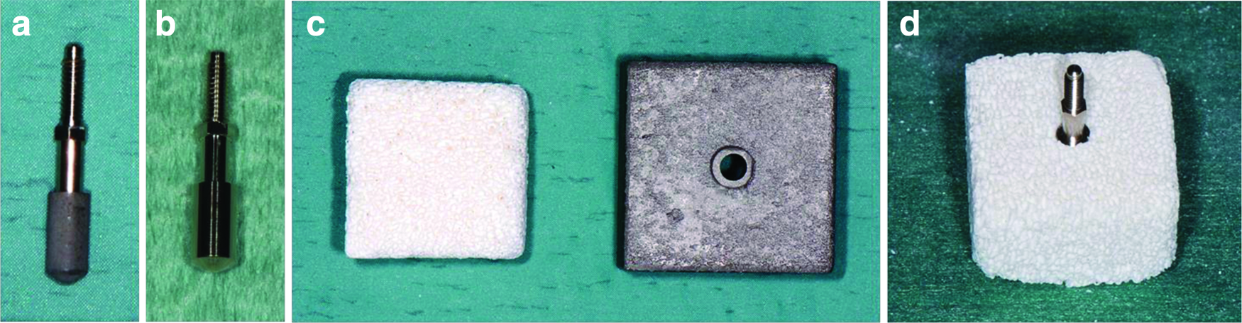
Customized cylindrical
Bone matrix
Orthoss® Blocks 2 × 2 × 1.3 cm (Geistlich Pharma AG, Wolhusen, Switzerland) were used in this study. Orthoss is a natural carbonated hydroxyapatite of bovine origin. It is a highly osteoconductive material because of its particular structure, which is very similar to human spongy bone, with interconnected macropores (100–300 μm), micropores, and nanopores (10–20 nm), resulting in a higher inner surface and excellent hydrophilic properties.
Dental implants were inserted in the bone blocks with the aid of a customized drilling guide (Loripadova Tecnologia, Padova, Italy) (Fig. 1c) using a twist drill (FFT3 300; Sweden & Martina) and dedicated drill stops. For each experiment, one implant was inserted on the 2 × 2 cm side of one bone block (Fig. 1d).
Human stem cells isolation
Human ADSCs were isolated from the adipose tissue of healthy patients (35–58 years old) undergoing cosmetic surgery procedures. All patients gave their written consent. The adipose tissues were digested and the cells were isolated, expanded, and seeded according to our previously described protocol. 25 Briefly, the fat was washed with phosphate-buffered saline (PBS; EuroClone, Milan, Italy) and digested using a solution of 0.075% collagenase from Clostridium histolyticum type II (Sigma-Aldrich, St. Louis, MO) in Hank's balanced salt solution (Lonza S.r.l., Milano, Italy), for 3 h at room temperature and in slow agitation. After digestion, collagenase activity was blocked with an equal volume of complete Dulbecco's modified Eagle's medium (cDMEM; Lonza) supplemented with 10% fetal bovine serum (Bidachem S.p.A., Milano, Italy) and 1% penicillin/streptomycin (EuroClone). After centrifugation for 4 min at 1200 rpm, the pellet was washed in PBS and filtered with a 70 μM cell strainer (BD Biosciences, Mississauga, Canada). The cell suspension was resuspended in cDMEM, transferred to a 25-cm2 tissue culture flask, then incubated at 37°C and 5% CO2 for 3 days.
Cell counts and 3D cultures
The viable cells were counted using the Trypan blue exclusion test. At the confluence point, ADSCs were detached from the flasks with a solution of 0.25% trypsin and 0.02% EDTA (EuroClone). After adding cDMEM, the cells were centrifuged for 4 min at 1200 rpm. The pellet was resuspended in cDMEM, then, 20 μL of the resuspended cell suspension was added to 80 μL of Trypan blue for each culture. Cell counting was done using a Burker's chamber.
Cells were seeded at a density of 106/cm2 around the dental implants on the bone matrix blocks in the presence of osteo-endothelial differentiation medium (cDMEM supplemented with 10 nM dexamethasone; 50 μg/mL
Immunofluorescence staining
Dental implants seeded with ADSCs for 3 days in cDMEM were fixed in 4% paraformaldehyde (Sigma-Aldrich) in PBS for 30 min, then incubated with 5 μg/mL Phalloidin–Tetramethylrhodamine B isothiocyanate in PBS for 40 min. Nuclear counterstaining was performed with 2 μg/mL Hoechst H33342 (Sigma-Aldrich) for 5 min. All images were obtained using a Leica DMI4000 (Leica Microsystems GmbH, Wetzlar, Germany).
Real-time PCR
Total RNA was extracted from the ADSCs 30 days after their seeding around the sand-blasted acid-etched dental implants on the bone blocks using the TRIzol® Reagent (Invitrogen, Carlsbad, CA). The samples were quantified with the NanoDrop™ spectrophotometer (NanoDrop 1000; Thermo Scientific, Waltham, MA). For the first-strand cDNA synthesis, 500 ng of total RNA was reverse transcribed using M-MLV RT (Moloney Murine Leukemia Virus Reverse Transcriptase; Invitrogen) according to the manufacturer's protocol.
Human primers were selected for each target gene using the Primer 3 software (Table 1). Real-time PCRs were run using the chosen primers at a concentration of 300 nM and FastStart SYBR Green Master (Roche Diagnostics, Mannheim, Germany) on a Rotor-Gene 3000 (Corbett Research, Sydney, Australia). The thermal cycling conditions were as follows: 15 min denaturation at 95°C; 40 cycles of 15 s denaturation at 95°C; annealing for 30 s at 60°C; and 20 s elongation at 72°C. Differences in gene expression were assessed with the 2ΔΔCt method 27 using ADSCs cultured in cDMEM on tissue culture polystyrene as a control. Values were normalized to the expression of the GAPDH internal reference, the abundance of which did not change under our experimental conditions. All experiments were conducted using three different preparations and repeated at least three times.
ALPL, alkaline phosphatase, liver/bone/kidney; CD31, PECAM-1, platelet/endothelial cell adhesion molecule 1; COL1A1, collagen, type I, alpha 1; GAPDH, glyceraldehyde-3-phosphate dehydrogenase; KDR, kinase insert domain receptor; PPARG, peroxisome proliferator-activated receptor gamma; RUNX2, runt-related transcription factor 2.
Mechanical pull-out test
The pull-out test measures the force needed to extract an insert embedded in a mass. The test was performed in quadruplicate and under three different conditions:
a) dry, uncultivated bone blocks; b) immersed, uncultivated bone blocks in osteo-endothelial differentiation medium for 30 days; c) cultivated, bone blocks loaded with ADSCs in osteo-endothelial differentiation medium for 30 days.
Dry (a) and immersed (b) samples were considered as controls.
Pull-out (tensile) tests on the implants in the bone blocks were performed using a universal testing machine (Galdabini SUN 2500, Varese, Italy) adopting a test speed of 1.5 mm/min. The force was measured by a multiaxial load cell28,29 and signals were acquired by a Cronos-PL 16 data acquisition system (IMC Dataworks, Madison, WI), adopting UNI-8 channels with a sampling time of 20 ms and a sampling rate of 100 kHz. The load cell was calibrated before the mechanical tests and balanced before each measurement. Immediately before the test, a mounter was attached to the extension pin of each implant. The implants were loaded at room temperature, adopting a custom fixture system (Fig. 2).

Pull-out tests on the implants in the bone blocks (tensile test). Immediately before the test, a mounter was attached to the extension pin of each implant. Implants were loaded after aligning and positioning the samples in the test machine. Color images available online at
Scanning electron microscopy
Scanning electron microscopy (SEM) analyses were performed on the sand-blasted acid-etched dental implants extracted from dry bone blocks and from cultivated blocks. The samples were fixed with 2.5% glutaraldehyde (glutaraldehyde solution Grade I, 25% in H2O, G5882; Sigma-Aldrich) in 0.1 M cacodylate buffer (sodium cacodylate trihydrate, C0250; Sigma-Aldrich) for 1 h before critical-point drying followed by gold–palladium coating. All micrographs were obtained using a JEOL 6360LV SEM microscope (JEOL, Tokyo, Japan). The SEM analysis was performed at the Padova University Interdepartmental Service Center (CUGAS).
Statistical analyses
One-way analysis of variance (ANOVA) was used for the gene expression data analyses. The repeated measures ANOVA with a post hoc analysis was performed using Bonferroni's correction for multiple comparisons, and t-tests were used to ascertain significant differences (p < 0.05). Repeatability was calculated as the standard deviation (SD) of the difference between measurements.
For the mechanical pull-out tests, the resultant force was computed as the maximum force reached during the test. Mean ± SD was calculated for each condition. One-way ANOVA was used to check for significant differences between the conditions, assuming statistical significance at p < 0.05. All analyses were performed using the SPSS 16.0 software (SPSS, Inc., Chicago, IL).
Results
Design of the in vitro experiments
Drawing from our experience in stem cell biology and having developed a tissue engineering strategy for the in vitro reconstruction of a bone-like tissue, 25 we here proposed a novel method for assessing the osseointegration properties of dental implants in vitro.
ADSCs were seeded onto a scaffold containing the implant prepared as shown in Figure 3, and maintained in culture in osteo-endothelial medium for 30 days (cultivated bone blocks). Control conditions were represented by bone blocks without cells, either let dry (dry bone blocks) or immersed in the culture medium (immersed bone blocks) over the same period.

Design of the in vitro experiments. Step 1: customized cylindrical (sand-blasted acid-etched or machined) implants were inserted into bone blocks with the aid of a customized drilling guide, using a twist drill and dedicated drill stops. Step 2: Three different conditions were tested: bone block seeded with adipose-derived stem cells (ADSCs) in osteo-endothelial differentiation medium (cultivated bone block), bone block let dry (dry bone block), and bone block immersed in osteo-endothelial differentiation medium (immersed bone block). Step 3: mechanical pull-out test was performed after 30 days to measure the force needed to extract the implants from the described bone blocks.
IF analyses on dental implants
IF analyses were carried out to compare the morphology and distribution of the cells on the two types of titanium implants before performing the pull-out tests. Both the sand-blasted acid-etched and the machined implants were able to support cell adhesion and growth. Nevertheless, ADSCs in contact with the sand-blasted acid-etched surface showed a polygonal morphology with extensions in multiple directions (Fig. 4a). On the contrary, cells adhering to the machined implant surface appeared with a spindle-like morphology and a parallel orientation (Fig. 4b).
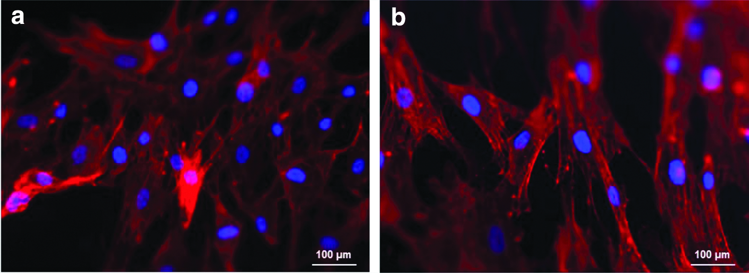
Immunofluorescence analysis of the dental implant surface.
Osteogenic differentiation of ADSCs onto bone blocks
The commitment of cells to an osteogenic phenotype was assessed by means of gene expression analyses (Fig. 5). The expression of osteoblast markers in ADSCs seeded around the sand-blasted acid-etched dental implants inserted in the bone blocks was significantly higher than in the control condition, represented by ADSCs seeded on tissue culture plates in cDMEM.
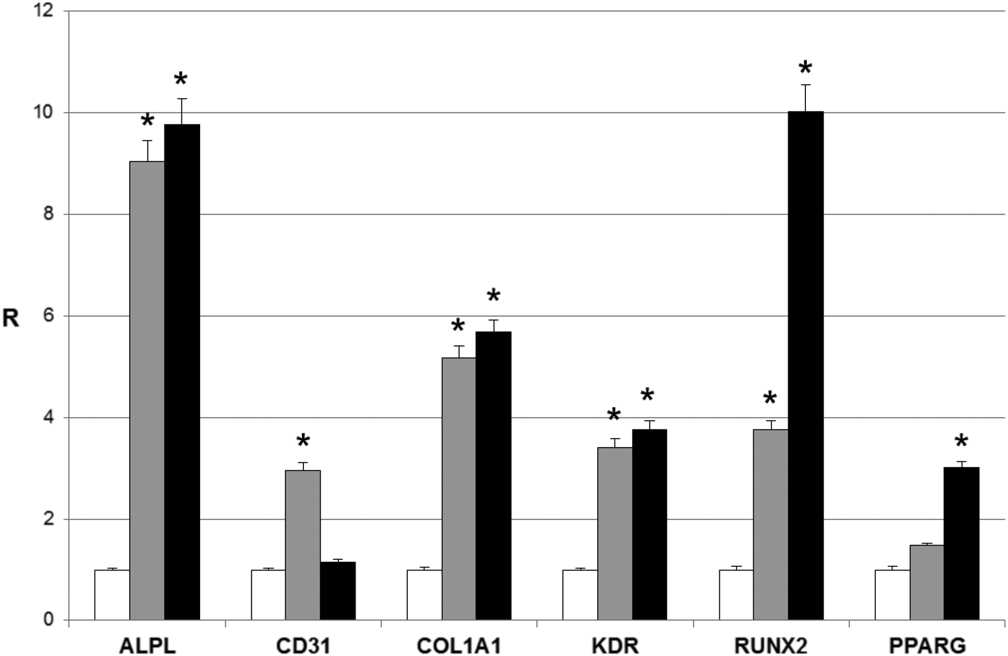
Real-time PCR analysis of alkaline phosphatase, liver/bone/kidney (ALPL), CD31, COL1A1, kinase insert domain receptor (KDR), runt-related transcription factor 2 (RUNX2), and PPARG. Gray bars indicate the expression level of the selected genes in the ADSCs seeded around the sand-blasted acid-etched dental implants in the bone blocks in the presence of osteo-endothelial differentiation medium for 30 days. Black bars represent the gene expression level of the same markers in the ADSCs seeded onto tissue culture polystyrene in osteo-endothelial differentiation medium for 30 days. Gene expression levels are given as ratios (R) with respect to the mRNA expression in the ADSCs seeded onto tissue culture polystyrene in cDMEM for 30 days (control condition, white bars). Data are mean and standard deviation (SD) (n = 3). Statistically significant differences are indicated as *p < 0.05 and compared with the control condition.
Similar results were obtained by comparing the expression of the same markers in ADSCs seeded on tissue culture plates with osteo-endothelial differentiation medium to the control condition. In detail, we found a significant (p < 0.05) increase in the expression of alkaline phosphatase, liver/bone/kidney (ALPL), in both the ADSCs cultured for 30 days onto bone blocks and on tissue culture plates (9.05 ± 0.41 and 9.77 ±0.51, respectively) in the presence of osteo-endothelial differentiation medium. Similarly, a significant (p < 0.05) upregulation was observed in the expression of COL1A1 (collagen, type I, alpha 1) (5.18 ± 0.23 and 5.68 ± 0.24), kinase insert domain receptor (KDR) (3.42 ± 0.16 and 3.76 ± 0.17), and runt-related transcription factor 2 (RUNX2) (3.76 ± 0.18 and 10.02 ± 0.53) in both the conditions described above. A significant (p < 0.05) increase in the expression of CD31 (PECAM-1, platelet/endothelial cell adhesion molecule 1) was detectable only in cells seeded onto bone blocks, but not in those cultivated on tissue culture plates (2.95 ±0.17 and 1.16 ± 0.05, respectively). On the contrary, PPARG (peroxisome proliferator-activated receptor gamma) expression was significantly (p < 0.05) higher in ADSCs seeded on tissue culture plates, but it did not change in cells seeded onto bone blocks (3.01 ± 0.12 and 1.48 ± 0.05, respectively).
Mechanical pull-out test
The results of the mechanical tests are summarized in Figure 6. Regarding the sand-blasted acid-etched titanium implants, the highest mean pull-out strength was reached with the cultivated blocks (7.66 ± 1.30 N), whereas the results obtained with the dry and immersed uncultivated samples were similar (1.52 ± 0.70 N and 1.96 ± 0.56, respectively). There was an approximately fourfold increase in pull-out strength after cultivation. The differences observed between the cultivated and uncultivated specimens were statistically significant (p < 0.05), whereas the differences between the dry and immersed uncultivated samples were not (p > 0.05).
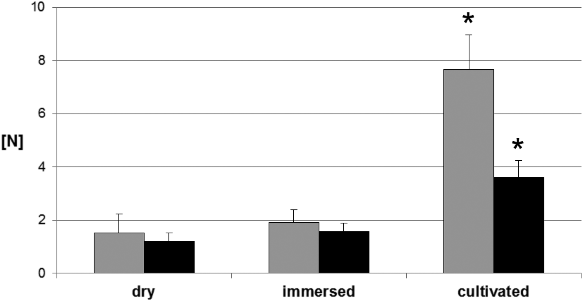
Pull-out tests. Average value and SD of force Newton (N) measured with the sand-blasted acid-etched implants (gray bars), and with the machined implants (black bars) in the three different experimental conditions (dry, immersed, cultivated). Statistically significant differences are indicated as *p < 0.05 and compared with the dry bone blocks.
The results of the pull-out test obtained with the machined titanium implants had the same trend observed for the sand-blasted acid-etched implants; that is, the pull-out strength measured on implants extracted from the cultivated bone blocks (3.61 ± 0.64 N) was significantly (p < 0.05) higher than that recorded on uncultivated samples (1.21 ± 0.31 N and 1.56 ± 0.32 N, respectively for the dry and immersed blocks). When comparing the pull-out values between the sand-blasted acid-etched and the machined implant surfaces in the cultivated groups, higher pull out values were measured for the first type of implants. The difference observed between the two groups was found to be significant (p < 0.05).
Morphological analyses of ADSCs with SEM
To confirm whether the improvement in pull-out strength was due to the presence of the cells, we performed SEM analyses on the sand-blasted acid-etched dental implants extracted from the dry and seeded bone blocks. The analyses of the sand-blasted acid-etched titanium surface extracted from uncultivated blocks revealed high level of structural organization, characterized by sharp ridges of titanium forming a honeycomb pattern (Fig. 7a). The sand-blasted acid-etched titanium implant retrieved from cultivated blocks showed a layer of ADSCs adhering to the surface of the implant with an osteoblast morphology (Fig. 7b).

Scanning electron microscopy analysis of the dental implants surface after the pull-out test.
Discussion
The present study aims at the development of an efficient experimental in vitro model for the analysis of dental implant stability, which is directly related to successful osseointegration.
The method proposed in this study is based on a tissue engineering approach, consisting in the insertion of a dental implant into a bone scaffold loaded with human ADSCs. The mesenchymal stem cells seeded onto bone blocks were committed to an osteoblast phenotype with the purpose of stimulating the bone integration process in vitro. The initial cell seeding number is a critical factor that should be strongly taken into account when designing a construct intended for tissue engineering. Indeed, cell seeding density influences cell proliferation, distribution, differentiation, and extracellular matrix synthesis.30–32 The optimal seeding density of a scaffold depends on the biomaterial type, its structure, and the seeding technique.33,34
The number of ADSCs that we seeded onto bone blocks around dental implants was chosen according to the results described in our previous work, 25 reporting that the selected cell seeding density positively influenced the osteogenic differentiation of ADSCs. Also in this work, the expression of the well-known osteogenic markers RUNX2, ALPL, and COL1A135–37 was clearly detectable in ADSCs cultivated onto bone blocks for 30 days. RUNX2 is a transcription factor whose expression is considered a prerequisite for osteoblast differentiation. 38 By contrast, PPARG is a transcription factor having antiosteoblastic effects on the osteogenic differentiation of ADSCs. 39 RUNX2 expression was followed by that of ALPL and COL1A1, both important for the synthesis of bone matrix and subsequent mineralization thereof. 40 In addition, the presence of endothelial growth factors in the differentiation medium strongly contributed to the simultaneous expression of both the surface endothelial marker CD31 41 and KDR, 42 one of the two receptors of VEGF. These results are very interesting because, since bone is a highly vascularized tissue, a good vascularization is crucial for improving the osseointegration process. 43
Pull-out tests were then used to assess implant stability, which relates to the contact between the dental implant surface and the cellularized mineral matrix. These tests are mainly applicable for unthreaded cylindrical implants, whereas most of the clinically available fixtures have a threaded design, and their interfacial failure depends on shear stress. 44
Over the years, several materials have been selected for performing the mechanical tests of dental implants.45,46 Due to the difficulties in obtaining, preparing, and preserving human cadaver bone, different substrates have been proposed as substitutes for the human bone. Pull-out tests have been widely carried out with polyurethane blocks used to simulate the mechanical properties of human bone. 47 These have been used for mechanical tests on both orthopedic implants 48 and dental implants.49–52 While solid, rigid polyurethane blocks can be used as substitutes for human jaw bone to measure the primary stability of implants, they are not suitable for assessing secondary stability relating to the dynamic process of osseointegration. Hence, the need for in vivo investigations to detect the extent of bone formation over the implant surface. Ex vivo mechanical tests (torque, pull-out, push-out) usually measure the amount of force or torque needed to induce interface failure of implants in bone.
Dental implant materials and surface treatments have been widely tested in the rabbit long bone model, since rabbit is the smallest animal that can accept commercially available dental implants in long bones.53,54 Seong et al. found that failure loads reached with the pull-out test correlated significantly with secondary implant stability in a rabbit tibia dental implant healing model. 55 Their findings are consistent with studies conducted by Baker et al., 56 who tested two different implant surfaces in short-term healing in the rabbit tibia: a dual-etched surface gained in pull-out strength more rapidly than a machined surface, and remained significantly stronger throughout the 8 weeks of the study, although the pull-out strength increased over time in both groups. In addition to the rabbit model, other studies describe the use of larger animals for performing pull-out tests; for example, pull-out measurements have been made after placement of dental implants in the mandible and maxilla of minipigs 57 or in the femur of rats. 58
To the best of our knowledge, there are no studies published to date that have used a 3D tissue-engineered bone model to assess dental implant osseointegration in vitro. Consequently, the mechanical pull-out data obtained with the novel method presented here cannot be compared with the findings of other reported in vitro and in vivo studies.
In our work, two types of titanium dental implants were tested and their pull-out strength measurements were compared to evaluate the effects of different implant surface treatments in the implant stability. The stability of moderately rough, flat cylindrical implants increased over time, thanks to the diffusion, proliferation, and differentiation of ADSCs cultured on the 3D scaffolds in the presence of osteo-endothelial differentiation medium. The mean pull-out strength was about four times higher after cultivation than under control conditions (dry and immersed bone blocks). This result confirmed the in vitro osseointegration achieved in the test samples, consistently with the presence of osteoblasts identified by gene expression analyses. Similarly, the pull-out strength calculated on machined dental implants after cultivation of ADSCs onto bone blocks was approximately two times higher than under control conditions. Nevertheless, when comparing the two types of dental implant surfaces in the cultivated samples, the machined implants presented significantly lower pull out values than the sand-blasted acid-etched implants.
Consistent with pull-out data, IF staining showed differences in cell morphology among the two groups. Although ADSCs showed adherence on both the sand-blasted acid-etched and machined titanium surfaces, morphological differences reflecting the cells' attempt to adapt and anchor to the substrate were evidenced. ADSCs grown on the sand-blasted acid-etched surface showed a polygonal morphology with multiple extensions, suggesting a dynamic adhesion state for these cells. On the contrary, ADSCs in contact with the machined implant surface appeared mainly spindle-shaped, indicating an overall weaker adhesion.
Taken together, these data would suggest that the sand-blasted acid-etched titanium surface provides a better substrate for cell adhesion than the smoother machined surface. This probably allows cells to establish more stable contacts, which are in turn responsible for a better osseointegration, as confirmed by the higher pull-out strength observed for the sand-blasted acid-etched cultivated samples.
These results are in agreement with published data relative to BIC obtained from in vivo studies. The histomorphometric measurement of the BIC percentage (BIC%) of osseointegrated implants is the standard procedure for the evaluation of bone formation on an implant surface. 59 High BIC% values are considered to be a prerequisite for implant stability, which is essential for successful implant treatment.60,61 In an experimental study in dogs, Sivolella et al. placed the same sand-blasted acid-etched implant used in this work in dog's mandible, and measured the BIC% after 3 months of healing. 62 They found that the implants were osseointegrated in mature bone presenting a BIC% of 46.1%. In a similar study conducted in dogs, Schliephake et al. observed a significant reduction in the BIC% in the machined dental implants compared to other modified implant surfaces after 3 months of healing. 63 More generally, implants with moderately rough surfaces have been described in the current literature having a more intensive BIC% than implants with smooth machined surfaces,64–66 resulting in higher mechanical retention when implanted in humans.
Considering our results, if an implant is inserted into a scaffold enriched with ADSCs committed to an osteoblast-like phenotype, the cells colonizing the implant surface have an osteoblast morphology. Pull-out tests confirmed that implants inserted in live scaffolds withstand a significantly greater tensile force than control implants extracted from scaffolds not loaded with ADSCs. Nonetheless, the pull-out strength seems to be influenced by the surface treatments of dental implants, being significantly higher for implants with a moderately rough surface than for machined implants. It is, therefore, likely that the sand-blasted acid-etched surface used in the present study improved cell adhesion, which in turn promoted cell differentiation, thus providing a structure more conducive to osseointegration when compared with the machined implant surface. Clearly, our in vitro studies were unable to reproduce the dynamic environment of the bone–implant interaction in vivo, and our results would need to be confirmed in animal models. We can nonetheless assume that osseointegration, defined as a close structural and functional contact between bone and implant, 67 was achieved in our 3D in vitro tissue-engineered bone model.
Conclusions
The results derived from this study suggest that the 3D tissue-engineered model described could be adopted for assessing in vitro the dynamics of bone–implant interactions that lead to osseointegration. The routine use of 3D models is not necessarily cheaper compared to animal experiments. Human-derived tissues used under controlled test conditions offer several advantages. Issues regarding ethical questions arising from the use of experimental animals and species extrapolation are avoided. 3D models have great potential in controlling regulatory risk assessment and reducing problems related to the reproducibility, quality control, and interpretation of results. We thus believe that our approach may represent a novel biomimetic alternative to in vivo animal models, and could be important for clinical knowledge that constitutes a reference point for specialists who have to evaluate, plan, and subsequently perform dental implant procedures.
Footnotes
Acknowledgment
This work was partially supported by Sweden & Martina (Due Carrare, Padova, Italy), which provided the customized implants.
Disclosure Statement
No competing financial interests exist.