




Abstract
The European Union (EU) directive 2010/63/EU on the protection of animals used for scientific purpose focused on reducing the number of animals and refinement of breeding. Animal studies are necessary to protect human health. Lots of animal models exist to study bone regeneration, but a reliable, well reproducible, and relatively inexpensive model with the possibility for multiple testing in the same animal is still missing. Rats may serve as good models for this due to the small animal size and good cost/benefit ratio. The present study aimed to develop a novel rat caudal vertebrae critical size defect model for bone regeneration and implant osseointegration studies The study was performed using Wistar rats with weight from 380 to 450 g. An incision was made on the dorsal side of the tail. After skin and muscles retractions, the vertebrae were exposed. Critical size defects for bone tissue engineering or implant placements for titanium body experiments were possible in each of the first four caudal vertebrae. Micro-computed tomography (CT) and histology were used to detect bone growth. There was no bone formation in the defects after 1 or 2 months of healing. When a calcium phosphate biomaterial was used (Bio-Oss®; Geistlich Pharma AG, Wolhusen, Switzerland), a good stability of the material in the defect was noted and bone growth was visible after 1 or 2 months. Results based on implant placement showed good primary stability after 3 months of healing. MicroCT showed integrated implant position through the vertebra. These results suggest that the rat caudal vertebrae may serve as a good new model for studying bone regeneration and implant osseointegration with the possibility of multiple testing within the same experimental animal and the potential to decrease number of experimental animals.
Introduction
T
In the field of bone grafting, many animal models have been used. Desirable attributes, such as relative similarity to humans, must be defined clearly before selecting the animal. Animal selection factors include: cost to acquire and care for animals, availability, acceptability to society, tolerance to captivity, and ease of housing. Sheep, goats, dogs, pigs, or rabbits are suitable for testing implantation of materials in bone.3,4 Monkeys, mainly rhesus monkey, cynomolgus monkey, and baboons are sometimes used despite their costs. Rodents such as mice, rats, and hamsters have been used widely for periodontal and bone research because of specific advantages such as small size, low cost, known age and genetic background, controllable microflora, and ease of handling and housing. 5 Rat models are suitable to assess histological bone regeneration, providing sufficient statistical significance reached by using numerous animals, and to provide preclinical relevance. 6 Different rat models have been developed based on reproducible defects in different bone locations. 6 Calvarial, tibial, femoral, and mandibular critical size bone defects in rats have been used in various studies to investigate the effectiveness in bone defect repair of bone regenerative agents such as growth factors, biomaterials, cell or tissue implantation, or any combination of these.7–10 Unfortunately, none of these models combine surgical access facilities, number of defect sites, and histological relevance.
Following the pioneer work of Blazsek et al. (2009) on the rat tail model to study implant osseointegration, we have developed an original rat caudal vertebrae model for bone tissue engineering. 11 This method allows the local filling of biomaterials delivering or not delivering drug or stem cells. Moreover, it permits the measure of neoossification, osseointegration, and bone regeneration in four defects per animal. This number of defects per animal increases its ethical acceptability, decreases the number of animals, and allows multiple comparisons of treatments within the same animal.
Materials and Methods
Experimental setup
The study was approved by the committee for animal welfare of Montpellier University with the referral number 1083 16/06/2014.
Three groups (3 rats per group) of male Wistar rats [(Crl:(Wi)Br) from Charles River France]. Animals with weight ranging from 380 to 450 g were used for an adequate vertebrae size. All animals were kept in light controlled air-conditioned rooms and fed ad libitum. The first two groups of rats were implanted with Bio-Oss® (Geistlich Pharma AG, Wolhusen, Switzerland) material for 1- and 2-month periods. In each rat, two vertebrae were used as a control (defect empty of materials) and two other vertebrae were used for Bio-Oss implantation.
The third group was treated with impacted-type titanium (Ti6Al4V) implant manufactured by FullTech Ltd (Budapest, Hungary). Implant shape was designed to allow bone growth as presented on Figure 1. Implantation time was 3 months. In each rat, four vertebrae were used for implantation.

Surgical procedure
Animals were anesthetized with an intraperitoneal injection of ketamine and xylazine (Alcyon, Pau, France) (40 and 9 mg/kg, respectively). The tail was disinfected and a dorsal incision was made approximately from Cd31 to Cd35 vertebrae. The skin and the muscles were retracted with buffered saline solution irrigation and the vertebrae were exposed. For Bio-Oss implantation and the empty cavity control, an intraosseous defect preparation of 3 × 3mm was performed in the exposed surface of the vertebrae. This size of the defect was chosen based on tests for critical size defect evaluation. The critical size defect was previously described in multiple studies, but particularly for the rat calvaria bone by Spicer et al., 12 as a defect without healing by itself after different periods (1–6 months) of observations.
In the group of titanium body application, one implant was placed into each vertebra after drilling with pointer and twist drills with 1.3- and 2.9-mm diameter drill for the neck part of the implant, and the last step of the drilling was performed with 2.9. The impacted-type implants have two parts, the apex is 0.8 mm in diameter and the coronal part is 2.9 mm wide. This part is fixing the implant in the drilled cavity. The space between the apex and the coronal part can be left empty or be filled with biomaterial (Fig. 1).
After the hard tissue treating, the muscles were repositioned over the defects and sutured together with resorbable sutures (Vicryl 4/0; Ethicon, Issy-les-Moulineaux, France). Then, the skin was tightly sutured with resorbable sutures (Vicryl 3/0; Ethicon). Following surgery, buprenorphine SR-LAB (1 mg/mL), (Wildlife Pharmaceuticals, Windsor, CO) at a dose of 1.2 mg/kg was used for systemic relief and to provide 72 h analgesia.
Rats were kept in individual cages and the wound healing was controlled daily for the first week and twice per week during the following healing periods. Every second day, the tails were disinfected using povidone–iodine solution (Betadine; Mundipharma, Paris, France).
Samples collection
At the end of the different long experimental periods, the rats were sacrificed by intraperitoneal injection of Pentothal (Alcyon) with a suitable dosage (200 mg/1.5 kg). The tails were harvested between Cd31 and Cd32. The soft tissues were carefully removed mechanically, and the samples were fixed in 5% formaldehyde solution in 4°C for 24 h. 11
MicroCT analysis
A three-dimensional (3D) radiographic evaluation was performed using an X-ray micro-computed tomography (CT) instrument (SKYSCAN 1172 X-ray Microtomograph; Microphotonics, Inc., Allentown, PA) with a 3D reconstruction software (Aviso, FEI company, Hillsboro, OR). Implanted samples were planned to be scanned at 360° rotation at 0.7° intervals. Measurements were made on the region of interest (ROI) ×1.5 mm tissue volume (TV) on the computer-reconstructed 3D samples. These measurements were done before the other analyses.
Histology
After micro-CT scanning, the specimens were decalcified in 4% EDTA for 3 weeks in microwave (Histos 5 Rapid, Tissue processor; Milestone, Birmingham, Italy). Samples were washed in water before processing for sucrose-gelatin impregnation and freezing in isopentane at −80°C. Then, slices were cut (55 μm thick) along the coronal plane through the center of each defect with a cryostat (Leica 3050S, Nussloch, Germany). The sections were stained with hematoxylin and eosin. Finally, after three washes in distilled water, the samples were passed through a graded sequence of alcohols (95°, 75°, and 95°) and mounted on slide with a mounting medium.13,14
For implant experiment, after micro-CT scanning, the pieces were dehydrated in a series of graded ethanol solutions and embedded in epoxy resin blocks (Epoxy embedding medium kit; Sigma-Aldrich, Lyon, France). Transverse sections (0.30 mm) were stained with toluidine blue and also mounted on slide with a mounting medium.
Results
Figure 2 shows the dorsal incision realized between Cd31 and Cd35 caudal vertebrae. After skin and muscles retraction, the vertebrae are visible. The incision permits to access three or four operative sites (vertebrae) in the same animal. Figure 2D represents a scheme of possible implantation of different type of materials in a different vertebra on the same animal. In this proof-of-concept study, the first two vertebrae were used as controls with a defect without filling material, and the second two served as experimental sites with Bio-Oss filling material or implant (see also in the Materials and Methods section).

Surgical procedure.
Figure 3 shows the correspondence between the 3D reconstruction of micro-CT scanned vertebra and the anatomy. The mean length of caudal vertebrae is 9.80 mm and the mean diameter is 3.70 mm. Figure 3C shows the position of the tissue, blood vessels, and nerves around the vertebra. The soft tissue arrangement of the vertebra, particularly the location of main vessels and nerve, are in favor for preparing the defects on the dorsal side of the rat tail. In Figure 4, vertebra reconstruction shows the bone defect (Fig. 4A) and implant (Fig. 4B) 1 month after the surgical procedure, and the vertebra bone architecture is presented in Figure 4C.

Rat caudal vertebrae anatomy.
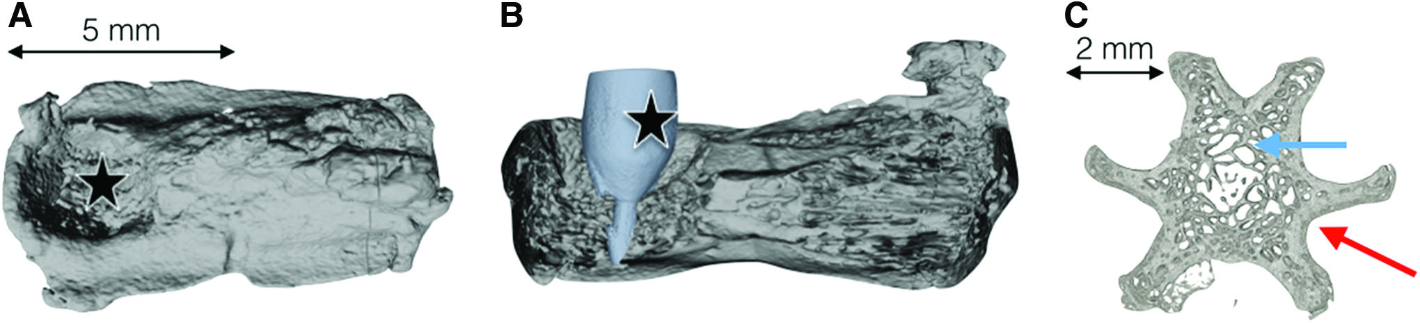
μCT 3D reconstruction of rat caudal vertebra.
During all of these experimentations, no infection of the operative site was observed. The behavior of the rat did not change during the experiments and the postsurgical care period.
MicroCT and histology analyses did not show bone formation when the defect was left empty after either 1 month (Fig. 5A, B) or 2 months (Fig. 6A, B). Bone regeneration within the applied size of the defect (2.9 × 3 mm) was never observed after either 1 or 2 months.

Rat caudal vertebra with an empty defect
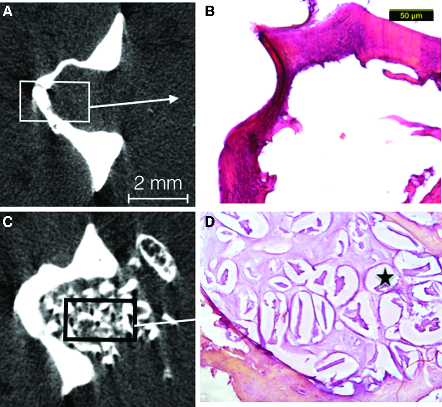
Rat caudal vertebra with an empty defect
When we used biomaterials like calcium phosphate (Bio-Oss) to fill the defect, we obtained a good retention of the material in the defect after 1 and 2 months. Bone formation around the particles after both 1 month (Fig. 5C, D) and 2 months (Fig. 6C, D) was well visible.
Figure 7 demonstrates the implant position through the vertebra after implantation. This titanium implant was specifically designed to be impacted into the cavity and to preserve a space between bone and implant in its apical part. Three months after implantation of this new design implant, microCT scanning showed the implant position through the vertebra (Fig. 7B). The histology analysis showed implant osseointegration with bone-implant contact after 3 months (Fig. 7C). During our study, no implant was lost.
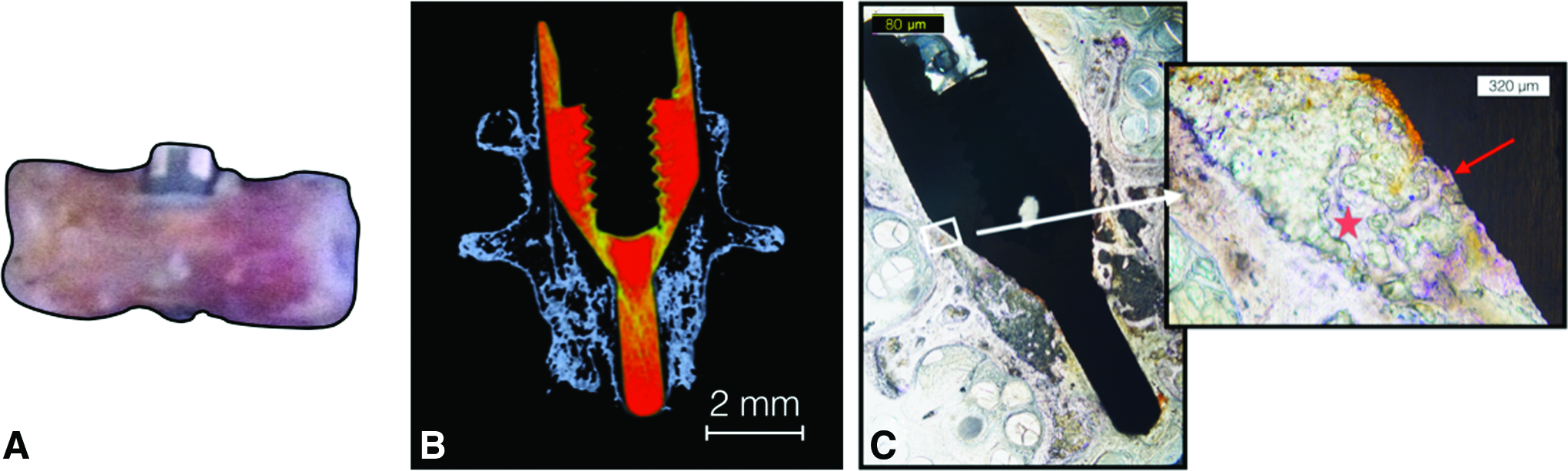
Implant placement through vertebra.
These proof-of-concept results validate our original assumption that the rat caudal vertebrae may serve as a good model for bone reconstruction. Both bone grafting biomaterials for new bone formation and various implants for osseointegration can be studied using this rat tail model.
Discussion
A number of animal models have been used to study the effectiveness of regenerative agents in bone tissue engineering. Still, the development of novel, more reproducible experimental animal models are required for testing and validating bone regenerative therapies. The best animal models should use the minimum number of animals that would provide reliable results and require the use of species with the lowest capacity to experience pain, suffering, distress or lasting harm that are optimal for extrapolation into target species. 15 The new European directive for the protection of experimental animals put animal welfare demands into the focus of related research strategies.1,15 Rodents, particularly rats, represent one of the most reliable target for such studies because of their relatively easy breeding and housing, genetic uniformity, and superior cost-effectiveness ratio. In laboratory rats, the most frequently used sites of experimentation for critical size defects and bone regeneration are the tibia, femur, calvaria, and the mandible.4,12,13,16 In rats, difficulty in the small size of the grafting site and the use of high number of animals to meet the statistical requirements are also challenging.
Our study was designed to establish a new rat model allowing to decrease the number of animals and to ease the surgical and care procedures. The possibility to use four or five caudal vertebrae at the same time represents the greatest advantage of our new model. One vertebra can be used for the control site and the others for different kind of treatments to compare them on the same rat at the same experimental time. From a statistical point of view, this new model allows decreasing the number of animals two to four times in comparison with models with only one or two sites per animal. Even more importantly, in the presented model, control sites and multiple experimental ones can be compared in the same animal in a standard manner, decreasing the bias due to interindividual variability.
For pain control, it is important to note that the rat tail primarily receives its innervations ventrally, and we use dorsal access to the vertebrae. It was also reported that even in case of caudal nerves injury, the tail movement is preserved. 17 Therefore, this model is ethically acceptable with the pain control and tail movement preserved. In addition, in comparison with the model developed by Blazsek et al., 11 this model seems to be superior since it is possible to construct a bone defect without cutting off the rat tail, which creates suffering and disturbed space localization of the rat. Another advantage is that the surgical procedure is easy to carry out in the dorsal face of the rodent tail, and it is easy to control postoperative cares with an easy view and manual access to the wound.
In the context of regenerative medicine using biomaterials in dentistry and maxillofacial surgery, large animal models have been preferred due to the reproducibility and surgical accessibility of experimental defects, as well as structural similarities to human situations. 6 However, the caudal vertebrae of the rat are highly similar to human jaw bones both from histological and anatomical points of view. The rat caudal vertebrae are similar to human jaws, in the lack of hematopoiesis, 18 a feature which is different in other bones, which are frequently used as an implant bed in animal models. This makes it a perfect model for evaluating bone regeneration methods in dental and maxillofacial research.
Based on the previous research results 11 and also on our findings, the rat tail is an easily accessible site for bone regeneration and implant osseointegration studies and allows safe and reproducible surgical procedure. An abundant cancellous bone delimited with an important cortical bone thickness constitutes the rat tail vertebra. This anatomy permits to study different sorts of biomaterials for bone regeneration or implant for osseointegration. Moreover, our data show that the tail vertebrae may provide an ideal tissue support for preclinical implant studies. The stability and longevity of integration of foreign materials into the bone represent a major problem in tissue engineering. Because of the difficult accessibility of the mandibular and maxilla, different research teams have succeeded in measuring osseointegration mainly in anatomically accessible bone compartments, such as hematopoietic femur.11,19
The tail vertebra appears to be a useful preclinical model where the strength of integration of titanium implants can be evaluated during osseointegration by mechanical measurements and structural analysis of newly formed bone by micro-CT or histology. The empty cavity we created around the implant allows to study the effects of biomaterials, stem cells, and growth factors on dental implant healing. 11 Moreover, this model allows to test the effect of various biomaterials on implants stability at different endpoints. The use of this model may also allow the investigation of different periods to build kinetic models of healing and then, to account for the local or systemic effect of the biomaterial implanted. Depending on the chosen experimental set up, the researcher can use only one, or more vertebrae by animal. In addition, in vivo microCT could permit to follow the new bone formation through the calculation of bone density using only a single group of animals. In future, our model may also allow to study different types of biomaterials, combined or not with stem cells and growth factors, on the same tail, decreasing the necessary number of experimental animals. Taken together, our model may become very useful at the preclinical level for both implantation and osseointegration studies.
Conclusion
The rat tail vertebrae model is promising for both bone tissue engineering and implant osseointegration studies. A critical size defect of 2.9 × 3 mm can be drilled, which allows to follow bone regeneration in response to various treatments. The model also permits 2.9-mm-wide implant placements with a great stability. The main advantage of our model is that four or five caudal vertebrae can be studied in the same animal at the same time, hereby achieving a decrease in the number of rats involved. The model is inducing minimal pain and suffering. These arguments follow the 3Rs recommendation for animal experiment.
Footnotes
Acknowledgments
The authors thank the histology platform RHEM (INSERM, Montpellier). This work was supported by the French–Hungarian TET-Balaton Program (TET_12_FR-2-2014-0010).
Disclosure Statement
No competing financial interests exist.