




Abstract
Decellularized tissue allografts are paving the way as an alternative to cellular tissue transplantation. Effective sterilization or decontamination of tissue allografts is paramount for the safety of the allograft; however, some of the current sterilization procedures have a detrimental effect on the tissue scaffold. The bactericidal and virucidal activity of copper (II) ions and hydrogen peroxide (H2O2) have been widely reported, however, their effect on the biology, biochemistry, and biocompatibility of decellularized tissue have yet to be elucidated. In this study, decellularized human dermis (dCELL human dermis) was treated with copper (II) chloride (CuCl2) and H2O2; both singly and in combination, and parameters, including concentration, pH, and synergy between CuCl2 and H2O2, were evaluated to identify conditions where any detrimental effects on the tissue scaffold were observed. Skin from 13 human donors was retrieved with appropriate consent and processed into dCELL human dermis. The dCELL human dermis was then treated for 3 h with 0.1 mg/L–1 g/L (w/v) CuCl2 and 0.01–7.5% (v/v) H2O2 and combinations of both of these in the same concentration range. dCELL human dermis treated with solutions of 0.1 mg/L–1 g/L CuCl2 or 0.01–7.5% H2O2 caused no detrimental effects on gross histology, collagen denaturation, collagen orientation, and biomechanical properties of the tissue or cytotoxicity. The highest combined concentration of CuCl2 and H2O2 demonstrated an increase in ultimate tensile strength, loss of collagen type IV immunostaining at the dermal-epidermal junction, and in vitro cytotoxicity. Combinations within the range of up to 10 mg/L CuCl2 with up to 0.5% H2O2 had no effect. The data identify the concentrations of CuCl2 and H2O2 solutions that have no effect on the biological, biomechanical, and biochemical properties of dCELL human dermis, while retaining biocompatibility. These treatments may be suitable for use as sterilization/decontamination agents on human decellularized tissues.
Introduction
W
Decellularized tissue allografts have been used for reconstructive surgical applications and transplantation for a number of years. The removal of cellular components from tissue allografts renders these decellularized grafts nonimmunogenic, allowing recellularization of the scaffold by the recipient's own cells.15–17 Decellularized tissue eliminates the concern of effect the sterilization procedure has on the graft cells, however, the effect of sterilants on the extracellular matrix scaffold needs to be taken into consideration. Studies have shown that PAA has a negative effect on type IV collagen at the basement membrane of acellular blood vessels and dCELL human dermis.18,19 Gouk et al. demonstrated that gamma irradiation of decellularized dermis reduced the elasticity, increased susceptibility to enzymatic degradation, and induced structural modifications that subsequently compromised the tensile strength of the tissue. 20 Thus, the need for the development of alternative sterilization methods is evident.
The sterilization activity of metal-based and other formulations, for example, cupric [copper (II)] ions or hydrogen peroxide (H2O2), is widely recognized. Several experimental studies have documented the antimicrobial and virucidal activity of cupric ions.21,22 Such reagents are utilized in water sanitization processes and the healthcare industry to disinfect reusable medical equipment.23,24 Copper-impregnated contact surfaces for example, trolleys or bed rails, have been shown to significantly reduce the bacterial bioburden compared to noncopper surfaces and reduce the rate of hospital-acquired infections.25,26 H2O2 in both liquid and gaseous states is used in the medical, food, and environmental industry as a broad-spectrum biocide, including efficacy against bacterial spores. 27 NHS Blood and Transplant (NHSBT) incorporates H2O2 into wash solutions during bone allograft processing as a means to reduce pathogen contamination without compromising the osteoinductivity and biomechanical stability of the bone.28–30 The potentiation of the bactericidal and virucidal activity of copper ions by H2O2 is well documented.21,22,31,32 In addition, Solassol et al. showed that combinations of cupric ions and H2O2 reduced the levels of prion protein (PrPsc) in infected brain homogenates to a greater degree than individual treatments. 33
To date, no chemical formulation containing a combination of copper (II) chloride (CuCl2) and H2O2 has been used as a sterilization agent in tissue banking or tissue engineering. Development of a new tissue sterilant must first establish that the candidate sterilant itself does not have any adverse effect on the tissue. In this study, using decellularized (dCELL) human dermis developed by NHSBT,19,34 we investigate a range of CuCl2 and H2O2 concentrations and pH values, used individually and in combination, to identify those treatments that do not damage dCELL human dermis. Evaluation of the effects of CuCl2 and H2O2 was determined by conventional histology and biomechanical analysis. Dermal collagen was assessed by immunohistochemistry, collagen birefringence, and hydroxyproline content. Cytotoxicity of treated dCELL dermis was evaluated by an in vitro contact assay. Our findings identify those CuCl2 and H2O2 treatments/concentrations that preserve the morphology, functionality, and biocompatibility of dCELL human dermis. These treatments/concentrations can subsequently be evaluated for their potential as a tissue sterilant.
Methods
Tissue
Cryopreserved skin from 13 deceased donors (males, aged 50–83 years old) was used in this study. All donor families had given full consent for R&D use. The donor was shaved and then washed with 0.5% chlorhexidine (EcoLab) and phosphate-buffered saline (PBS; Sigma Aldrich). Strips of skin (5–6 × 30 cm) were retrieved by trained Tissue Services Donation staff from the legs and the back and sides of the torso using a dermatome (Braun) set to a nominal depth of 0.33 mm. The skin was treated with an antibiotic cocktail (Cambridge Antibiotics; Source BioScience), cryopreserved in 20% v/v glycerol (Source BioScience), and stored at −80°C until use.
Dermis decellularization
Skin was thawed, washed in PBS, and decellularized as described in Hogg et al. Briefly, the epidermis was removed by incubating skin in 1 M sodium chloride (Source BioScience) for 18 h (37°C); the dermis was then washed in PBS and incubated in hypotonic buffer (Source BioScience) for 16 h (4°C). Dermis was washed in PBS and incubated for a further 24 h (25°C) in a 0.01% (w/v) sodium dodecyl sulfate detergent buffer (Source Bioscience). After another PBS wash, dermis was transferred to a nuclease buffer (Source BioScience) containing 1 U/mL benzonase (Merck Millipore) for 3 h (37°C). The proteinase inhibitor aprotinin (10 KIU/mL) and the antibiotics penicillin–streptomycin (1% v/v) (Sigma Aldrich) were used in the incubations. After decellularization, the dCELL human dermis was washed in PBS +0.1% EDTA (VWR) and frozen at −40°C until use.
Stability of copper chloride and hydrogen peroxide solutions
Solutions of 0.1, 1, 10, and 100 mg/L and 1 g/L (w/v) CuCl2 (Sigma Aldrich) at pH 3, 4, and 5 were prepared in distilled water and stored at 4°C. Reagent grade (30%) H2O2 (Sigma Aldrich) was used to prepare solutions of 0.01, 0.1, 0.5, 1.0, 3.0, and 7.5% (v/v) in distilled water at pH 3, 4, and 5 and were stored at 4°C. Solutions containing a combination of CuCl2 and H2O2 were made in the concentration range of 0.1–100 mg/L CuCl2 + 0.01–3.0% H2O2 and stored similarly. Combining these concentrations of CuCl2 and H2O2, the pH was maintained below pH 5 and therefore there was no need for adjustment (Table 1). To establish if there was any degradation of copper or hydrogen peroxide over time (44 days), the concentration of copper (II) was determined photometrically using an AQUANAL™-plus copper (Cu) kit (Sigma Aldrich) according to the manufacturer's instructions. H2O2 levels were measured photometrically using a quantitative peroxidase kit (Thermo Scientific) according to the manufacturer's instructions.
At 44 days, due to a reduction in the measured concentrations of CuCl2 when combined with 3% and 7.5% H2O2, the pH of these solutions were not determined (ND). Values are means ± 95 CI.
Copper chloride and hydrogen peroxide treatment
dCELL human dermis was treated with 0.1 mg/L–1 g/L (w/v) CuCl2 at pH 3, 4, and 5 and 0.01–7.5% (v/v) H2O2 at pH 3, 4, and 5 for 3 h (5 mL/cm2 dermis). dCELL human dermis was treated with a combination of CuCl2 and H2O2 at a concentration range of 0.1–10 mg/L and 0.01–1%, respectively, between pH 3.1–4.9 for 3 h. All treatments were carried out at 25°C at 200 rpm. After chemical treatment, dCELL human dermis was washed in distilled water (Baxter) thrice for 20 min and frozen at −40°C until further use. Control tissue was nontreated dCELL human dermis.
Histology
After CuCl2 and H2O2 treatments, a sample of dCELL human dermis (∼1 cm2) was cut and fixed in 10% formal saline (VWR). Dermis was embedded in paraffin wax and 5-μm sections were cut using a microtome (Finesse 325; Thermo Scientific) and mounted on slides. Sections were stained with hematoxylin and eosin (H & E) for general morphology. Van Giesons staining was performed for visualization of collagen fibers.
Collagen birefringence
Wax-embedded CuCl2 and H2O2-treated dermis were mounted on glass microscope slides. Sections were dewaxed in xylene (VWR) and dehydrated in ethanol (VWR). Sections were placed in Harris hematoxylin for 8 min followed by picro-sirius red solution (Direct Red 80; Sigma Aldrich) for 1 h. Slides were washed in acidified and distilled water before mounting with DPX (VWR). Sections were viewed under polarizing light microscopy (Leica DMLA). Upon polarization, the stained collagen fibers disperse light displaying colors ranging from green (thinner fibers) to red (thicker, mature fibers). To quantify the proportion of fibers displaying red interference, using ImageJ software, the background was subtracted to allow the total tissue area to be measured (in pixels). The hue was resolved to select the red fibers, which was calculated as a percentage of the total tissue area (in pixels).
Collagen type IV immunohistochemistry
Wax-embedded sections were mounted on silanized coated slides (Dako) and dewaxed by dipping into xylene followed by rehydration in absolute ethanol, 95% ethanol, and distilled water. Sections underwent heat-induced epitope retrieval in an antibody retrieval solution (Dako) at 95–99°C for 5 min. The endogenous peroxidase activity was quenched by incubation with peroxidase block solution (Dako). Sections were incubated with monoclonal mouse anti-human collagen IV antibody (Dako) for 60 min. Sections were then incubated with peroxidase-labeled polymer for 30 min followed by incubation with diaminobenzidine (DAB+) substrate chromagen solution (Dako). To ensure there was no nonspecific binding of the primary antibody, negative control tissue was treated with IgG (Dako). All sections were viewed with a Leica DMLA light microscope.
Biomechanical analysis–ultimate tensile strength
Lloyds Instruments Universal Testing Machine (LRX Plus) was used to determine the structural strength of cellular and dCELL dermis treated with CuCl2 and H2O2. Tissue was cut into universal dumbbell shapes (35-mm length ×2-mm minimal width) using a Ray Ran die cutter (DIN-5304 Type S3). The thickness of the dermis was measured using Digimatic Vernier calipers (Mitutoyo). Dumbbells were pulled to breaking point with a 100N load cell at a speed of 100 mm/min. Ultimate tensile stress (N/m2) was determined by dividing load (Newtons) at break by the cross-sectional area, using Nexygen software.
Biochemical analysis–collagen denaturation
Five millimeter biopsies from CuCl2- and H2O2-treated dermis were treated with 10 mg/mL α-chymotrypsin (Sigma Aldrich) for 24 h (37°C) to digest denatured collagen, releasing hydroxyproline. Positive and negative controls were achieved by treating dermis with 2.5 M sodium hydroxide and 50 mM glutaraldehyde, respectively, for 16 h (25°C). The supernatant was acid hydrolyzed (12 M HCl; 121°C and 18 psi for 1 h) followed by treatment with chloramine T (Sigma Aldrich) and 4-(Dimethylamnio)-benzaldehyde (Sigma Aldrich). The level of hydroxyproline in the supernatants was obtained by measuring the absorbance at 595 nm on an Ultra Microplate reader (BioTek).
In vitro cytotoxicity
Five millimeter biopsies of CuCl2- and H2O2- treated dermis were attached to the base of wells of a 24-well tissue culture plate using steristrips (3M Healthcare). Human skin fibroblasts (HSF) and MG63 cells (an osteosarcoma cell line; ECACC) were grown to 70% confluency, harvested, counted, and seeded into the wells containing dermis and incubated for 24 h in a 5% CO2 and 37°C incubator. Cells were fixed with 50%, 70%, and 100% ethanol and stained with 20% Giemsa solution (VWR). Growth of cells up to and around the skin biopsies was visualized using an inverted microscope (Leica, 090-135-002).
Statistical analysis
All numerical data were analyzed using GraphPad Prism 5 software. All numerical data are shown as mean values ±95% confidence limits. Statistical significance was assessed using one-way analysis of variance (ANOVA). Values of p < 0.05 were considered to be statistically significant.
Results
Stability of CuCl2 and H2O2 solutions
Solutions of 0.1 mg/L CuCl2 were below the lower sensitivity threshold of the assay; so the stability of this solution was not tested. In solutions containing either only CuCl2 (1 mg/L–1 g/L) or only H2O2 (0.01–7.5%), there was no change from the initial concentrations of either reagent over a 44-day period, irrespective of the pH (data not shown).
When combinations CuCl2 and H2O2 were mixed (Day 0), there was a significant reduction in the detectable concentration of copper, within minutes, in 1, 10, and 100 mg/L CuCl2 solutions with 7.5% H2O2 (Fig. 1 and Supplementary Table S1; Supplementary Data are available online at
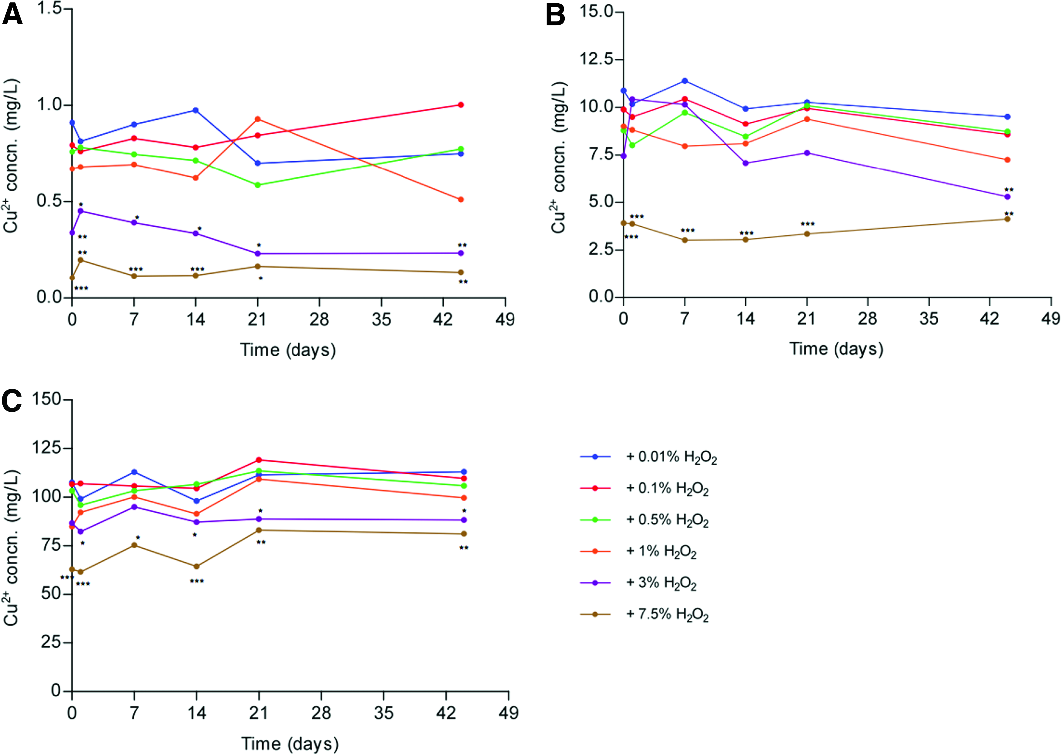
Concentration of Cu2+ ions in solutions of 1 mg/L (
Over 21 days, all concentrations of H2O2 (0.01–3%) were maintained in the presence of 0.1, 1, and 10 mg/L CuCl2 (Fig. 2). However, an increase in CuCl2 to 100 mg/L demonstrated a significant reduction in the measured concentrations of 0.5%, 1%, and 3% H2O2 solutions from 14 days, which continued to decline over a further 30 days (Fig. 2C–E and Supplementary Table S2). The pH of solutions containing a mixture of CuCl2 and H2O2 was maintained between pH 3–5 over a 44-day period and therefore there was no need to adjust the pH (Table 1).

Concentration of H2O2 in solutions of 0.01% (
Based on these findings, the data presented in this article is of dCELL dermis treated with solutions containing 0.1 mg/L–1 g/L (w/v) CuCl2 or 0.01–7.5% (v/v) H2O2 at pH 3, 4, and 5. dCELL human dermis was also treated with combinations of CuCl2 in the concentration range of 0.1, 1, and 10 mg/L together with H2O2 at concentrations of 0.01%, 0.1%, 0.5%, and 1%.
Histological analysis
H&E staining of decellularized human dermis treated with 0.1 mg/L–1 g/L CuCl2 between pH 3–5 and 0.01–7.5% H2O2 between pH 3–5 showed no differences in gross histology compared to nontreated dCELL human dermis (data not shown). Similarly, there were no differences in the extracellular matrix structure when dCELL human dermis was treated with a mixture of CuCl2 and H2O2 up to the maximum concentrations of 10 mg/L and 1.0%, respectively, (Fig. 3).

H & E staining of dCELL human dermis
Ultimate tensile strength
There was no significant difference in the ultimate tensile strength of dCELL human dermis treated with 0.1 mg/L–1 g/L CuCl2 or 0.01–7.5% H2O2 compared to nontreated dCELL human dermis at pH 3, 4, and 5 (Fig. 4A, B). Similarly, 0.1 mg/L CuCl2 combined with all concentrations of H2O2 and 1 mg/L CuCl2 combined with 0.01%, 0.1%, and 0.5% H2O2 did not affect the ultimate tensile strength of dCELL human dermis (Fig. 4C). However, addition of 1.0% H2O2 to solutions of 1 mg/L CuCl2 resulted in a significant increase in the ultimate tensile strength of dCELL human dermis (Fig. 4C). A 10-fold increase in CuCl2 to 10 mg/L combined with H2O2 (0.1–1%), however, did not result in any significant change in dCELL dermis ultimate tensile strength (Fig. 4C).
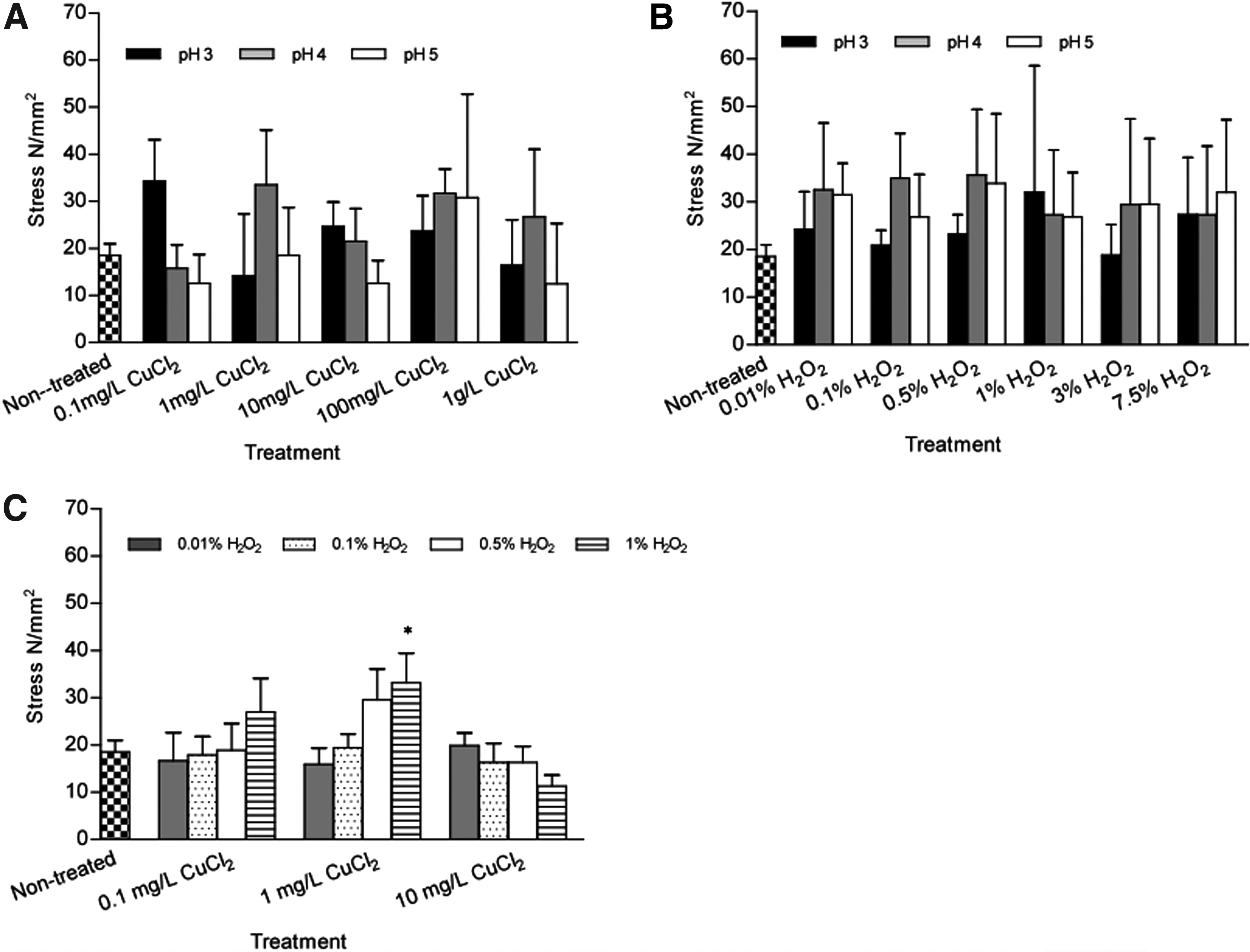
Ultimate tensile strength of dCELL human dermis treated with 0.1 mg/L–1 g/L CuCl2 (
Collagen analysis
To assess if treatment of dCELL human dermis with CuCl2 and H2O2 caused any damage to the collagen fibers, Van Giesons stain, collagen birefringence, and collagen type IV immunohistochemistry was performed. In addition, any denaturation of collagen fibers was evaluated by measuring hydroxyproline levels in solubilized dCELL human dermis.
Collagen appeared tightly packed and uniformly organized; Van Giesons staining showed that CuCl2 in the concentration range of 0.01–10 mg/L together with H2O2 in the concentration range of 0.01–1.0% caused no visual disruption to the collagen network (Fig. 5). In addition, to identify if there was any loss of mature collagen fibers, dermis was stained with picro-sirius red and viewed under polarized light resulting in birefringence of the collagen fibers in which mature collagen fibers were viewed as red in color, which were quantified as a percentage of the total tissue area. There were no significant differences in the quantity of stained mature collagen fibers of dCELL dermis treated with single and combined CuCl2 and H2O2 solutions (Tables 2 and 3).
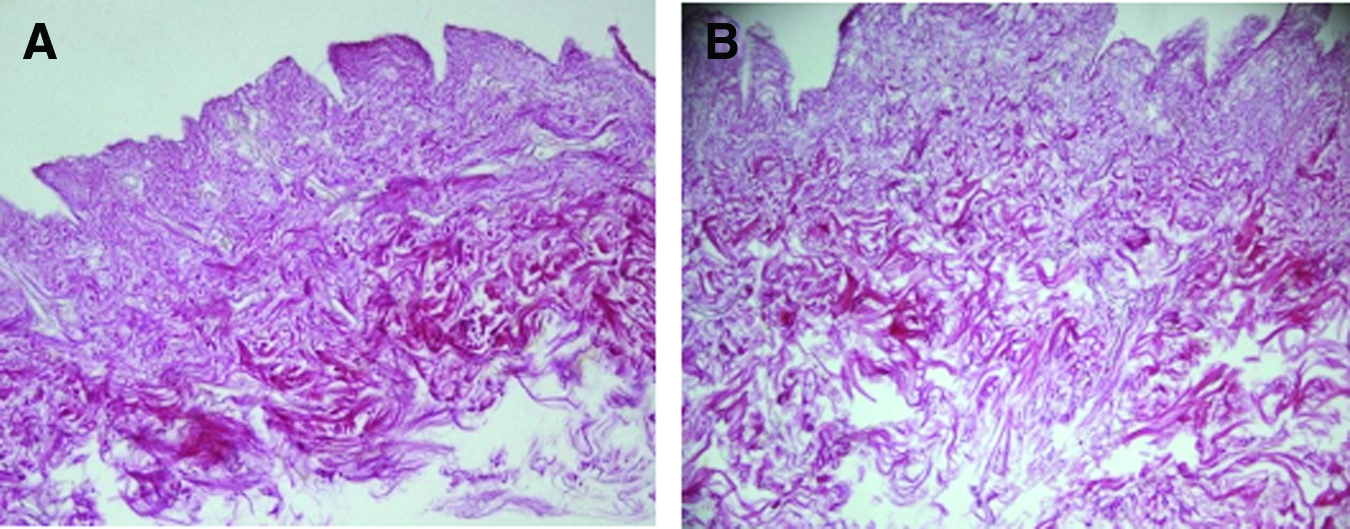
Van Giesons staining of dCELL human dermis
Red interference of nontreated dCELL dermis was 55.18% ± 0.82. There was no significant difference in the percentage of red interference of treated dermis compared to nontreated dCELL dermis. Values are means ±95% CI.
There was no significant difference in the percentage of red interference of treated dermis compared to nontreated dCELL dermis (55.18% ± 0.82). Values are means ±95% CI.
Collagen type IV immunostaining was primarily located in the basement membrane of the dermal–epidermal junction and blood vessels (Fig. 6). Immunohistochemical staining showed that collagen type IV remained intact after treatment of dCELL human dermis with 0.1 mg/L–1 g/L CuCl2 (pH 3, 4, and 5), 0.01–7.5% H2O2 (pH 3, 4, and 5), or combinations of 0.1 mg/L CuCl2 with 0.01–1% H2O2 and 1 mg/L CuCl2 with 0.01–1% H2O2 (Fig. 6). An increase in CuCl2 concentration to 10 mg/L together with 1% H2O2 caused staining for collagen type IV to be lost at the epidermal–dermal junction; however, positive staining was still seen at the basement membrane of blood vessels of these samples (Fig. 6).
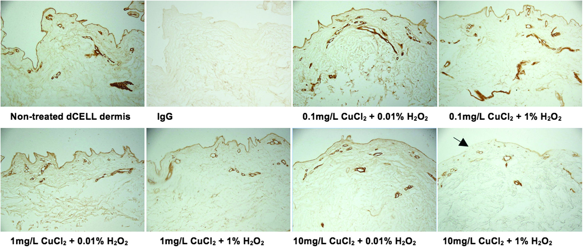
Collagen IV immunohistochemical staining of dCELL human dermis treated with CuCl2 in combination with H2O2 at the concentrations stated. Loss of collagen IV staining was evident at the dermal–epidermal junction of dCELL dermis treated with 10 mg/L CuCl2 + 1% H2O2 (black arrow). Negative control tissue was treated with IgG. Original magnification ×20. Color images available online at
Treating dCELL dermis with CuCl2 (1 mg/L–1 g/L), H2O2 (0.01–7.5%) at pH 3, 4, and 5 and 0.1 mg/L, 1 mg/L, and 10 mg/L CuCl2 in combination with 0.01–1% H2O2 did not induce a significant change in hydroxyproline release compared to nontreated dCELL dermis (Fig. 7).

Hydroxyproline release from dCELL human dermis treated with 0.1 mg/L–1 g/L CuCl2 (
Biocompatibility
The combination of 10 mg/L CuCl2 with 1% H2O2 was observed to induce some cytotoxicity in HSF cells with sparse growth around the dCELL dermis (Fig. 8). All other combinations were not cytotoxic to either HSF or MG63 cells (not shown).
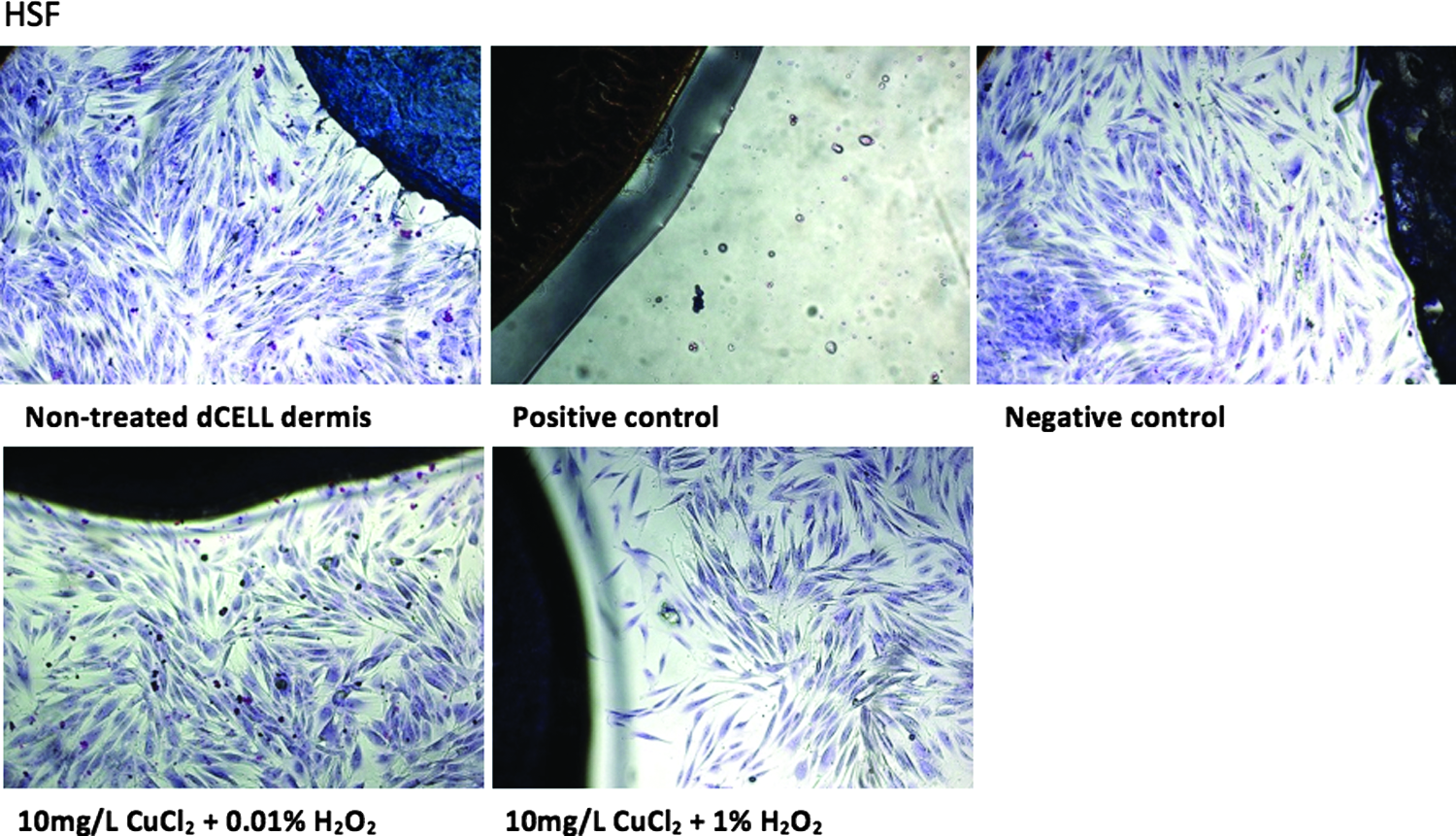
In vitro contact cytotoxicity of combined CuCl2- and H2O2 -treated dCELL human dermis with HSF, original magnification ×10. HSF grew around and in contact with dCELL human dermis treated with all concentrations and combinations of CuCl2 and H2O2 except for 10 mg/L CuCl2 with 1% H2O2 where HSF made sparse contact with the dCELL dermis. HSF grew when cultured with steristrips (negative control). There was no evidence of growth around the cyanoacrylate glue (positive control). Color images available online at
Discussion
The purpose of this study was to determine the concentrations of CuCl2 and H2O2, either singly or in combination, which did and did not elicit changes in morphology, functionality, and biocompatibility of dCELL human dermis. Concentrations of CuCl2 were chosen, which are below the toxic levels for humans–mean plasma copper concentration, in a healthy adult range between 80–100 mg/dL (800–1000 mg/L). 35 Unbound copper ions exhibit greater toxicity compared to complexed copper. 36 H2O2 lacks toxicity following its rapid decomposition to H2O and O2, making it a suitable decontamination reagent where human application is concerned. For example, 3–6% (v/v) H2O2 is commonly used as an antiseptic and a contact lens cleaning solution, dental disinfectants often contain up to 1% H2O2, and 3% H2O2 is used routinely during processing of human bone.28,29,32,37 In this study, concentrations up to 7.5% (v/v) H2O2 were found to have no detrimental effect on the structural or functional properties of dCELL human dermis. pH, temperature, and metal ions all affect the stability of H2O2. H2O2 is stable at low pH; so in this study, all solutions containing H2O2 were made between pH 3–5. Increases in temperature accelerate the decomposition of H2O2. While a rise in ambient temperature to 30°C has little effect on H2O2 stability, every further 10°C increase in temperature causes approximately a 2.2-fold increase in the rate of decomposition. 38 In this study, all treatments were performed at 25°C. Metal ions, particularly manganese, iron, and copper also have a negative effect on the stability of H2O2. The stock H2O2 used in this study contained stabilizers in the form of metal chelators. At 3% and 7.5% H2O2, these chelators may have been in sufficient quantity to chelate the copper ions (Fig. 1). In addition, any free copper ion at the higher CuCl2 concentrations would have induced breakdown of H2O2, explaining the reduction in the amount of measurable H2O2 (Fig. 2).
Histological visualization revealed that single and combined CuCl2 and H2O2 treatment did not cause any alteration to the morphological architecture of dCELL dermis. In this study, dermis was obtained from donors over the age of 50 years. The collagen fibers appeared tightly packed, uniformly aligned, and thicker, presumably mature fibers were evident in the reticular layer. Neither the single or combined treatments of CuCl2 and H2O2 had any effect on collagen structure or orientation. Hydroxyproline is a major component of collagen, having a key role in stabilizing the triple-helical collagen structure. 39 Measuring free hydroxyproline levels indicates the proportion of denatured collagen. In this study, CuCl2 and H2O2 treatments were not associated with any alteration in hydroxyproline content. Collagen type IV is a major component of the basement membrane at the dermal–epidermal junction where it maintains structural integrity and functionality of the membrane as well as having an important role in wound healing.40–42 NHSBT produces dCELL dermis for topical applications such as treating burn injuries and nonhealing ulcers and for internal injuries such as rotor cuff injuries. Reepithelialization of the decellularized graft is an essential part of the wound healing process when used topically. 17 Collagen type IV has a key role in inducing keratinocyte migration to the wound site, an early and crucial part of the reepithelialization process. 43 A loss in collagen type IV immunoreactivity at the dermal–epidermal junction of dermis was observed with the maximum combined concentration of CuCl2 (10 mg/L) and H2O2 (1%). Oxidative damage to collagen is well documented. Metal ions, notably cuprous [copper (I)] and cupric ions when mixed with H2O2 mediate site-specific damage through binding to collagen, resulting in cross-linking, decarboxylation, and fragmentation of collagen. 44 These mechanisms may have accounted for this loss of collagen type IV staining.
Biomechanical properties of skin depend on the body location. Collagen and elastin are responsible for the tensile strength and viscoelasticity of skin, respectively. Changes in the orientation of dermal collagen fibers during deformation minimize the strain of the skin. 45 In this study, tensile strength of dCELL dermis was measured by application of a constant slow increase in load (100N at 100 mm/min). Treating dCELL human dermis with CuCl2 or H2O2 individually did not compromise the ultimate tensile strength. However, 1 mg/L CuCl2 with 1% H2O2 induced a significant increase in ultimate tensile strength. Interestingly, a further increase in CuCl2 to 10 mg/L with H2O2 did not have a significant effect on the dCELL dermis ultimate tensile strength. No denaturation of collagen occurred with CuCl2 and H2O2 individual treatments, which may account for the maintenance of the ultimate tensile strength
After CuCl2 and H2O2 treatments, dCELL human dermis was washed with distilled water to remove any residual reagents. In vitro contact cytotoxicity identified that the maximum combined concentration of CuCl2 (10 mg/L) and H2O2 (1%) had slight cytotoxic effects on HSF. The cytotoxic effect of copper ions on mouse skin fibroblasts has previously been reported by Cao et al. (2012). 46
This study aimed to identify the maximum concentration range of CuCl2 and H2O2 that, when applied to dCELL human dermis would not have an adverse effect on the tissue. The data indicate that single treatment of CuCl2 in the concentration range of 0.1 mg/L–1 g/L or a single treatment of H2O2 in the concentration range of 0.01–7.5% had no effect on the morphology of the acellular scaffold. The collagen network remained intact, consistent with the hydroxyproline analysis that suggested there was no collagen denaturation associated with these treatments and the biomechanical data that showed there was no change in the tensile strength. The treated dermis was biocompatible in vitro with both primary cells and a cell line, having no effect on their morphology or proliferation. Combining both 10 mg/L CuCl2 with 1% H2O2 did cause damage to the decellularized dermal scaffold; notably, there was a loss of collagen type IV at the dermal–epidermal junction and induced cytotoxicity in primary HSF. Based on the findings presented here, application of CuCl2 up to a concentration of 1 g/L or H2O2 up to 7.5% individually or combining 10 mg/L CuCl2 with 0.5% H2O2 are the maximum doses of these chemicals that do not have an adverse effect in terms of morphology, functionality, and biocompatibility. Further work should investigate the effects of these treatments on other tissue allografts and the bactericidal effects of the combined reagents.
Footnotes
Acknowledgment
We acknowledge the support of NHSBT for the funding of this research.
Disclosure Statement
No competing financial interests exist.
References
Supplementary Material
Please find the following supplemental material available below.
For Open Access articles published under a Creative Commons License, all supplemental material carries the same license as the article it is associated with.
For non-Open Access articles published, all supplemental material carries a non-exclusive license, and permission requests for re-use of supplemental material or any part of supplemental material shall be sent directly to the copyright owner as specified in the copyright notice associated with the article.
