
Abstract
Globally millions are blind due to corneal disease, yet tissue for transplantation is a limited resource. This study characterizes the physical and biological properties of a novel collagen-based scaffold. Transparency, optical coherence tomography (OCT), and scanning electron microscopy (SEM) were used to analyze the structure of the scaffold, synthesized using rat tail collagen I. Water content was determined. The tensile strength was assessed using a micro-mechanical analyzer. In vitro biocompatibility was assessed by culturing the scaffold with epithelial or keratocyte spheres. The mean scaffold transmittance was 0.72 at 358 nm, 0.88 at 570 nm, and 0.92 at 900 nm. OCT imaging confirmed that the scaffold maintained a corneal shape, with a central thickness of 502 μm and a reflectivity profile comparable to that of a normal human cornea. SEM of the scaffold revealed multiple lamellae on cross section. The mean water content was 88.7% ± 0.7%. Ultimate tensile strength for the noncross-linked scaffold was 1.23 ± 0.27 MPa compared with 2.21 ± 0.70 MPa for the cross-linked scaffold (human corneal anterior stroma 1.53 ± 0.86 MPa) at a strain rate of 0.5%/s. Epithelial cells migrated over the scaffold to confluence. Keratocytes populated the scaffold and maintained a lamellar arrangement. The properties of this novel scaffold suggest that it has potential to be developed into a corneal tissue substitute for human transplantation.
Introduction
C
Although artificial corneas (keratoprostheses) such as the Boston KPro or AlphaCor are available for clinical use, they suffer a number of limitations. Common complications of keratoprostheses include formation of a retro-prosthetic membrane, extrusion, chronic inflammation, infection, melting at the cornea prosthesis interface, and leakage.3–7
The ideal corneal tissue substitute would be transparent and biocompatible, closely resemble the structure of the cornea, have elasticity and tensile strength similar to that of native tissue (thereby enabling surgical manipulation), have high optical transmittance, allow the diffusion of nutrients, induce minimal inflammatory response, and support cell attachment, proliferation, and differentiation.8–10
Scaffolds that have synthetic constituents often meet the biomechanical demands required but do not readily support cell attachment, proliferation, and differentiation. 11 Researchers have therefore attempted to produce tissue substitutes using biological components. Collagen is the major protein constituent of the extracellular matrix and so has been a commonly used component in tissue scaffold construction. 12
Here, we describe the development of a novel collagen scaffold that may potentially be used as a corneal substitute.
Materials and Methods
Synthesis of scaffold
Centrifugal ultrafiltration was performed by placing 10 mL of 5 mg/mL Cultrex 3D culture matrix rat collagen I (Trevigen, Gaithersburg, MD) in a Vivaspin Turbo 15 (Sartorius AG, Goettingen, Germany) with a 10 kDa molecular weight cut off polyethersulfone (PES) membrane. The solution was spun in a centrifuge unit (Sigma 3K15) at 4°C at 2500 rpm for 30 h, thereby increasing the concentration of the solution to 125 mg/mL (volume 0.4 mL). The resulting firm collagen gel was divided into eight pieces of equal weight. Each piece was placed in a separate standard centrifuge tube and spun at 4°C at 2500 rpm for 10 h in 50 mL Milli-Q water to rehydrate the collagen. Two pieces were then combined by centrifuging in a single centrifuge tube for an additional hour. The combined rehydrated gel was then removed and placed in a mould. Two types of moulds were used to produce either flat or cornea-shaped scaffolds. The flat mould consisted of a plastic ring (internal diameter of 12 mm and a height of 8 mm) placed on a glass slide with a glass cover slip placed on top of the mould. The cornea-shaped mould consisted of two rigid contact lenses spaced 8 mm apart. After the collagen gel was placed in the respective mould, 8 μL of 1 M NaOH was applied to the surface of the gel. The mould was then placed in a humid chamber at 37°C for 4 h. The plastic ring was removed, and the resulting cylindrical gel (placed on a glass slide and covered by a glass coverslip) was placed in a dry incubator at 37°C for at least 12 h to allow complete dehydration. The dehydrated gel was subsequently rehydrated by immersion in Milli-Q water for 4 h.
Cross-linking of scaffold
Ultraviolet/riboflavin
Dehydrated scaffolds were soaked in 0.1% riboflavin in phosphate buffered saline (PBS). Light irradiation was then commenced using an ultraviolet A (UVA) double diode 370 nm light source located ∼10 mm above the scaffold. This produced a radiant energy of 3 mW/cm2 or 5.4 J/cm2. Irradiation was performed for 30 min, then the sample was turned over and the irradiation repeated for a further 30 min. The sample was then flushed and soaked in 800 mL of Milli-Q water for 24 h to remove the riboflavin from the scaffold.
Genipin
Hydrated scaffolds were soaked in 1 mM Genipin in PBS for 4 h at room temperature, followed by washing in PBS and Milli-Q water, 1 h each.
EDC
Hydrated scaffolds were immersed in 33 mM 1-ethyl-3-(3-dimethylaminopropyl)carbodiimide hydrochloride (EDC, in NaH2PO4 0.05 M, pH 5.5) for 4 h at room temperature, followed by washing for 15 min in Na2HPO4 0.1 M, pH 9.1, 15 min in 1 M NaCl, and 1 h in Milli-Q water.
Transmittance
Each flat scaffold sample was cut into a 6.5 mm diameter circular button using a corneal trephine and placed in a well in a 96-well plate with 200 μL MilliQ water. The absorbance of three samples to light at 358, 420, 570, 750, and 900 nm was measured using a microplate spectrophotometer (BioTek Synergy HT Multi-Mode Microplate Reader; BioTek Instruments, Inc., Winooski, VT), with the absorbance of Milli-Q water control subtracted from each reading. The transmittance of light through the sample at a particular wavelength was calculated as 10e(−absorbance). The transmittance of the three samples before and after UV/riboflavin collagen cross-linking was compared.
Thickness and shape
En-face and cross-section images of scaffold samples were obtained using the Spectralis anterior segment optical coherence tomography (OCT; Heidelberg Engineering GmBH, Heidelberg, Germany) to assess their thickness and shape.
Water content
Three scaffolds were dried at room temperature for at least 12 h and the dry weight measured. The dehydrated samples were subsequently hydrated to equilibrium by soaking in Milli-Q water for 4 h and the wet weight measured. The following formula was used to calculate the percentage water content of the hydrated scaffolds:
Degradation in vitro
The degradation profile of scaffolds before and after UV/riboflavin cross-linking (three samples each) was assessed using a collagenase degradation assay. A 72 h Corneal shield (Oasis, San Dimas, CA) with a weight of 45 mg was also included for comparison. Samples were individually weighed and soaked in PBS with saturated CaCl2 for 3 days to allow swelling before degradation was commenced. One unit collagenase (collagenase from Clostridium histolyticum, type 1A, Sigma C9891) in 0.5 U/mL PBS with saturated CaCl2 was applied per 50 mg wet weight scaffold. The scaffolds were then weighed five times a week for 2 weeks. The results were expressed as percentage weight remaining.
Light microscopy analysis of concentrated gel
Light microscopy was performed to analyze the macrostructure of the collagen gel immediately after centrifugation. The concentrated collagen gel (final volume 0.4 mL) was removed from the vivaspin tube and dehydrated at 37°C for 24 h in a dry incubator and then viewed using light microscopy.
Scanning electron microscopy
Scanning electron microscopy (SEM) was performed to analyze the microstructure of the scaffold. A scaffold was fixed in 4% paraformaldehyde for 20 min, washed thrice for 15 min in PBS, then transferred through 15 min washes of water, 30%, 50%, 70%, 90%, and 100% ethanol. The sample was cut in half with a tissue chopper (Starrett 263M) for cross-sectional and en-face views. The samples were critical point dried and platinum coated 1 h before images were taken with a scanning electron microscope (Philips FEI XL30 S-FEG).
Tensile strength
Six scaffold samples were bisected and one half of each sample was cross-linked using UV/riboflavin (n = 2), Genipin (n = 2), or EDC (n = 2) to investigate whether one method was superior to the others in terms of enhancement of biomechanical properties. The remaining half of each sample remained noncross-linked (n = 6).
Tensile strength of the scaffolds and for comparison, a 76-year-old healthy human donor cornea, was assessed using a micro-mechanical analyzer similar to that described by Taberner et al. 13 First, dried flat scaffold samples were cut into rectangular strips (∼100 μm × 300 μm × 3 mm). Each strip was hydrated in Milli-Q water for a period of no less than 3 h before being transferred into the mechanical analyzer. For the human donor cornea, epithelium, Descemet's membrane, and endothelium were removed by gentle scraping, the cornea was divided into anterior and posterior portions using a lamellar dissector then cut into rectangular strips (∼100 μm × 300 μm × 3 mm). Two anterior and two posterior strips were tested. The corneal sections were kept in organ culture medium before being transferred to a measurement chamber, at which time they were hydrated in Milli-Q water. The individual specimen strips were attached end on between two glass capillaries using cyanoacrylate. At one end the capillary was attached in turn to a linear motor. At the other, it was attached to a cantilever force transducer that was carried on a second linear motor. Distilled water was continuously passed through the measurement chamber superfusing the sample to maintain hydration.
To quantify force, the deflection of the stainless steel cantilever was measured using a heterodyne laser interferometer with a resolution of 3.1 × 10⁻¹⁰ m and a measurement bandwidth of 20 MHz, down sampled to 500 kHz. The apparent stiffness of the cantilever, for the laser configuration used in these experiments, was calibrated at 1400 N/m using a strain-gauge force transducer (FUTEK LSB200). The first resonant frequency of the unloaded cantilever and collagen mounting system was measured at ∼1120 Hz and the noise equivalent force, as quantified by the standard deviation (SD) of force noise over a measurement bandwidth of 1 kHz, was 2.23 μN. The heterodyne interferometer was also used to measure the displacement of the two linear actuators attached to either end of the sample, one of which carried the force transducer. Sample cross section was determined using an OCT integrated into the mechanical tester. 14 Young's modulus, and tensile strength, was computed by dividing the force by the mean cross-sectional area. A series of cyclic loading experiments were performed for each sample, first by varying the target strain between 0.1% and 2% and maintaining a fixed strain rate of 0.01%/s. Subsequently, target strain was held constant at 1% and strain rate was varied between 0.01%/s and 0.1%/s. A full-volume scan was acquired using the OCT during the load and unload step of the cyclic loading pattern at a combination of 1% target strain and 0.1%/s strain rate. Finally the samples were strained to failure at a rate of 0.5%/s.
Cellularization of the scaffold
To investigate whether the scaffold supports corneal keratocyte growth, porcine keratocyte spheres were cultured and seeded onto scaffolds.
Whole porcine eyes were sterilized by 4% povidine iodine and 4% sodium thiosulfate. Corneal epithelial cells were removed by scraping using a keratome before and after immersion for 40 min at 37°C in 1.2 U/mL of dispase (Gibco; Life Technologies, Carlsbad, CA) in minimum essential medium (MEM; Gibco; Life Technologies). The de-epithelialized cornea was incubated at 37°C for 20–24 h in 1 mL of MEM (Gibco; Life Technologies) containing 2 mg/mL Collagenase (Sigma-Aldrich, St Louis, MO) and 0.5 mg/mL Hyaluronidase (Sigma-Aldrich). A single cell suspension was obtained and cultured in Advanced Dulbecco's modified Eagle medium (DMEM; Gibco) supplemented with 1% GlutaMAX (Gibco), 1% antibiotic/antimycotic (Gibco), 2 ng/mL recombinant human EGF (Invitrogen, Waltham, MA), and 1 ng/mL recombinant human bFGF (Invitrogen). Cells were cultured on a 22 mm round glass coverslip in a well of a six-well cell culture tray. Keratocytes cultured in this way were known to aggregate and form spheres. 15
After 10 days in culture, keratocyte spheres were pipetted onto each noncross-linked scaffold in individual wells of a six-well cell culture tray. Scaffolds were incubated overnight to allow the spheres to settle down onto the scaffold surfaces. Remaining medium was removed and replaced with Advanced DMEM (Gibco) supplemented with 1% GlutaMAX (Gibco), 1% antibiotic/antimycotic (Gibco), and 25 ng/mL recombinant human IGF-II (Gibco). Medium was replaced twice weekly and keratocytes on scaffolds monitored using a Leica DMIL IL LED inverted microscope (Leica Microsystems, Wetzlar, Germany). Digital images were acquired with a Leica MC120 HD microscope camera (Leica Microsystems), controlled by a PC computer running Leica Application Suite version 4.4.0 (Leica Microsystems).
After 42 days, scaffolds were fixed in 4% paraformaldehyde in PBS for 19 h at 4°C, washed in PBS, and cryo-protected by 20% sucrose for 4 h at room temperature and 30% sucrose for 24 h at 4°C. Scaffolds were embedded in Tissue-Tek Compound (Sakura, Tokyo, Japan), frozen in liquid nitrogen, and stored at −20°C until cryosectioning. Fixed frozen scaffolds were cut into 16 μm thick sections with a Microm HM550 cryostat (Thermo Fisher Scientific, Bremen, Germany) and stored on glass slides at −20°C before immunohistochemical labeling. Slides were labeled with rabbit anti-keratocan primary antibody (sc-66941, 1:100; Santa Cruz, Dallas, TX), as a marker for keratocytes, and mouse anti-alpha smooth muscle actin (α-SMA) primary antibody (Novocastra, NCL-SMA, 1:50), as a marker for myofibroblasts, followed by goat anti-rabbit Alexa568 secondary antibody (A11011, 1:400; Life Technologies,) and goat anti-mouse Alexa488 secondary antibody (A10680, 1:400; Life Technologies). Slides were mounted with ProLong-Gold Antifade Mountant with DAPI (P1014; Life Technologies). Images were taken with an Olympus FV1000 confocal laser scanning microscope (Olympus, Tokoyo, Japan).
To investigate whether the scaffold supports corneal epithelial cell growth, a corneal limbal rim from a 74-year-old human donor with prior research consent was obtained from the New Zealand eye bank postsurgery. A 3 mm × 3 mm × 200 μm piece of limbus was dissected and placed epithelial-side down onto the flat scaffold. The scaffold and the epithelial explant were cultured in advanced DMEM supplemented with 1% Glutamax and 1% antibiotic/antimycotic, with medium changed twice a week. After 28 days, light microscope images were taken. The scaffold was fixed and cryosectioned as described. Slides were incubated with mouse anti-cytokeratin antibody (DAKO M3515, 1:50) for epithelial cells followed by goat anti-mouse Alexa568 antibody (A-11004, 1:400; Life Technologies). Slides were mounted with ProLong-Gold Antifade Mountant with DAPI. Images were taken with an Olympus FV1000 confocal laser scanning microscope.
Statistical analysis
Transparency and water content before and after cross-linking were compared using paired Student's t-tests. Degradation of noncross-linked scaffolds compared to cross-linked scaffolds were compared using unpaired Student's t-tests. A p-value of <0.05 was considered statistically significant. Tensile strength data were subject to histogram analysis for normality, and the data were found to be not normally distributed, therefore a log transform was applied. The transformed data was examined for the effect of cross-linking, strain-rate, and strain on apparent stiffness using analysis of variance, and the effect of cross-linking between at least one pair of cross-linking treatments was found to be highly significant (p < 0.0001). To determine the relative effects of cross-linking, pairwise t-tests were performed between all cross-linking groups while controlling for the effects of the strain and strain-rate, the subsequent results are presented as ratios of mean tensile strength in Table 2.
Results
Transparency
The scaffold was transparent and uniform upon diffuse illumination and illumination with a slit-beam (Fig. 1). The scaffolds consistently showed a high degree of transparency at visible wavelengths. The mean scaffold transmittance was 0.72 ± 0.04 (mean ± SD, average of three scaffolds) at 358 nm, 0.81 ± 0.03 at 420 nm, 0.88 ± 0.02 at 570 nm, 0.91 ± 0.01 at 750 nm, and 0.92 ± 0.01 at 900 nm (Fig. 2). Cross-linking resulted in decreased transmittance of ultraviolet light (0.55 ± 0.06 at 358 nm, p = 0.03), and 0.78 ± 0.04 at 420 nm, p > 0.05), and greater transmittance of visible light (0.92 ± 0.02 at 570 nm, p = 0.04, 0.95 ± 0.01 at 750 nm, p = 0.01, and 0.95 ± 0.01 at 900 nm, p = 0.01). There was no significant difference in scaffold transmittance after 7 days of incubation in phenol red-free culture medium (DMEM/F12, Gibco) at 37°C.
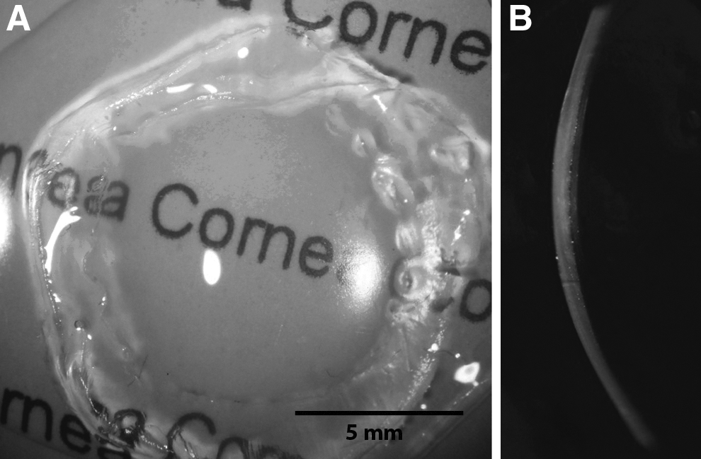
The scaffold has a high degree of transparency and uniformity of curvature on diffuse illumination
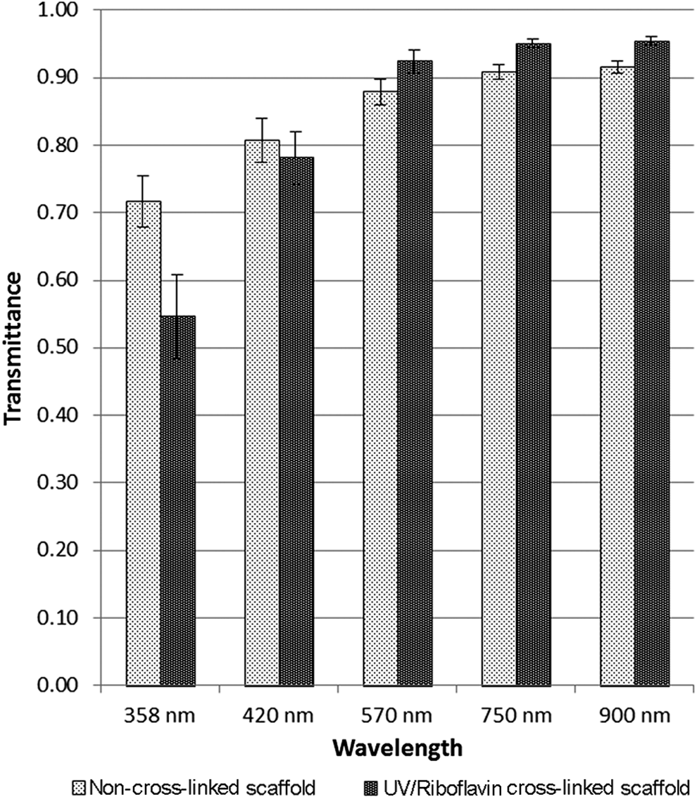
The mean ± standard deviation (SD) transparency of three samples before and after ultraviolet (UV)/riboflavin cross-linking. Collagen cross-linking resulted in decreased mean transmittance of UV light and increased transmittance of visible light.
Thickness and shape
OCT imaging confirmed that the scaffold maintained a corneal shape with a central thickness of 502 μm and reflectivity profile comparable to that of a normal human cornea (Fig. 3).

Anterior segment optical coherence tomography images of a normal human cornea (upper) and the scaffold (lower) show similar reflectivity profiles and thickness with maintenance of scaffold shape when removed from mould. Color images available online at
Water content
The mean water content of the scaffold was 88.7% ± 0.7% (mean ± SD, average of three scaffolds), and was 83.2% ± 2.8% (p = 0.07) after collagen cross-linking.
Degradation in vitro
Fifty percent of the original weight of noncross-linked scaffolds (n = 3) remained after 5 days compared to complete degradation of Oasis 72 h corneal collagen shields after 3 days (Fig. 4). Collagen cross-linking significantly improved the stability of the scaffold, with 72% of the original weight of cross-linked scaffolds (n = 3) remaining after 14 days. The difference between the cross-linked and noncross-linked scaffolds was statistically significant at all time points (p < 0.01).
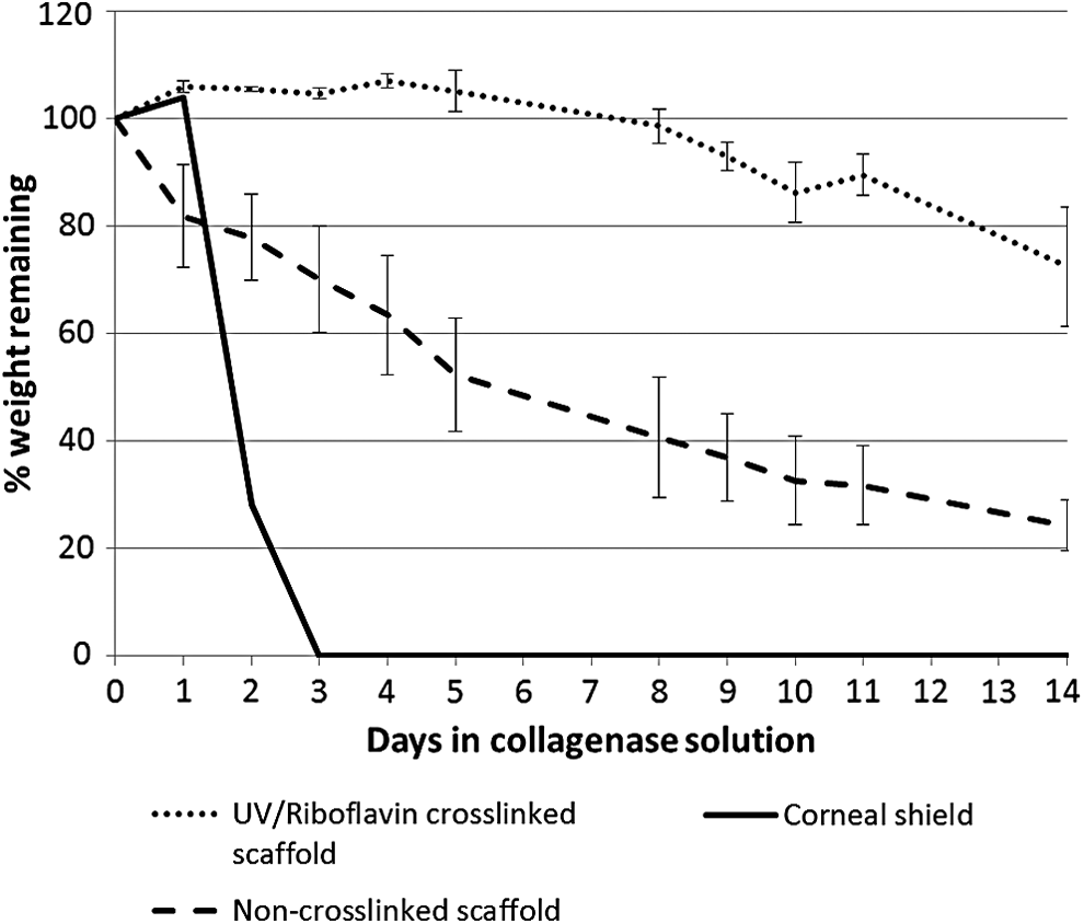
Degradation profile of noncross-linked scaffolds, UV/riboflavin cross-linked scaffolds, and a corneal collagen shield. Values shown are mean ± SD of three UV/riboflavin cross-linked scaffolds, three noncross-linked scaffolds, and one corneal shield.
Light microscopy analysis of concentrated gel
Light microscopy demonstrated that the collagen concentrated by centrifugation was arranged in multiple lamellae, seen at the cutting edge (Fig. 5, arrows); a high degree of alignment was observed in a collagen piece concentrated by centrifugation then dehydrated (Fig. 5).
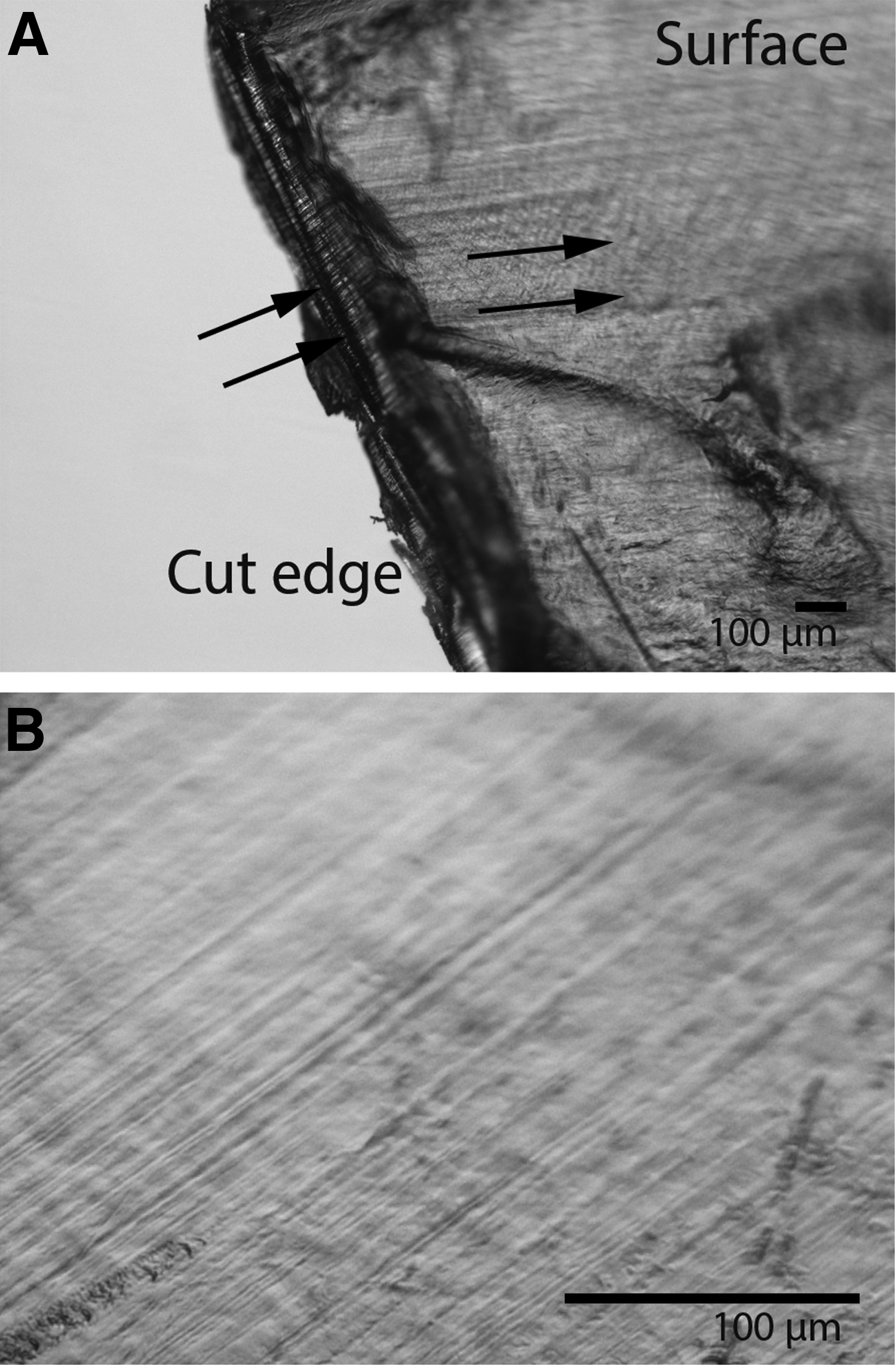
Light microscope images of concentrated collagen.
Scanning electron microscopy
SEM of the scaffold showed that the surface of the scaffolds was amorphous and that the scaffold was composed of multiple lamellae on cross section (Fig. 6).
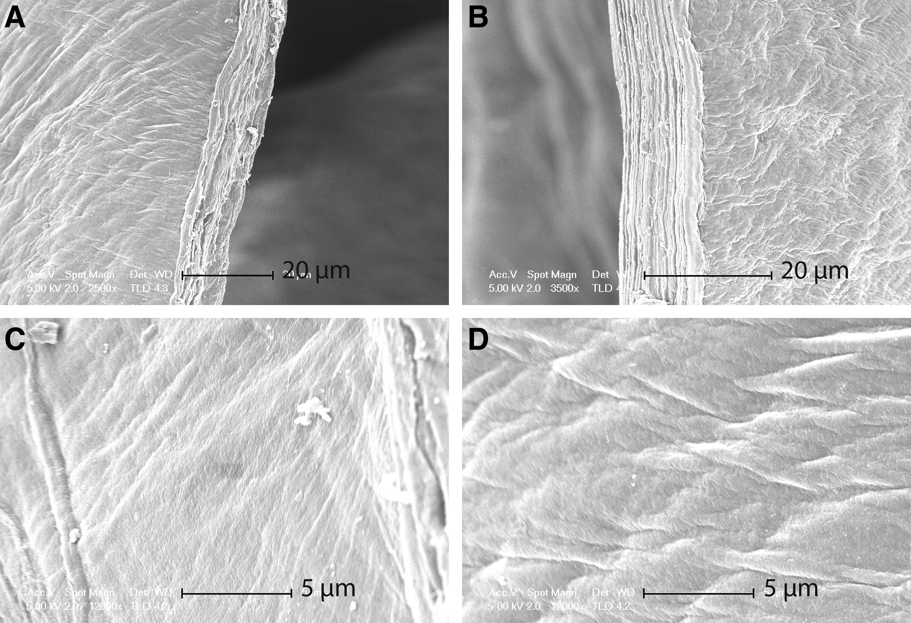
Scanning electron microscopy showing
Tensile strength
The mean Young's modulus and ultimate tensile strength of the corneal stroma (two anterior and two posterior strips) and noncross-linked (n = 2) and cross-linked scaffolds (n = 2 each, UV/riboflavin, Genipin, and EDC) are shown in Table 1. Statistical significance between pairs was tested after log-transformation of tensile strength data; the ratios of means of tensile strength are shown in Table 2. (Ratios are presented as columns over rows, e.g., the log-transformed mean tensile strength of UV/riboflavin cross-linked gel/noncross-linked gel equals 1.824, and the differences between UV/riboflavin cross-linked and noncross-linked gels were statistically significant at **p < 0.01.) The ultimate tensile strength of noncross-linked scaffolds (average 1.23 MPa) falls between the values of the anterior (1.53 MPa) and posterior (0.55 MPa) stroma. Cross-linking using UV/riboflavin (2.21 MPa), Genipin (1.96 MPa), and EDC (5.38 MPa) increased the ultimate tensile strength of scaffolds (all statistically significant compared to noncross-linked scaffolds). UV/riboflavin cross-linking produced an ultimate tensile strength that was significantly different from EDC, but not Genipin.
EDC, 1-ethyl-3-(3-dimethylaminopropyl)carbodiimide; SD, standard deviation; UVA, ultraviolet A.
p < 0.01; bp < 0.0001.
Epithelial and keratocyte cell growth on the scaffold
The scaffold supported epithelial cell growth on the surface and keratocyte growth both on the surface and within the scaffold itself (Fig. 7). Light microscopy showed that keratocytes from porcine keratocyte spheres covered the surface and infiltrated the interior of the scaffold from 17 days in culture. Confocal microscopy of cross sections of the scaffold after 42 days of culture showed that the cells covering the surface of the scaffold were fibroblastic and immunopositive for α-SMA, while the cells within the scaffold had retained the keratocyte phenotype, being immunopositive for keratocan. Epithelial cells from a human donor limbal rim migrated and fully covered the surface of a scaffold in multiple layers after 28 days of culture. Immunohistochemistry showed that the cells were immunopositive for cytokeratin, a marker for epithelial cells.
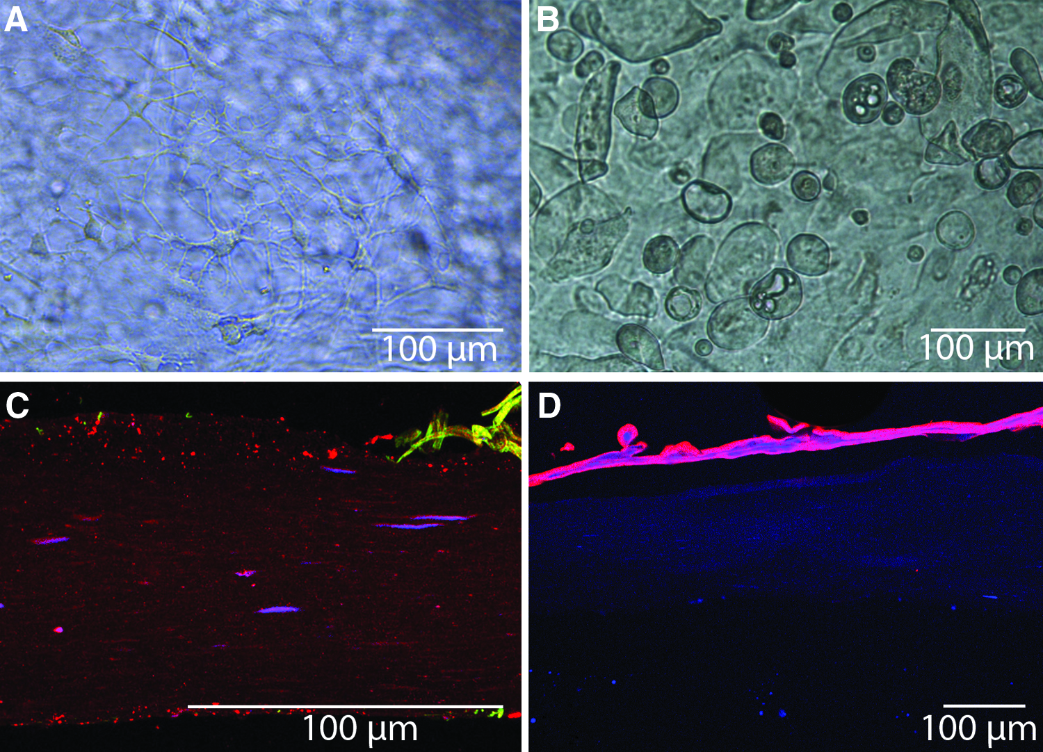
Light and confocal microscopy images showing porcine keratocyte infiltration into the scaffold
Discussion
Type I collagen is the most dominant collagen type in the cornea. 16 We have shown that a novel scaffold composed purely of type I collagen has high water content, high clarity, relatively slow degradation that can be prolonged by UV cross-linking, strong mechanical strength, and supports epithelial cell and keratocyte growth. The transmittance of the noncross-linked scaffold in the UV range (0.72 at 358 nm) is comparable to that of the human cornea, as reported in previous studies (0.70 at 358 nm at age younger than 45, 17 0.54 at age older than 45, 17 and 0.72 for all ages 18 ). The transmittance in the visible light range, for example, 0.88 at 570 nm and 0.92 at 900 nm, is slightly less than that of the human cornea (0.91–0.92 at 570 nm and 0.96 at 900 nm at all ages17,18). Cross-linking of the scaffold decreased transmittance of UV light from 0.72 to 0.55 at 358 nm, akin to the effect of aging, 17 and increased transmittance in the visible light range from 0.88 to 0.92 at 570 nm, and from 0.92 to 0.95 at 900 nm, comparable with the human cornea. This is also consistent with the observation of increased transparency of porcine corneas after UV/riboflavin cross-linking. 19
We were able to create a cornea shaped scaffold with a thickness (502 μm) comparable to that of the human cornea. 20 Bovine corneal stroma has a water content of 75–85%21,22 and human corneas have a water content of ∼80%. 23 The water content of the scaffold (88.7% before cross-linking, and 83.2% after cross-linking) was slightly higher than that of the cornea, possibly related to the lack of proteoglycans and endothelial cell pump regulation of hydration.
During the degradation assay, a small increase in wet weight (% weight remaining) was initially seen in the cross-linked scaffolds and the corneal shield (Fig. 4). We attribute this to continued osmotic swelling, despite presoaking the samples in a control solution for 3 days before the degradation assay. Degradation of the noncross-linked scaffold was slow compared with the 72 h corneal collagen shield. The corneal shield was fully degraded after 3 days, whereas more than 60% of the noncross-linked scaffold remained and 100% of the cross-linked scaffold remained at this time point. Collagen cross-linking has previously been shown to increase resistance to collagenase degradation. 24 Therefore, the scaffold was relatively stable and can be further stabilized by collagen cross-linking.
Light microscopy, scanning and transmission electron microscopy showed that the scaffold was composed of layers of collagen, consistent with the lamellar organization of collagen in the corneal stroma. 25 Therefore, this substrate has the potential to be repopulated by keratocytes in a layered organization as in native cornea.
Noncross-linked scaffolds have a mechanical strength in between the posterior and anterior strom; collagen cross-linking significantly improved the mechanical strength of the scaffolds beyond that of the anterior stroma. This was not surprising as UV/riboflavin cross-linking of human corneas has been shown to increase the biomechanical strength of the cornea by 300%. 26 Although the sample sizes in this study were small, the magnitude of the effect of cross-linking was large. Future studies with larger sample sizes will further elucidate the significance of these differences. The scaffold supported reepithelialization and keratocyte cellularization, demonstrating superior biological activity compared with artificial materials used in artificial corneas, which typically do not support cellularization. Future development of the scaffold will include testing in animal models of corneal transplantation to assess in vivo toxicity, degradation, immunogenicity, bio-integration, clarity over time, resistance/susceptibility to infections, and immunologic processes with the ultimate goal being development of a corneal substitute.
Footnotes
Acknowledgments
This study was funded by the Health Research Council of New Zealand. Tensile tests were conducted with support from the Marsden Fund, from government funding, administered by the Royal Society of New Zealand. The funding sources had no involvement in the collection, analysis, and interpretation of data, in the writing of the report, or in the decision to submit the article for publication.
Disclosure Statement
No competing financial interests exist.