
Abstract
Adipose-derived stem cells (ASCs) are typically expanded to acquire large numbers of cells for therapeutic applications. Diverse stimuli such as sphingosylphosphocholine and vitamin C have been used to increase the production yield and regenerative potential of ASCs. In the present study, we hypothesized that ZnO nanorods have promising potential for the enhancement of ASC proliferation. ZnO nanorods were prepared using three different methods: grinding and boiling at low temperature with and without surfactant. The physicochemical properties of the nanorods such as their crystallinity, morphology, size, and solvent compatibility were evaluated, and then, the ability of the synthesized ZnO nanorods to enhance ASC proliferation was investigated. Scanning electron microscopy images of all of the ZnO powders showed rod-shaped nanoflakes with lengths of 200–500 nm. Notably, although ZnO-G produced by the grinding method was well dispersed in ethanol, atomic force microscopy images of dispersions of both ZnO-B from boiling methods and ZnO-G indicated the presence of clusters of ZnO nanorods. In contrast, ZnO-B was freely dispersible in 5% dextrose of water and dimethyl sulfoxide, whereas ZnO-G and ZnO-M, produced by boiling with ethanolamine, were not. All three types of ZnO nanorods increased the proliferation of ASCs in a dose-dependent manner. These results collectively suggest that ZnO nanorods have promising potential for use as an agent for the enhancement of ASC proliferation.
Introduction
Z
Although ZnO nanorods are considered as safe materials, their potential toxicity has been investigated.6,7 One possible mechanism by which ZnO nanorods may exhibit toxic effects is via oxidative stress caused by reactive oxygen species (ROS) produced from metastable nanoparticles.8–10 Free electrons and holes formed from ZnO nanorods after light absorption as a photocatalyst may also lead to the production of free hydroxyl radicals, which are strong oxidants. At the cellular level, the structure and surface chemistry of ZnO nanorods are critical factors for controlling toxicological responses.3,11–13 Current methods for the synthesis of ZnO nanorods result in the formation of clusters and macroscopic agglomerates. 14 For example, when commercial ZnO nanoparticles of various sizes (10, 30, 60, and 200 nm) were dispersed in water, significant clusters and nondispersed aggregated particles were observed in addition to dispersed ZnO nanoparticles.14,15 Therefore, the physicochemical properties of ZnO nanorods should be modified to improve their dispersibility.
Adipose-derived stem cells (ASCs) exist in adipose tissue and can be isolated from lipoaspirates during elective surgical procedures. ASCs can be obtained in large quantities using a simple isolation procedure and have the potential for utilization in tissue repair and regeneration.16–18 For example, we demonstrated that ASCs exhibit wound-healing, anti-wrinkle, and hair-regenerative potential through building-block functions and paracrine effects.19–22 ASCs are typically expanded to acquire large numbers of cells for therapeutic applications. Pharmacological stimuli, such as sphingosylphosphocholine, lysophosphatidic acid, and vitamin C, have been used to increase the production yield and regenerative potential of ASCs.23–25 However, stimulation of ASC proliferation by ZnO nanorods has not yet been demonstrated.
In the present study, we hypothesized that ZnO nanorods have promising potential for the enhancement of ASC proliferation. ZnO nanorods were prepared using three different methods: grinding and boiling at low temperature with and without surfactant. The physicochemical properties of the ZnO nanorods, including their crystallinity, morphology, size, and solvent compatibility, were evaluated, and then, the ability of the synthesized ZnO nanorods to enhance ASC proliferation was investigated.
Materials and Methods
Chemicals
ZnO and zinc acetate were purchased from Duksan Pharmaceutical Co. Ltd. Absolute ethanol (99.9%) was obtained from OCI company Ltd. (-)-Epigallocatechin gallate (EGCG) was obtained from Sigma-Aldrich. Dulbecco's modified Eagle's medium (DMEM) and minimum essential medium-alpha (MEM-α) were purchased from Hyclone (Thermo Scientific). Fetal bovine serum (FBS) and antibiotics were obtained from GIBCO (Invitrogen). Antibodies recognizing ERK (1:1000) and phosphor-ERK (1:1000) were purchased from Cell Signaling Technology, while α-Tubulin (1:10,000) was purchased from Santa Cruz Biotechnology. Horseradish peroxidase (HRP)-conjugated secondary mouse antibody (1:10,000) and HRP-conjugated secondary rabbit antibody (1:10,000) were purchased from Cell Signaling Technology. All chemicals used were of reagent grade. Deionized water (DW) was obtained using a Milli-Q water purification system (Millipore).
Synthesis of ZnO nanorods
ZnO nanorods were synthesized using three different methods: grinding and boiling with and without surfactant (Fig. 1). Ground ZnO nanorods (ZnO-G) were prepared by grinding ZnO using a mortar and pestle for 10 min.
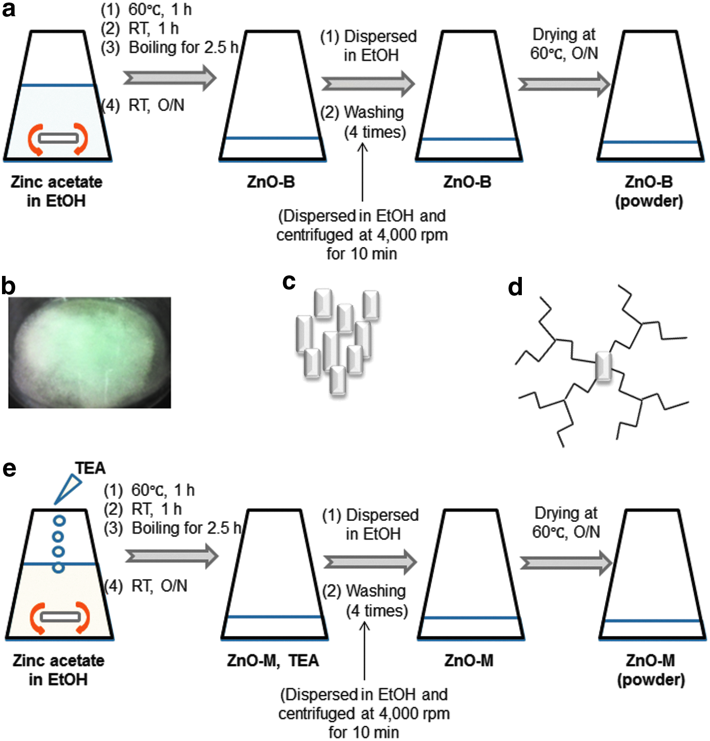
Schematic diagram of the synthetic methods used to prepare ZnO nanorods, along with their particle morphologies:
Boiled ZnO nanorods without surfactant (ZnO-B) were synthesized by boiling zinc acetate in absolute ethanol (Fig. 1a). Briefly, ground zinc acetate (350 mg) in ethanol (80 mL) was stirred at 60°C for 1 h to afford a clear, homogenous solution that was then incubated at room temperature for an additional hour. After incubation, the solution was boiled for 2.5 h and then cooled to room temperature. At this point, the solution comprised a white precipitate dispersed in ethanol (Fig. 1b). The precipitate was separated via centrifugation at 4,000 rpm for 10 min, washed with ethanol three times, and then dried in an oven at 60°C overnight (Fig. 1c).
ZnO nanorods prepared in the presence of a surfactant (ZnO-M) were micellar ZnO nanorods (Fig. 1d) and were synthesized using triethanolamine (TEA) following the literature procedure with a slight modification (Fig. 1e). 26 TEA served as a polymerization agent and controlled the micellar structure of the ZnO nanorods. Briefly, a zinc acetate solution was prepared by dissolving ground zinc acetate (350 mg) in ethanol (80 mL). This solution was stirred at 60°C, and then, TEA was added all at once at a 1:1 TEA/Zn molar ratio. The resultant mixture was stirred at 60°C for 1 h to afford a clear, homogenous solution, which was incubated at room temperature for an additional hour. The solution was then boiled for 2.5 h and subsequently cooled to room temperature. The white precipitates formed in the ethanol solution were separated via centrifugation (4,000 rpm for 10 min) at room temperature. The washing steps were repeated three times to remove any free polymeric TEA. The white precipitates were then dried in an oven at 60°C overnight. Three batches of ZnO nanorods were prepared and characterized.
Characterization of the ZnO nanorods
Powder X-ray crystallography
The crystallinity of the ZnO nanorods was analyzed via powder X-ray diffractometry (PXRD). The XRD patterns of the powdered ZnO nanorod samples were recorded from 20 to 80 2θ/θ using a high-resolution X-ray diffractometer (HR XRD, SmartLAB, Rigaku) with CuKα radiation.
Scanning electron microscopy
The size distributions and morphologies of the ZnO nanorods were analyzed using scanning electron microscopy (SEM, MIRA-3-FEG-SEM, Tescan).
Particle size determination
The particle sizes of the ZnO nanorods were analyzed using a light scattering spectrophotometer (ELS-Z; OTSUKA Electronics) with a standard cell. For the measurements, the ZnO nanorods were diluted with filtered 5% dextrose (Dex) before analysis to obtain the appropriate intensity. The mean diameters (nm) of the ZnO nanorods were determined at room temperature.
Solvent screening assay
To determine the dispersibility of the ZnO nanorods, various solvents were screened: 5% Dex, 0.9% NaCl, phosphate-buffered saline (PBS), dimethyl sulfoxide (DMSO), and DW. The concentrations of each of the ZnO nanorod solutions were fixed at 1 mg/mL. The colloidal behavior of the ZnO nanorods was evaluated by taking photographs immediately after each colloidal solution was prepared and after incubating each solution for 7 days at room temperature in a dark room.
Ultraviolet–visible spectroscopy
To confirm the optical absorption properties of the ZnO nanorods, ultraviolet–visible (UV-Vis) spectroscopy was performed. ZnO nanorods were dispersed in 5% Dex at 1 mg/mL, diluted with 5% Dex to 0.03125 or 0.0625 mg/mL, and then placed into standard cuvette cells, and the UV–Vis absorption spectra were recorded. Samples were scanned at room temperature in the wavelength range from 200 to 800 nm. A Varian UV-Vis spectrophotometer (Cary 100 Conc, Varian) was used with Cary scan applications.
Cell culture
Human subcutaneous adipose tissue samples were acquired following elective liposuction of healthy females, and informed consent was received and approved by the institutional review board. The obtained samples were digested with 0.075% collagenase type II (Sigma) under gentle agitation for 45 min at 37°C and then centrifuged at 300 g for 10 min to obtain the stromal cell fractions. Each pellet was filtered through a 70-μm nylon mesh filter and resuspended in PBS. Each cell suspension was then layered onto histopaque-1077 (Sigma) and centrifuged at 840 g for 10 min. The supernatant was discarded, and the cell band that was buoyant over the histopaque was collected. Each retrieved cell fraction was cultured overnight at 37°C under 5% CO2 in a control medium (DMEM with 10% FBS and 100 U/mL penicillin). The ASCs were characterized via transdifferentiation and analysis of cell surface markers using flow cytometry. 18 The ASCs were then grown in MEM-α with 10% FBS, 1% penicillin, and streptomycin at 37°C in humidified air containing 5% CO2 because MEM-α enhanced the growth rate of ASCs comparing with DMEM. 27 ASCs from one donor were used in the following experiments.
Cell proliferation assay
ASCs were seeded in a 48-well plate at a density of 5 × 10³ cells/well. After 24 h, the medium was replaced with a medium containing 0.2% FBS to starvation. The following day, cells were treated with ZnO nanorods (1.56, 3.1, 6.25, 12.5, 25, 50, and 100 μg/mL) for 48 h. The medium was then removed, and the cell numbers were determined using a CCK-8 assay kit (Dojindo). The cells were treated with 10% CCK-8 solution in the medium for 4 h, and then, the absorbance was determined at 450 nm using a microplate reader (TECAN, Gordig).
Western Blotting
The ASC total proteins were isolated using a sodium dodecyl sulfate (SDS) lysis buffer containing protease inhibitors. The lysates were then separated using 10% SDS-polyacrylamide gel electrophoresis (PAGE) and transferred to a polyvinylidene (PVDF) membrane (Millipore), which was subsequently blocked using 5% nonfat milk for 1 h at room temperature. After blocking, the membrane was washed with TBS-T (0.1% Tween-20 in Tris-buffered saline) and then incubated with the primary antibody overnight at 4°C. On the following day, the membrane was incubated with an HRP-conjugated secondary antibody for 1 h at room temperature in ECL solution (Millipore) and then exposed.
Statistical analysis
All results are expressed as mean ± SD. Data are representative of triplicate independent experiments. The statistical significance of the differences among the groups was tested using the Student's t-test and analysis of variance (ANOVA), with p < 0.05 considered to be significant.
Results
XRD analysis
The XRD patterns of all of the obtained ZnO nanorod powders are shown in Figure 2. The diffraction peaks for ZnO-G are well indexed to the wurtzite structure. The diffraction patterns for ZnO-G, ZnO-B, and ZnO-M all exhibited ZnO-crystal peaks in the 2θ/θ scanning range from 30 to 40, although the peak intensities were not as high in the spectra for ZnO-B and ZnO-M because these samples were not subjected to a calcination step at high temperatures. In particular, the diffractogram for ZnO-B showed two peaks in the 2θ/θ scanning range from 20 to 30, suggesting complexation of ZnO nanorods with zinc acetate besides washing with absolute ethanol four times.

XRD patterns of the ZnO nanorods obtained using the different synthesis methods.
Microscopic analysis
The morphologies of the ZnO nanorods were analyzed using SEM and AFM. SEM images of each of the ZnO powders revealed rod-shaped nanoflakes with a size distribution of 200–500 nm in length (Fig. 3). In contrast, AFM images of each of the dried ZnO nanorod ethanol dispersions showed different morphologies. The ZnO-G nanorods were well dispersed in ethanol, whereas the ZnO-B and ZnO-G nanorods formed clusters, suggesting different physicochemical properties (Supplementary Fig. S1; Supplementary Data are available online at
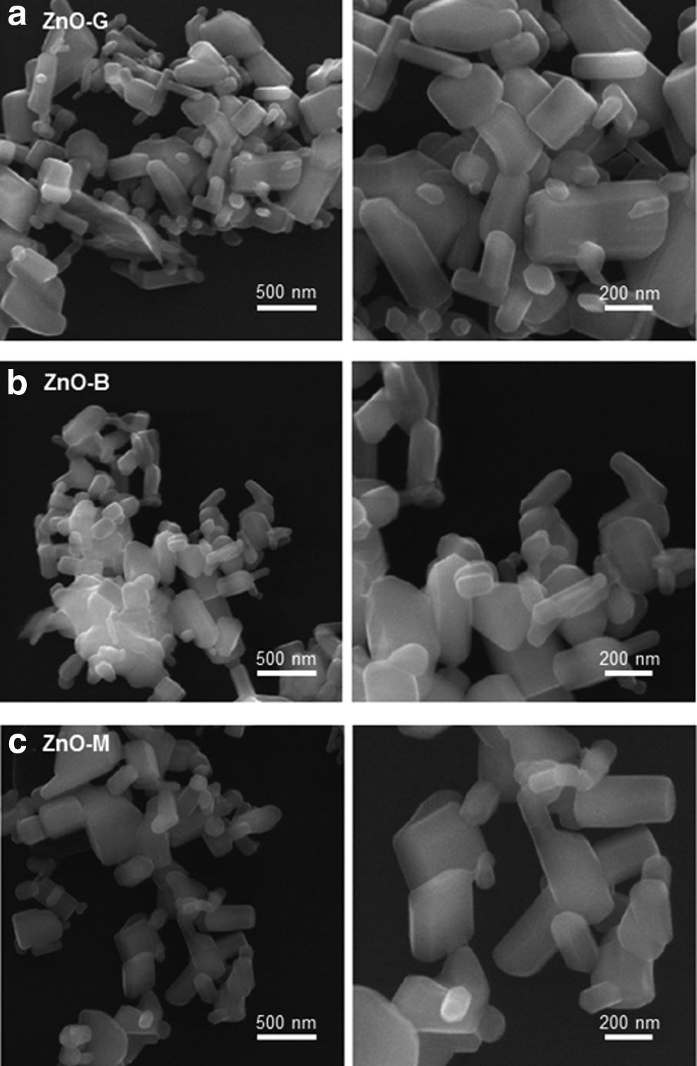
SEM images of ZnO nanorods:
Size analysis of the ZnO nanorods
The sizes and particle size distributions of the ZnO nanorods were also determined in 5% Dex (Fig. 4). All of the ZnO nanorod samples contained nanorods with lengths of 200–500 nm, which agreed well with the SEM results. Interestingly, in Dex, the size distributions for the ZnO-B and ZnO-M nanorods were narrow Gaussian distributions without any large aggregates.
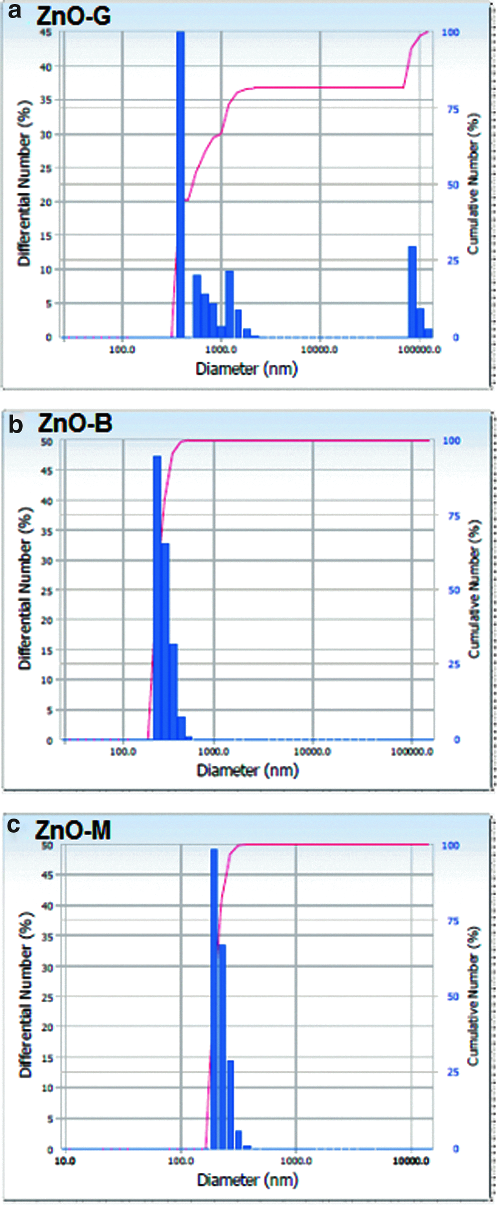
Particle size distributions of the ZnO nanorods in 5% Dex:
Solvent compatibility
The dispersibility of ZnO nanorods was evaluated in aqueous solutions: 5% Dex, 0.9% NaCl, PBS, DMSO, and DW (Fig. 5). Unlike the ZnO-G and ZnO-M nanorods, the ZnO-B nanorods were freely dispersible in 5% Dex and DMSO (Fig. 5a). In addition, this dispersibility of the ZnO-B nanorods was maintained for 7 days (Fig. 5b). In contrast, all of the ZnO nanorods, including ZnO-B, gently precipitated when dispersed in PBS because of the presence of various electrolytes in the PBS solution.
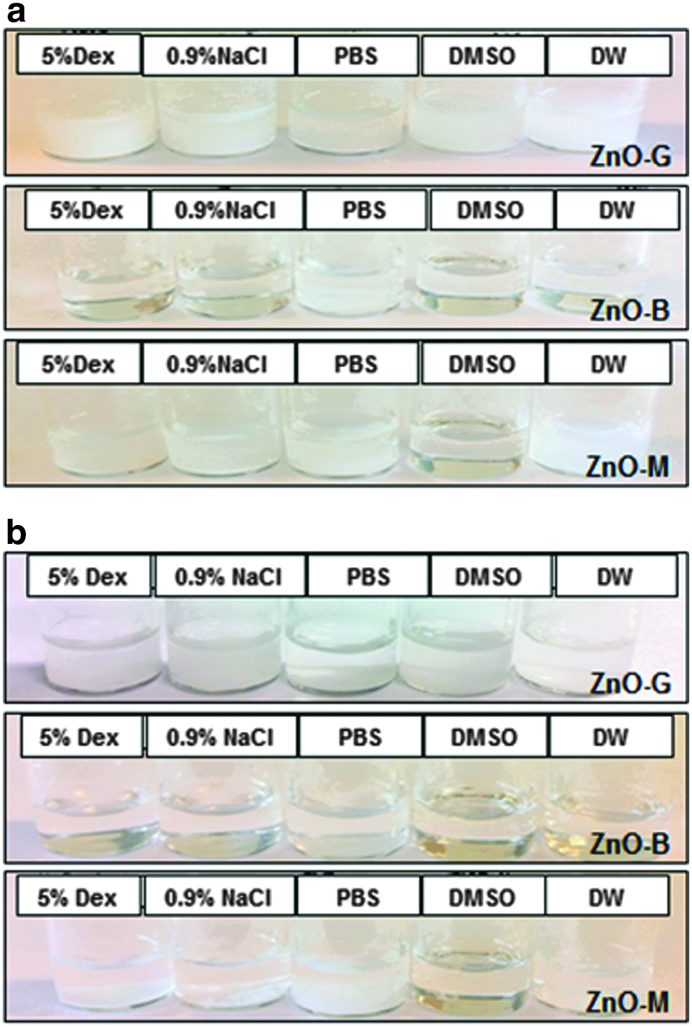
Solvent compatibilities of ZnO nanorods using 5% Dex, 0.9% NaCl, PBS, DMSO, and DW:
UV-Vis spectroscopic analysis
The optical absorption properties of the ZnO nanorods were characterized using UV-Vis spectroscopic analysis (Fig. 6). The UV-Vis absorption spectra for each of the ZnO nanorod samples were recorded from 200 to 800 nm. All of the spectra for the ZnO nanorods exhibited strong absorbance below 400 nm. ZnO-B (Fig. 6b) and ZnO-M (Fig. 6c) did not exhibit any absorption above 400 nm, suggesting the scattering of the UV-Vis light.
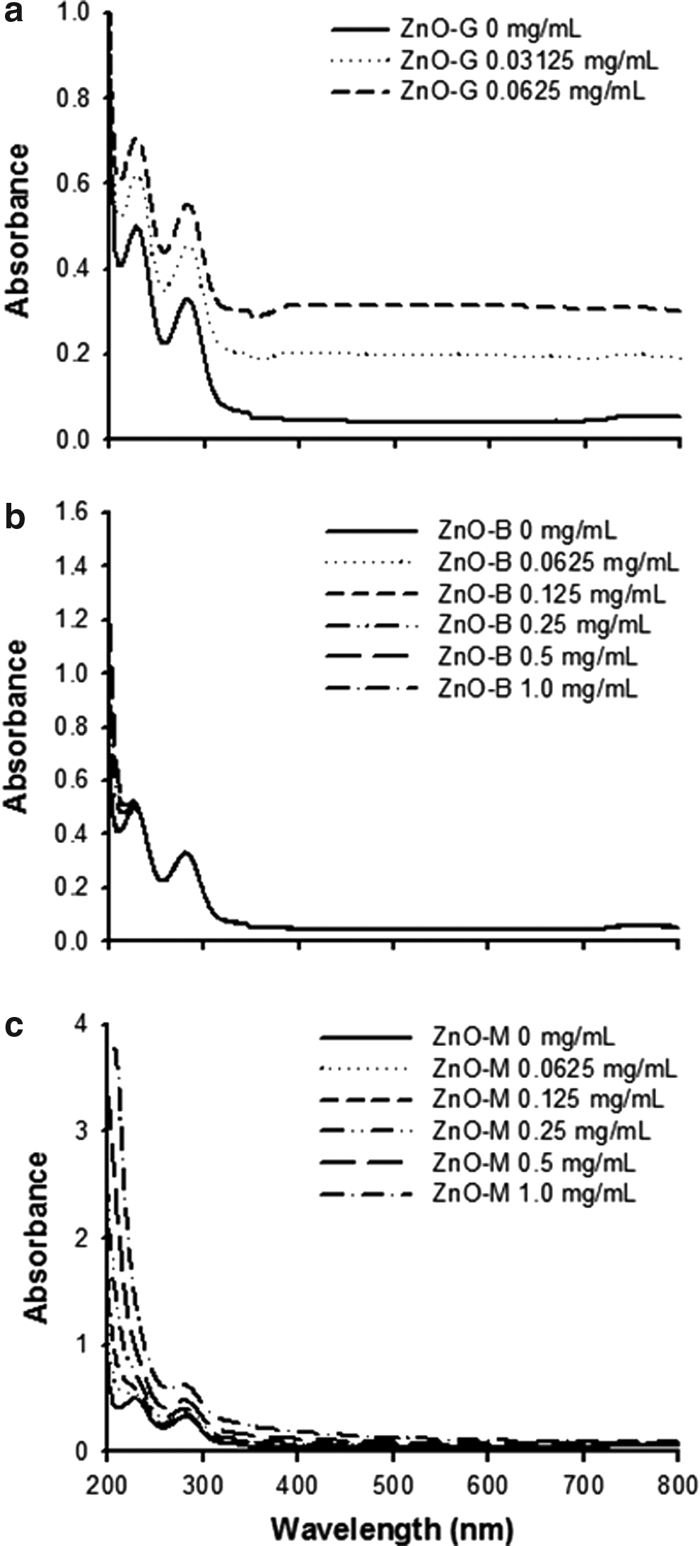
UV-Vis spectra of ZnO nanorods at various concentrations.
Influence of ZnO-B nanorods on ASC proliferation
All ZnO nanorods increased the proliferation of ASCs according to the results of the CCK assays (Fig. 7a). However, the ZnO-G nanorods were toxic at a high concentration of 25 μg/mL. Both the ZnO-B and ZnO-M nanorods significantly increased the proliferation (>130%) in a dose-dependent manner (p < 0.01). The mitogenic effect of the ZnO nanorods was also investigated by determining the level of activation of extracellular signal-regulated kinase (ERK), and it was found that the ZnO nanorods increased the phosphorylation of ERK1/2 in the ASCs (Fig. 7b).
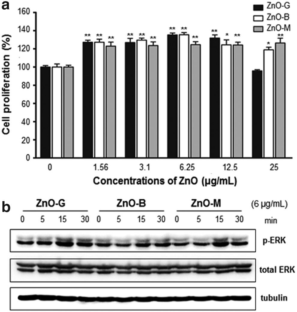
ZnO nanorod enhancement of ASC proliferation based on the activation of ERK.
Based on proteomic analyses of the ZnO-treated ASCs using 2D-PAGE analysis with matrix-assisted laser desorption/ionization-time-of-flight (MALDI-TOF) mass spectrometry and bioinformatics tools, including MASCOT [National Center for Biotechnology Information (NCBI) database], a 2.5-fold increase in the expression of as many as 18 proteins was revealed by comparing the results for the treated samples with those of a nontreated control group (Supplementary Table S1). Among the proteins of increased expressions, thioredoxin reductase I (TrxR I), a known redox system enzyme, was confirmed using the pharmacological TrxR inhibitor, EGCG (Supplementary Fig. S2a). After ASCs with added ZnO-B nanorods were treated with EGCG (10 μM) for 40 h, the EGCG was observed to significantly reduce TrxR I gene expression. ZnO-B enhanced the ASC proliferation, whereas cotreatment of ZnO-B and EGCG reduced the ASC proliferation potential (Supplementary Fig. S2b).
Cell surface markers and differentiation potential of ASCs were analyzed to investigate the ASC characteristics after ZnO-B treatment. In control ASCs, CD 73, CD90, and CD105 except for CD34 were expressed. ZnO-B-treated ASCs were comparable to control ASCs (Supplementary Fig. S3). For the differentiation potential of ASCs, we checked the adipogenic, osteogenic, and chondrogenic differentiation of ZnO-B-treated ASCs comparing with control ASCs treated with 5% Dex. After ZnO-B treatment, ASCs still had a differentiation potential (Supplementary Figs. S4 and S5). Comparing with negative and positive controls in chondrogenesis, Col II and Sox-9 mRNA expression levels were significantly upregulated after ZnO-B treatment (Supplementary Fig. S5d–f).
Discussion
ZnO is extensively used in industrial fields, such as food packaging, cosmetics, and pharmaceutics. Because of its antibacterial activity and tissue regeneration potential, nanosized ZnO that is dispersible in aqueous solutions has been synthesized using various methods such as mechanochemical processes, precipitation with or without a surfactant, sol–gel methods, and solvothermal, hydrothermal, and microwave techniques. 28 ZnO with different particle structures, shapes, and sizes, and thus wide-ranging physicochemical properties, is obtained using these various production methods. Therefore, the modification of ZnO production methods is an effective strategy for preparing novel ZnO-based nanomaterials for use in biomedical applications.29–32
In this study, we used three simple methods for the preparation of ZnO nanorods: grinding of ZnO using a mortar and pestle and boiling zinc acetate in ethanol with and without a surfactant (Fig. 1). The synthesized ZnO nanorods were then evaluated to determine their ability to enhance the proliferation of ASCs. Grinding is one of the simplest methods used for the commercial-scale downregulation of the particle size of ZnO. Boiling with and without a surfactant at room temperature enables the synthesis of ZnO nanorods based on the interactions between zinc acetate and organic materials (e.g., the ethanol, surfactant). In particular, in the presence of a surfactant, micellar ZnO nanorods are expected. In all cases, white ZnO nanorods precipitated were obtained and then washing with ethanol. Each of the samples exhibited different ZnO structures in ethanol (Supplementary Fig. S1). The ZnO-B and ZnO-M also formed clusters of several nanorods with donut and globular shapes.
In the present study, the ZnO nanorods were synthesized at room temperature without calcination. Although several XRD peaks for ZnO nanorods were confirmed in the range from 30 to 40 2θ/θ, their peak intensities were relatively low because of the fact that the reactions were performed at a low temperature for a short time (Fig. 2). On the basis of these results for the ZnO crystallinity of the nanorods, it is recommended that calcination be included as a final step in ZnO nanorod syntheses. However, when the ZnO nanoparticles were calcined in the present study, they became hydrophobic and were not dispersible in aqueous solutions. There are several approaches for the preparation of hydrophilic ZnO nanoparticles using surface modification with hydrophilic polymers such as polyethylene glycol and its derivatives.3,11,26
The morphology, particular size, and size distribution of ZnO nanorods are important factors for determining their dispersibility in aqueous solutions.11,33 The morphology of ZnO is typically a nanoflower-like shape consisting of ZnO needle clusters. In the present study, the ZnO powders produced using the three methods were categorized as rod-type particles (Fig. 3). SEM analysis revealed that the lengths of the nanorods in the ZnO powders ranged from 200 to 500 nm. Three types of ZnO nanorods showed a similar morphology besides the use of different preparation methods. After dispersion of ZnO-B and ZnO-M nanorods in 5% Dex (a biocompatible solution), narrow distributions of 200–500 nm were again observed (Fig. 4). When determining the dispersibility of the ZnO nanorods, only solvents that are appropriate for biomedical use were screened to minimize cytotoxicity and avoid any toxicity concerns (e.g., 5% Dex, 0.9% NaCl, PBS, DMSO, and DW) (Fig. 5). ZnO-B was completely dispersible in various aqueous solvents except PBS, whereas ZnO-G and ZnO-M slowly settled in many of the solutions. Xiaoyong et al. reported that ZnO nanorods exhibit severe cytotoxicity because of aggregation in cell media 14 ; their hydrodynamic diameters increased 2.9- to 58.2-fold, even though the ZnO nanoparticles were sonicated.
One of the applications for ZnO nanoparticles is as a UV protector in personal care/cosmetic products because of their high transparency and ability to absorb and scatter UV light. The level of UV absorption was highest in ZnO-G, lower in ZnO-B, and lowest in ZnO-M at wavelengths below 400 nm (Fig. 6). In addition, the ZnO-B and ZnO-M nanorods showed minimum absorption levels at wavelengths greater than 400 nm (Fig. 6b, c), which were comparable with those reported by Kumar et al. 34 Among the UV-Vis lights, UVB (280–315 nm) is one of stimulators for ASCs due to the mediation of ROS generation. 35 Although high-dose UVB absorption may cause toxicity, low-dose UVB irradiation stimulated ASC survival and migration based on induction of growth factors (e.g., basic fibroblast growth factor, keratinocyte growth factor, hepatocyte growth factor, and vascular endothelial growth factor). However, low-dose UVB radiation did not affect the ASC proliferation.
Following evaluation of the properties of the ZnO nanorods, their potential for enhancing the proliferation of ASCs was investigated using CCK-8 assay. Although the detection mechanism of cells in CCK-8 assay is based on the mitochondrial metabolic activity, CCK-8 assay has been used in cytotoxicity and proliferation tests.36,37 All the ZnO nanorods did enhance the proliferation of ASCs at the range of 1.56–12.5 μg/mL (Fig. 7). While ZnO-G exhibited cytotoxicity at the highest nanorod concentration (25 μg/mL), ZnO-B and ZnO-M continued to provide ASC proliferation enhancement. The results are matched for those from UV-Vis spectroscopy. ZnO-B and ZnO-M of nanorods had low UV absorption levels compared with ZnO-G. Like conventional preconditioning agents, including vitamin C, ultraviolet B (UVB), platelet-derived growth factor, and ROS generator,25,35,38,39 ZnO nanorods clearly have the potential to increase the production and regeneration of ASCs.
To elucidate a possible mechanism of action, the influence of the ZnO nanorods on protein expression was investigated. Proteomics analyses using 2-PAGE, MALDI-TOF MS, and bioinformatics tools revealed that the expression of 18 proteins was increased by over 2.5-fold (Supplementary Table S1). Among the proteins of increased expression, the proliferation potential of ZnO nanorods should be related to the thioredoxin (Trx) system in the present study, 40 specifically TrxR I. TrxR is an enzyme to reduce Trx and Trx-like multiple proteins in the redox system. 41 In particular, TrxR I is one of subtypes for TrxR, which is essential for cell growth and survival against oxidative stress. 42 In ASCs, TrxR I gene expression increased after ZnO-B treatment and was blocked by EGCG, a TrxR inhibitor (Supplementary Table S1 and Supplementary Fig. S2a). ZnO-B treatment also enhanced the ASC proliferation comparing with control and cotreatment of ZnO-B and EGCG. In this test, we specifically used a live cell imaging system, IncuCyte™ (Essen Bioscience) to confirm the kinetics of ASC proliferation (Supplementary Fig. S2b). ASC proliferation levels are comparable to those from CCK-8 assay.
Furthermore, ZnO nanorod-treated ASCs still had their stem cell properties such as surface marker expressions (Supplementary Fig. S3) and differentiations (Supplementary Figs. S4 and S5). Differentiation effect of ZnO on human mesenchymal stem cells to osteoblast was already reported by Foroutan, et al. 43 From Foroutan, et al., its function of osteogenic differentiation was dependent on the size (30 nm) as well as the concentration of ZnO nanoparticles (30 μg/mL). These results indicate that ZnO nanorods may be effective as a preconditioning agent for the promotion of ASC proliferation for biomedical applications.
Conclusion
ZnO nanorods were prepared using simple methods of grinding and boiling without calcination. Despite their poor crystallinity, ZnO nanorods were dispersible in Dex showing UVB absorption. Screening organic materials or regulating sophisticated temperature control with long reaction time would improve the crystallinity of ZnO nanorods. They enhanced the ASC proliferation. ZnO nanorod-treated ASCs still had surface marker expression potential and differentiation potential for adipogenesis and osteogenesis. The results of the present study suggest that ZnO nanorods have a promising potential as a preconditioning agent for the enhancement of ASC proliferation.
Footnotes
Acknowledgments
We thank Jun-Eon Jin for the SEM and AFM analyses of the ZnO nanorods. This research was supported by a grant from the National Research Foundation (NRF) funded by the Korean government (2014054836).
Disclosure Statement
The authors have no conflicts of interest and have received no payment for the preparation of this article.
References
Supplementary Material
Please find the following supplemental material available below.
For Open Access articles published under a Creative Commons License, all supplemental material carries the same license as the article it is associated with.
For non-Open Access articles published, all supplemental material carries a non-exclusive license, and permission requests for re-use of supplemental material or any part of supplemental material shall be sent directly to the copyright owner as specified in the copyright notice associated with the article.