
Abstract
Photoencapsulation of cells inside a hydrogel system can provide a suitable path to establish a gel in situ for soft tissue regeneration applications. However, the presence of photoinitiators and blue or UV light irradiation can result in cell damage and an increase of reactive oxygen species. We here evaluate the benefits of an antioxidant pretreatment on the photoencapsulated cells. We study this by evaluating proliferation and viability of MG63 cells, which we combined with a gelatin methacrylate (GelMA) hydrogel system, using the photoinitiator, VA-086, cured with 440 nm blue light. We found that blue light irradiation as well as the presence of 1% VA-086 reduced MG63 cell proliferation rates. Adding a short pretreatment step to the MG63 cells, consisting of the antioxidant molecules N-acetylcysteine (NAC) and reduced glutathione (GSH), and optimizing the GelMA encapsulation steps, we found that both NAC and GSH pretreatments of MG63 cells significantly increased both proliferation and viability of the cells, when using a 15% GelMA hydrogel, 1% VA-086, and 1-min blue light exposure. These findings suggest that the use of antioxidant pretreatment can counteract the negative presence of the photoinitiators and blue light exposure and result in a suitable environment for photoencapsulating cells in situ for tissue engineering and soft tissue applications.
Introduction
H
There are many different hydrogel materials that can be used for cell encapsulation, offering a range of properties to select from for a specific application. In this study, we chose gelatin, a derivative of collagen that is formed by breaking the natural triple-helix structure of collagen into single-strand molecules. 3 Gelatin has found many uses in tissue engineering applications, primarily due to its biocompatibility and ease of gelation.4–6 Gelatin gels have also been utilized for delivery of growth factors to promote vascularization of engineered new tissue. 7 However, a downside of gelatin is possible variability from source to source, limited antigenicity, and instability at body temperature.8,9 Causing gelation with thermal methods is challenging because the temperature changes required make manipulation difficult. Photopolymerization offers a simple alternative that can control the gelation process by modifying the gelatin with methacrylic anhydride, adding several methacrylate groups at its side chains. This modification can be carried out by reacting methacrylic anhydride with the amine-containing side groups of gelatin, turning gelatin into a light-curable gelatin methacrylate (GelMA).10–12 GelMA has been gaining popularity as a biomaterial since it is inexpensive and has tunable physical and biochemical properties. However, the degradation rate of GelMA can only be modified by changing the concentration, making it difficult to use. 13 Some studies of gelatin preparations for regenerative medicine applications have been obtained with endotoxin. 14
The GelMA solution can be photopolymerized into a hydrogel at body temperature (37°C) in the presence of photoinitiators and light sources. 15 A photoinitiator is a compound that absorbs UV or visible light and converts the absorbed light energy into chemical energy. These changes occur in the form of initiating species, that is, free radicals or cations. The photoinitiation systems used in cell encapsulation include 2-hydroxy-1-[4-(hydroxyethoxy)phenyl]-2-methyl-1-propanone (Irgacure 2959), 2,2′-azobis[2-methyl-n-(2-hydroxyethyl)propionamide] (VA-086), and lithium phenyl-2,4,6-trimethylbenzoylphosphinate (LAP).16,17 We chose VA-086 as the photoinitiator because it was reported to be less toxic than Irgacure 2959. 16 VA-086 is a type I photoinitiator, which undergoes a unimolecular bond cleavage upon irradiation to yield free radicals that can cause cell damage. For example, free oxygen radicals contribute to oxidative damage to DNA and play an important role in the etiology of cancer and atherosclerosis and are also implicated in studies on aging. 18 VA-086 is typically cured with UV light between 365 and 385 nm wavelength. We decided to use 440 nm, which while off-peak for VA-086 photoinitiation, allows for more biocompatible irradiation. 16
Oxygen can be toxic to cells at higher concentrations. In its ground state, molecular O2 is relatively unreactive. Its partial reduction gives rise to reactive oxygen species (ROS) such as singlet oxygen, superoxide radical anions, and hydrogen peroxide. 19 The presence of intracellular oxygen can initiate a chain of reactions at the cellular level that causes damage to critical cell biomolecules. These radicals are atoms, molecules, or ions, which are highly toxic and have a single unpaired electron. Environmental stress, such as UV and free radicals generated during photopolymerization, can induce a higher intracellular ROS level through multiple mechanisms. Therefore, a trade-off exists between the need to supply light energy to the photoinitiator to generate the free radicals necessary to assist in the gelation and the need to maintain a safe environment for the cells embedded in the hydrogel. This trade-off motivated our choice for 440 nm blue light over the perhaps more commonly used higher energy UV radiation.
To further reduce the effects of free radicals generated during photopolymerization, antioxidants can be added to the culture medium. The antioxidant molecules can stabilize or deactivate free radicals and reduce cell damage. Antioxidants can be endogenous to the cell or they can be obtained exogenously as a part of the diet or growth medium. Under conditions of heavy oxidative stress, endogenous antioxidants may not offer sufficient protection, and dietary antioxidants may be added to maintain optimal cellular functions.
The antioxidant glutathione (GSH) is one of the most important nonenzymatic removal systems for exogenous and endogenous free radicals.
20
In cells, GSH plays an important role in maintaining the redox state. It is an abundant tripeptidyl molecule and plays important roles in protecting cells against oxidative stress-induced cellular damage, detoxifying xenobiotics, and in drug metabolism. GSH can maintain the exogenous antioxidants (such as vitamins C and E) in their reduced forms and help reduce the damage caused by UV light to skin. There are three amino acids in the GSH structure: glutamate, glycine, and cysteine.
21
The thiol group in the cysteine residue of the GSH acts as a reducing agent, donating the reducing equivalent (H+ plus e−) to other unstable molecules (e.g., ROS). Cysteine can limit the rate of GSH synthesis during times of oxidative stress. N-acetylcysteine (NAC) is the acetylated precursor of both the amino acid
In this study, we investigate how the presence of GSH and NAC can be of benefit when using a blue light-activated hydrogel system consisting of gelatin methacrylate and VA-086. We chose cells from the MG63 osteosarcoma cell line to be encapsulated because this cell line has previously been used as an osteoblast mode in vitro.23,24
Materials and Methods
Synthesis of methacrylated gelatin
GelMA was synthesized according to Benton et al. 10 ; 1 g of gelatin was added to 10 mL of phosphate-buffered saline (PBS) and heated at 50°C until the gelatin was dissolved completely. Methacrylic anhydride was added by stirring into the mixture until the target volume (1 mL) was reached at a constant rate of 0.1 mL/min. The reaction was then allowed to proceed for 3 h at 50°C, after which the solution was diluted fivefold with warm (55°C) PBS. This was followed by dialysis with tubular dialysis membranes (Orange Scientific) of 6000–8000 molecular weight for 1 week against 40°C distilled water to remove the methacrylic acid and salts. The solution was lyophilized for 1 week to obtain a white sponge-like foam, which was stored at −80°C for further reaction.
Culturing of the MG63 osteosarcoma cells
MG63 cells were cultured in Dulbecco's modified Eagle's medium (DMEM) supplemented with 10% fetal bovine serum and 1% Penicillin–Streptomycin–Amphotericin B antibiotics. Cells were kept in a humidified incubator at 37°C in the presence of 5% CO2.
Evaluation of the curing time using the upside-down method and viscometer
Hydrogels were prepared in either 12.5% or 15% (w/v) of gelatin in DMEM, with 1% VA-086 by weight. GelMA and VA-086 were weighed and then added into the DMEM and mixed. Dried glass vials were filled with 1 mL hydrogel solution and irradiated under a custom-made 10 W, 440 nm blue LED light source (60 mW/cm2). The light was placed one centimeter below the bottom of the vial at room temperature (25°C) for different periods of time. The gelation start time was defined as the time at which the mixture no longer floated when the vial was turned upside down. The viscosity change of the GelMA during blue light curing was also recorded using a Brookfield rheometer (DV3TRV™ Rheometer; Brookfield, Middleboro, MA). One milliliter of GelMA was loaded into a transparent glass container and light-cured from the bottom. A V-74 spindle with a rotation speed of 15 rpm was used for the viscosity test. Data were collected every 5 s, and the readings between 10% and 90% torque were recorded and expressed in centipoise (cP).
Compression test
Uniaxial compression measurements were performed on a universal testing machine at room temperature in compression mode. All measurements were tested at a crosshead speed of 1 mm/min until the samples were compressed by 70% in height.
MTT assay
An MTT assay was used to measure the proliferation rate of MG63 cells 24 h after they were exposed to blue light for 1, 2, and 4 min. The cells were pretreated with different concentrations of the antioxidants, GSH and NAC (0, 10, 20, and 30 mM), which were dissolved in DMEM and then incubated at 37°C for 20 min. The cells were then washed in PBS, 1% VA-086 was added, and exposed to blue light for 1, 2, and 4 min. The cell proliferation rate was then measured by MTT assay. For photoencapsulated MG63 cells, after the above steps, the cells were washed in PBS and isolated by trypsin, and then mixed with the GelMA solution in DMEM, with the photoinitiator, VA-086, included (see previous methods for details). Evaluation of cell growth was done by MTT assay at 1, 3, and 7 days after MG63 photoencapsulation, both for cells with and without the antioxidant pretreatment. Ten microliter MTT solution (5 mg/mL in sterile PBS) was added into each well and incubated for 3 h at 37°C. After incubation, the medium was removed and 200 μL DMSO (Sigma-Aldrich) was added to each well and incubated for 30 min; 100 μL of test sample taken from each well was added into a 96-well plate for absorbance measurements using an ELISA reader at 570 nm with a reference wavelength of 620 nm.
Antioxidant pretreatment and photoencapsulation of MG63 cells in hydrogel
Before cell encapsulation, a 1% solution of the photoinitiator, VA-086, in PBS was prepared and stored at −80°C. Fifteen percent of w/v GelMA was dissolved into the solution. To test the effect of NAC and GSH, MG63 cells cultured on tissue culture plastics were pretreated with 10 mM NAC, 20 mM NAC, 10 mM GSH, and 20 mM GSH for 20 min each. As experimental control, no antioxidant pretreatment was performed. Cells were then trypsinized, counted, and spinned down. The cell pellets were resuspended in the polymer solution with VA-086, until the cells were evenly distributed (at 106 cells/mL). Twenty microliters of cell-containing hydrogel solution in each 24-well cell culture dish was irradiated under blue LED light at room temperature (25°C) to initiate the photopolymerization. This step should be performed rapidly to minimize cell settling before light exposure. After the gels cross-linked, they were washed with PBS and cultured in DMEM. On days 1, 3, and 7 after seeding, samples were collected to conduct MTT assay and Live/Dead assay test.
ROS assay
ROS tests (Cell Biolabs, Inc.) were performed according to the manufacturer's protocol, using the cell-permeable fluorogenic probe, 2′,7′-dichlorodihydrofluorescein diacetate (DCFH-DA), which is first diffused into the cells and then deactelyated into nonfluorescent 2′,7′-dichlorodihydrofluorescein (DCFH). ROS rapidly oxidized DCFH into 2′,7′-dichlorodihydrofluorescein (DCF). The fluorescence intensity was measured using a fluorometric plate reader at 480/530 nm.
Live/Dead cell viability assay
To test for cytotoxicity after GelMA photoencapsulation, we used a Live/Dead assay. Live cells were detected through the presence of intracellular esterase activity. An enzymatic conversion of the nonfluorescent cell-permeant calcein AM into the intensely fluorescent calcein was conducted. Ethidium homodimer-1 (EthD-1) enters through damaged cell membranes and binds to nucleic acids. This produces a bright red fluorescence in dead cells. The solution consists of 2 μM calcein AM and 4 μM EthD-1 as a working solution, which was added directly to the GelMA hydrogel. The hydrogel was incubated at room temperature for 30 min, and cells were imaged under a fluorescence microscope and labeled.
Statistical analysis
All experimental results are expressed as mean ± standard error of the mean. Comparative studies of means were analyzed using one-way ANOVA with a statistical significance of p < 0.05. In all figures, the significance at p < 0.05, 0.01, and 0.001 was labeled as *, **, and ***, respectively.
Results
Viscosity change, gelation time, and compressive modulus of GelMA hydrogels cured by blue LED irradiation
The viscosity of 12.5% and 15% GelMA solution increases rapidly at 55 and 40 s, respectively (Fig. 1C). These numbers are longer than the gelation time of 33.3 ± 1.5 and 29.3 ± 1.1 s when measured by the upside-down method (Fig. 1B). The compressive modulus of 12.5% and 15% of GelMA hydrogels is 9.28 ± 0.47 and 16.46 ± 1.01 kPa, respectively (Fig. 1D). The compressive modulus of the GelMA hydrogel increases as the GelMA concentration increases, as expected. The 2.5% difference in the GelMA concentration results in a statistically significant increase in the compressive modulus of GelMA (p < 0.001).

The viscosity increase of the GelMA solution was nonlinear. Before light curing, the GelMA solution has a low viscosity, comparable with water. At the start of gelation, when the GelMA begins to photopolymerize, the viscosity increases. As the GelMA hardens, eventually the shear loading exceeds the maximum shear stress of GelMA gel, and the rotating spindle resulted in gel rupture. At this point, a zigzag curve was observed (Fig. 1C). Although the gelation starting time was determined using a viscometer, the change in viscosity does not fully represent the regular static photopolymerization process because the viscosity measurement was done in a dynamic environment. However, the viscosity measurement still can provide a simple way to evaluate the gelation behavior.
Cytotoxicity test of GelMA prepolymers
Cytotoxicity is one of the critical factors determining whether the prepared GelMA is pure enough for further cell encapsulation experiments. Figure 2 shows the live and dead images of MG63 cells cultured with 12.5% and 15% (w/v) for 24 h. No dead cells (red fluorescence) were found in the control group and the two experimental groups, suggesting that the prepared GelMA is not cytotoxic to MG63 cells.

Images of LIVE/DEAD assays of MG63 cells (6 × 10⁴/cm2) cultured for 24 h in different concentrations of GelMA solution medium.
The effects of blue LED light irradiation on MG63 proliferation and ROS levels
Some studies showed that blue light can induce mitochondrial DNA damage and produce free radicals in certain cells. 25 To test the effect of cells exposed to blue light, MG63 cells (2 × 10⁴/well) were irradiated for 0, 1, 2, and 4 min, rinsed with 1× PBS, and then cytotoxicity was evaluated by MTT assay. Figure 3A shows that 1, 2, and 4 min of blue light exposure causes a reduction of MG63 cell proliferation rates, which is most significant at a 4-min exposure time (p < 0.01). On the other hand, ROS levels rapidly increased after blue light exposure of 1, 2, and 4 min (Fig. 3B). This suggests that blue light exposure has a negative effect on cell proliferation rates and on ROS levels.

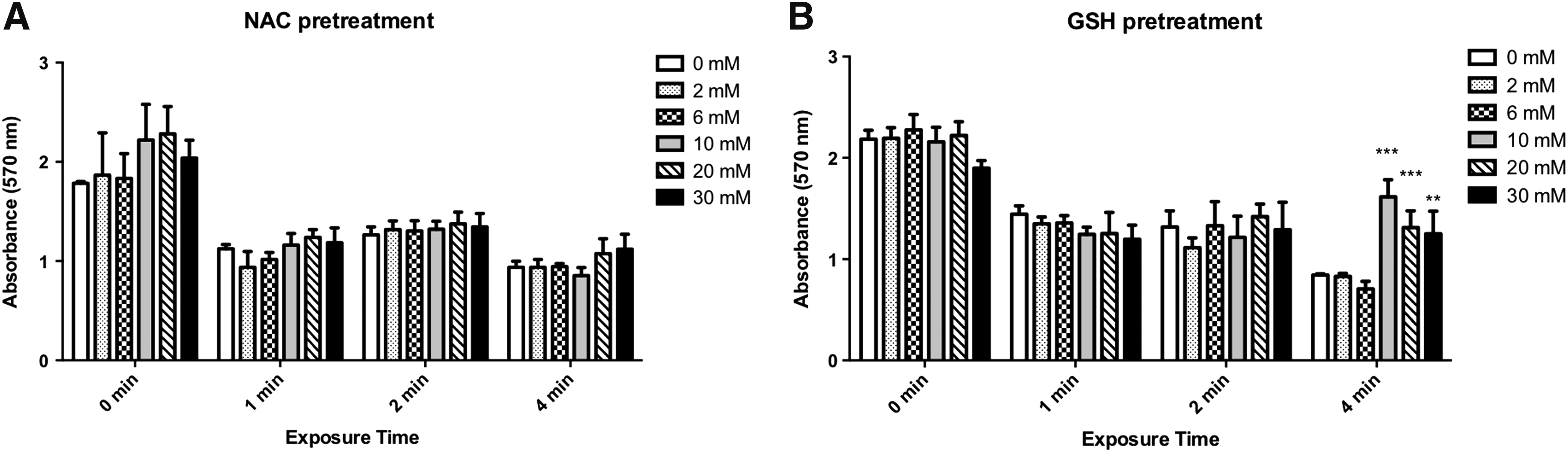
The effects of NAC and GSH pretreatment on MG63 exposed to VA-086 with blue LED light exposure
MG63 cells were seeded in a 24-well dish and pretreated with 0–30 mM of NAC (Fig. 4A) and 0–30 mM of GSH (Fig. 4B) using a volume of 300 μL for 20 min. Three hundred microliters of 1% VA-086 was added to each well, followed by 1–4 min of blue light exposure. The results showed no significant difference in the absorbance value of the MTT assay for the NAC pretreatment (Fig. 4A). In contrast, cells pretreated with 10–30 mM GSH proliferated significantly better than the controls after 4 min of blue light irradiation (Fig. 4B). The 10 and 20 mM GSH pretreatment showed the highest MG63 proliferation rates (p < 0.001). We also confirmed the ROS levels in MG63 cells under the same experimental conditions. The data showed that 10 mM of NAC pretreatment significantly decreased ROS levels after 2 and 4 min of blue light exposure (Fig. 5A). In addition, the level of ROS generation decreased for 10–30 mM GSH pretreatment and 2 and 4 min of blue light irradiation (Fig. 5B). The above results indicate that both NAC and GSH can reduce ROS levels on MG63 cells and that GSH is more effective than NAC for ROS level reduction.
Cell proliferation rate of MG63 cells treated with different concentrations of NAC
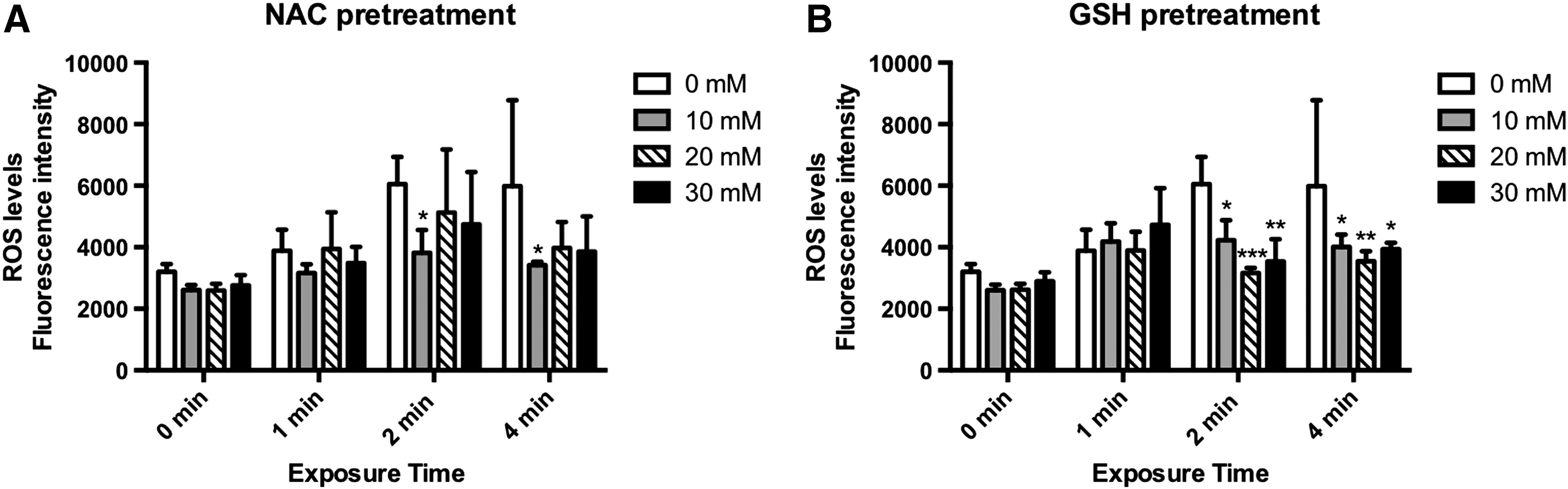
ROS levels of MG63 cells treated with different concentrations of NAC
Evaluation of untreated and antioxidant-pretreated MG63 cells photoencapsulated in hydrogel
Untreated MG63 cells and MG63 cells pretreated with NAC and GSH were encapsulated, and after 1, 3, and 7 days, tested with MTT (Fig. 6) and Live/Dead assays (Fig. 7). Cells were found to survive after photoencapsulation, and cells pretreated with NAC and GSH exhibited higher proliferation rates than controls (Fig. 6). During days 1 and 3, GSH and NAC showed very similar results in the MTT measurements for both 10 and 20 mM concentrations. On day 7, however, 20 mM of GSH induced slightly higher cell proliferation rates than 20 mM of NAC (Fig. 6).

Cell proliferation of MG63 cells photoencapsulated in GelMA, evaluated by MTT assay. In comparison with the control, all antioxidant pretreatments significantly reduced the ROS during photoencapsulation (***p < 0.001).
The images obtained by the Live/Dead assay show that most cells encapsulated in GelMA hydrogel survive, as seen by the green fluorescence throughout the 7-day period under all conditions (Fig. 7A–E). For the controls, we observed a higher occurrence of red fluorescent cells, that is, dead cells, than for any antioxidant-treated MG63 cells on day 1, 3, or 7 (Fig. 7A–E).

Fluorescent images of LIVE/DEAD assays of MG63 cells photoencapsulated in GelMA:
Discussion
The compressive strength of our light-cured GelMA is comparable with that reported in the literature. 26 The compressive strength of GelMA is suitable for applications such as regeneration of soft tissues, where the material does not bear higher physiologic loads. One such example is the regeneration of periodontal tissues in the periodontal pocket, where osteoblasts containing GelMA solution can be injected into periodontal pocket, followed by blue light curing. The GelMA solution can thus be turned into gel in situ and hold the osteoblasts in the periodontal pocket, and also act as a barrier that prevents gingiva cells from ingrowth into the periodontal pocket. For this example, a low compressive strength of GelMA does not affect its clinical potential.
Based on the results from the live and dead assay, which we used to test for the biocompatibility of the GelMA solution, it was found that GelMA did not kill cells (Fig. 2). However, the morphology of MG63 cells in the control group was slightly different from the cells incubated with GelMA, suggesting that our GelMA solution may have resulted in a slight increase of cytotoxicity.
There are many studies on the effects of visible and UV radiation and the photochemical damage in neural retina and retinal pigment epithelium (RPE).25,27–29 Some in vivo studies have shown that the death of blue light-irradiated RPE cells occurs through apoptotic mechanisms.30,31 Blue light damage may induce free radical production in some cells, resulting in phototoxicity. Our MTT assay tested for phototoxicity, and longer blue light exposure caused damage to MG63 cells (Fig. 3), with increases in light intensity or exposure duration both leading to increased cytotoxicity, as expected.
Supplementation of NAC (a precursor of GSH, the body's main antioxidant) increases GSH levels and supports the antioxidant and nitric oxide systems during infection, inflammation, stress, and exposure to toxins.32,33 High NAC concentrations (5 and 10 mM) can reduce cell death and restore mitochondrial activity after a 24-h cotreatment. 34 The duration of the antioxidant pretreatment is critical. MG63 cells are human osteosarcoma cells, and ROS may be necessary for tumor cells. 35 The antioxidant may therefore inhibit the generation and accumulation of intracellular ROS if the treatment is too long. In our experiments, 20 min of NAC and GSH pretreatment time was a suitable treatment duration, above which the NAC started to show certain levels of cytotoxicity (data not shown).
The choice of the photoinitiator concentration is important. In all our experiments, we chose 1% as the concentration for VA-086 because other studies with up to 1% (w/v) of VA-086 reported low cytotoxicity. 36 At these concentrations, sufficient VA-086 is present to enable photocross-linking, yet without significant cell death. 37 To generate radicals, the photoinitiator requires light irradiation. Therefore, we combined the VA-086 photoinitiator with the blue light source to evaluate the effects of NAC and GSH on reducing oxidative stress during the initiation step of the polymerization. Concentrations of 10 or 20 mM NAC did not protect MG63 cells against the radicals generated from VA-086 under blue light irradiation (Fig. 4A). However, concentrations of 10–30 mM GSH effectively protected MG63 cells from VA-086 and blue light exposures up to 4 min. The reduction of ROS is reflected in the increased absorbance value of the MTT assay (Fig. 4B). Because the VA-086/blue light exposure assay was performed immediately after the antioxidant treatment, we think that NAC may not have been converted to replenish the GSH in time, explaining why NAC did not help to reduce ROS. This hypothesis was supported by the results from the ROS tests (Fig. 5).
At the initiation of polymerization, radicals are created from initiators excited by UV or visible light. If there is no monomer present in the reaction solution, the radicals recombine. The recombination occurs on the microsecond timescale. However, the radicals generated from VA-086 during light exposure can also damage cells, especially for exposure durations of 4 min. Therefore, for our cell encapsulation experiments, we selected durations of 1 min. With a monomer present, free radical generation will continue beyond the blue light exposure time. This results in an increased free ROS damage to cells. However, with 20 min of antioxidant pretreatment, the encapsulated MG63 cells can survive and proliferate significantly better than experimental controls (Figs. 6 and 7). Among all samples, 20 mM of GSH showed the highest cell survival rates after 7 days, effectively reducing the free radicals generated during photoencapsulation. Additional experiments may be performed to further optimize the dosage, treatment time, and frequency of the antioxidant pretreatment for photoencapsulation.
Conclusion
In this study, we encapsulated MG63 cells using a GelMA/VA-086/blue light hydrogel system. It was found that blue light can replace UV light to initiate photoencapsulation, opening the possibility of enabling cell therapy in clinical settings using standard, dental, handheld blue light devices. We further discovered that NAC and GSH pretreatments can effectively reduce the oxidative stress damage created during photoencapsulation. This novel photoencapsulation protocol can be applied to additional cell types to increase cell survival and proliferation rates.
Footnotes
Acknowledgment
This project was funded by the Ministry of Science and Technology, Taiwan (Project no. NSC101-2314-B-010-037).
Disclosure Statement
No competing financial interests exist.