
Abstract
Functional monitoring of the fate of implanted templates, which restore the function of lost tissues, is still a challenge. Whereas histology can give excellent insight into material and tissue remodeling, longitudinal studies are hampered by the invasive character. Noninvasive imaging techniques, which allow longitudinal studies in the same individual and provide functional information, might be beneficial. In this study, magnetic resonance imaging (MRI) was applied as a noninvasive tool to monitor the progress of vasculogenesis and inosculation in in vitro prevascularized collagen/fibrin templates implanted in mice during a period of 4 weeks. MRI results were compared with histological findings to evaluate whether the two technologies were complementary and to evaluate the added value of MRI. When in vitro prevascularized templates were implanted in mice, histological analysis showed the presence of mouse blood cells in the engineered vessels 2 weeks after implantation. The MR images showed that template perfusion, a measure of vascularity, became significant at 3 weeks. For tissue engineering purposes, contrast-enhanced MRI appears to be an attractive tool to evaluate the vascular outcome longitudinally without the need to sacrifice animals and the functional information can be superimposed on the static histological information.
Introduction
N
Multiple imaging tools are available: single-photon emission computed tomography (SPECT), positron emission tomography (PET), ultrasound (US), X-ray computed tomography (CT), optical imaging (OI), and high-field magnetic resonance imaging (MRI). MRI does not involve ionizing radiation, has a high-spatial resolution without penetration problems, 2 and the outcome of preclinical studies can easily be translated to the clinic. 3
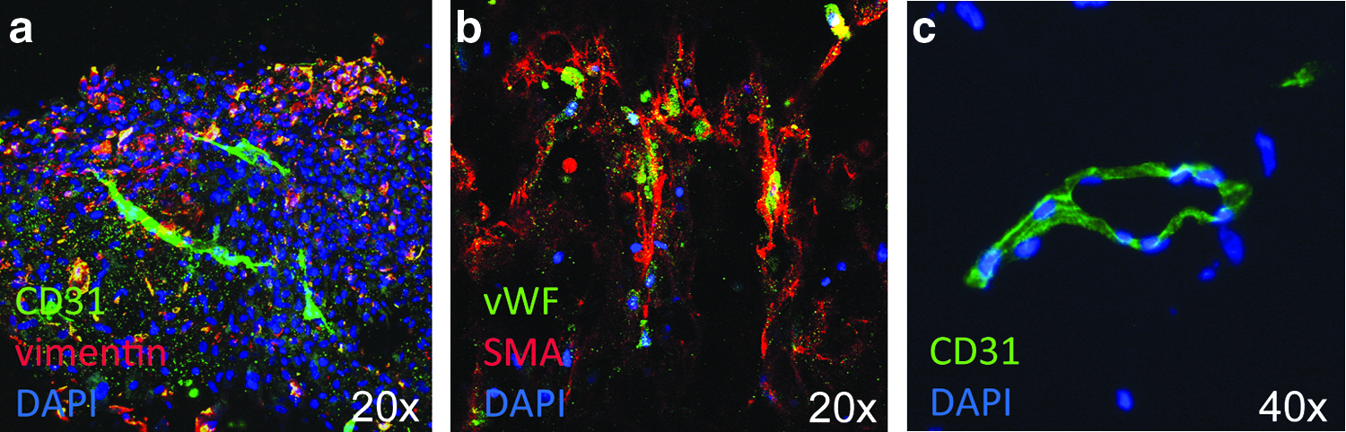
Prevascularization techniques have been studied to enhance the survival and to accelerate the remodeling of implanted materials, particularly for larger templates.4,5 After grafting, the progress of vascularization/inosculation has been visualized with histology; however, this provides only an anatomical snapshot of this highly dynamic process and functionality can only be surmised. Through MRI, different types of functional information can be obtained, which can be superimposed on the histological information (Fig. 1). For instance, it is possible to determine vascular functionality with administration of contrast agents. 6

The workflow of preparing materials, implanting in animal, and monitoring the implants with MRI. Smart materials with or without cells are grafted into animals to replace the lost tissues. The progress of tissue remodeling and vasculogenesis in smart materials can be monitored either with histology (invasive) or with imaging tools (noninvasive), MRI is particularly useful as it can be performed noninvasively and provides a high-resolution anatomy (2D resolutions down to about 80 μm). With this technique, signals of water protons are recorded and their intensities are displayed as a function of their location. Image contrast can be manipulated by T1 or T2 relaxation time weighting. In addition, contrast can be enhanced by using Gadolinium (Gd) or iron-labeled contrast agents. These contrast agents can be used to visualize tissue perfusion after contrast agent infusion or can be tagged to compounds such as those used in templates or implants. Using these MR methods, tissue vasculature or remodeling can be analyzed in live animals. Here, Gd contrast-enhanced T1 and T2 MR images are shown that were used to localize the implant in cadavers. Orange dotted line indicates the location of the template. MRI, magnetic resonance imaging. Color images available online at
The goal of this study was to monitor the fate of in vitro prevascularized collagen/fibrin templates with emphasis on (time of) inosculation and functionality of the engineered microvasculature in the implanted templates with MRI. MRI was compared with histology to investigate the added value and complementarity of MRI and histology.
Materials and Methods
Cell culture
Human umbilical vein endothelial cells (HUVECs, a kind gift from Prof. Otto C. Boerman, Department of Nuclear Medicine, Radboudumc) were expanded in microvascular endothelial cell growth medium-2 (EGM-2 MV; Lonza) with supplements (CC-4147; Lonza). Human bladder smooth muscle cells (hbSMCs; ScienCell) were expanded in smooth muscle cell medium (SMCM; ScienCell) with growth supplement, fetal bovine serum, and penicillin/streptomycin solution. hTERT-human urothelial cells (hUCs, a kind gift from Dr. M.A. Knowles, Leeds, United Kingdom) were expanded in keratinocyte serum-free media (Invitrogen) with supplements, including bovine pituitary extract, epidermal growth factor, and cholera toxin (Sigma-Aldrich). The media were replaced three times per week.
Prevascularization
Four types of hybrid templates were prepared: a blank template (Acellular), a urothelial cell (UC)-seeded template, a prevascularized template (Pre-V), and a prevascularized template with urothelial cells (Pre-V + UC); 0.4% (w/v) type I collagen templates were prepared as previously described. 7 Collagen templates were sterilized with gamma irradiation (Synergy Health). Fibrinogen solution (from bovine plasma; Sigma-Aldrich) was prepared to a final concentration of 10 mg/mL as described 8 and sterilized by passing through a filter (0.22 μm; Whatman).
To prepare prevascularized hybrid templates, a dry 0.4% (w/v) collagen template was placed in the insert ring of a Transwell system (0.4 μm polyester membranes, 12-well plate; Costar). HUVECs (2.5 × 10⁵ cells/mL) and hbSMCs (2.5 × 10⁵ cells/mL) were encapsulated in 500 μL fibrinogen (10 mg/mL) mixed with 11 μL thrombin (Sigma-Aldrich, 50 U/mL) and applied on the dry collagen sponge. The mixture of cells and gel was absorbed by the collagen sponge. The hybrid template was incubated at 37°C for 1 h in a humidified incubator containing 5% CO2 to stabilize. Thereafter, a mixture of EGM-2 MV: SMCM (1:1 [v/v]) was added to the top and bottom chambers of the Transwell plate. To inhibit the fibrinolysis process, 12 mM of ɛ-aminocaproic acid (Sigma-Aldrich) was added. 9 The medium was replaced three times weekly. Hybrid templates were cultured for 2 weeks before grafting.
To evaluate the influence of hUCs on the Pre-V templates, hUCs were seeded (2.5 × 10⁴ cells/mL) on the surface of Pre-V template 1 day before implantation. Additionally, UC templates were produced by seeding hUCs on top of Acellular templates. These templates were cultured for 1 day and implanted. These UC templates were included to study the possible influence of hUC coverage on the pre-V templates and served as control for the nonvascularized templates.
Scanning electron microscopy
Templates were fixed with 2% (v/v) glutaraldehyde, dehydrated by graded series of ethanol (30–100%), and dried using a critical point dryer (Polaron; Quorum Technologies). Samples were sputtered with gold for 60 s by a gold/palladium coater (Cressington 208HR sputter coater) and examined by scanning electron microscopy (SEM) (JSM-6310; JEOL).
Template grafting
Animal studies were conducted in accordance with the principles laid out by the revised Dutch Act on Animal Experimentation and performed after approval from the institutional Animal Welfare Committee of the Radboud University Nijmegen (DEC-nr. 2012-118). Eight-week-old male Balb/c nu/nu mice (N = 36) were purchased from Charles River. The template was grafted on the ventral abdominal wall with three sutures (Prolene, 6-0; Ethicon) on the edge of templates. Peritoneum and skin were closed using 4-0 Vicryl sutures (Ethicon) and sterilized staplers. Animals were evaluated 1, 2, and 4 weeks postimplantation using MRI and sacrificed for immunohistochemical analysis. Grafting was as follows: blank hybrid templates (Acellular, N = 6), hUC-seeded hybrid templates (UC, N = 10), prevascularized hybrid templates (Pre-V, N = 10), and prevascularized hybrid templates plus hUCs (Pre-V + UC, N = 10).
Magnetic resonance imaging
MR measurements were performed on an 11.7 T horizontal bore small-animal MR system (BioSpec; Bruker BioSpin). A circular polarized resonator was used for signal transmission, and a receiver surface coil (1.5 × 1.5 cm) was placed adjacent to the template. Mice were placed in the MR system under sedation in a supine position. Body temperature was maintained at 37°C. The numbers of animals analyzed from each group were Acellular, N = 4; UC, N = 5; Pre-V, N = 8; and Pre-V + UC, N = 5. The contrast agent, Gadomer-17 (InvivoContrast GmbH), was administrated (150 μmol/kg body weight) as a rapid bolus injection (<5 s) through the tail vein.
T1 relaxation time was measured by a rapid acquisition rapid echo (RARE) sequence with variable repetition time (TR) (400, 800, 1500, 3000, and 5000 ms) and a fixed echo time (TE) of 30 ms. Parameters were RARE factor = 2, field of view = 40 × 40 mm, matrix size = 256 × 256, and total acquisition time (TA) = 8 min.
Dynamic contrast-enhanced MRI was performed using a fast low-angle shot (FLASH) sequence.10,11 Imaging parameters were TE = 1.602 ms, TR = 52 ms, flip angle = 40°, field of view = 40 × 40 mm, matrix size = 256 × 256, slice thickness = 1 mm, and four slices in coronal directions. One baseline image was measured before contrast administration. Immediately thereafter, dynamic T1-weighted imaging was performed by repeating the same FLASH sequence of 5 s 499 times. The dynamic uptake of Gadomer-17 was monitored for TA = 42 min.
MR images were processed using a home-built program in MevisLab. The signal-to-noise ratio (SNR) was calculated on a manually drawn region of interest (ROI) at the location of the template. The SNR before and after injection of Gadomer-17 was recorded over time to track signal intensity (SI) changes. For semiquantification purposes (an indication of the amount of Gadomer-17 in each implanted template), the area under curve (AUC) was calculated from the SI-time curve up to 2000 s using GraphPad Prism 5 software.
Data are presented as mean (±standard deviation [SD]). Several ROIs were analyzed and the numbers of ROIs were determined based on the thickness of templates. Two-tailed t-test analysis was used to determine the significance of T1 relaxation time changes after Gadomer injection. The ability of MRI to identify different permeability levels was tested using two-way ANOVA. Post-test analysis of differences among means was based on Bonferroni. p < 0.05 was considered significant.
Immunohistochemistry
Antibodies are summarized in Table 1. Templates were fixed in O.C.T. compound (TissueTek) and snap-frozen. Specimens were cut at 5 μm and fixed with methanol for 10 min at 21°C. For laminin staining, slides were incubated in 0.15 mg/mL pepsin in 0.2 M HCl at 37°C for 10 min. Then, slides were washed three times for 10 min with Tris-saline. For human mitochondria staining, mouse on mouse blocking reagent (MOM; Vector Labs) was applied to reduce background noise from mouse tissue according to the manufacturer's instructions.
Slides were incubated with 10% normal goat serum (NGS) in 1% bovine serum albumin (BSA) in phosphate-buffered saline (PBS; pH 7) for 1 h at 21°C for blocking. Primary and Alexa Fluor-labeled secondary antibodies were incubated in 1% NGS 1% BSA in PBS for 1 h and 30 min at 21°C, respectively. Slides were mounted with Prolong Gold mount medium with DAPI (Invitrogen) and evaluated by fluorescence microscopy (LEICA DC 300F; Leica Microsystems) or high-content microscopy (LEICA DMI6000B).
For whole template staining, templates were incubated with antibodies as previously described and the nuclei were stained with Hoechst 33342 (Invitrogen). Complete templates were scanned by confocal laser scanning microscopy (FV1000; Olympus). Images were processed with Fiji (ImageJ, fiji.sc). The length of microvessels was estimated by comparing images with a scale bar.
Quantitative reverse transcription polymerase chain reaction
RNA was extracted with TRIzol (Invitrogen) according to the manufacturer's instructions. The central part of each template was evaluated and at every time point at least two templates were investigated. Primer sequences were designed to detect human vascular endothelial growth factor A (VEGF-A), mouse VEGF-A, human B2 microglobulin (B2M), and mouse B2M. cDNA was amplified with Q-PCR using Tag DNA polymerase. The primer sequences were as follows: Forward human VEGF-A (F) 5′-GAGGGCAGAATCATCACGA-3′ and Reverse (R) VEGF-A 5′-ATCTGCATGGTGATGTTGGA-3′; mouse VEGF-A (F) 5′-GCAGAAGTCCCATGAAGTGAT-3′ and VEGF-A (R) 5′-CTGCATGGTGATGTTGCTCT-3′. VEGF-to-B2M ratios were calculated and presented as mean (±SD). Occasionally, material was limited to one sample.
Results
Figure 1 shows the preparation of the hybrid collagen/fibrin templates, in vitro vascularization, implantation procedure, and different analyses.
Hybrid template
SEM analysis showed open honeycomb-like structures of 0.4% type I collagen templates after cross-linking and freeze drying (Supplementary Fig. S1a, b, and enlarged images in S1e–f; Supplementary Data are available online at
Cellular distribution in vitro
After 2 weeks of in vitro culture, HUVECs and hbSMCs occupied the template up to a depth of 300–400 μm. Small capillary-like structures were formed with hbSMCs supporting HUVECs and formation of a lumen (Fig. 2). Seeding of hUCs on top of prevascularized and unmodified templates resulted in a single cell layer (Supplementary Fig. S2a, b).
Prevascularization of hybrid templates and cellular distribution in vitro.
In vivo analyses
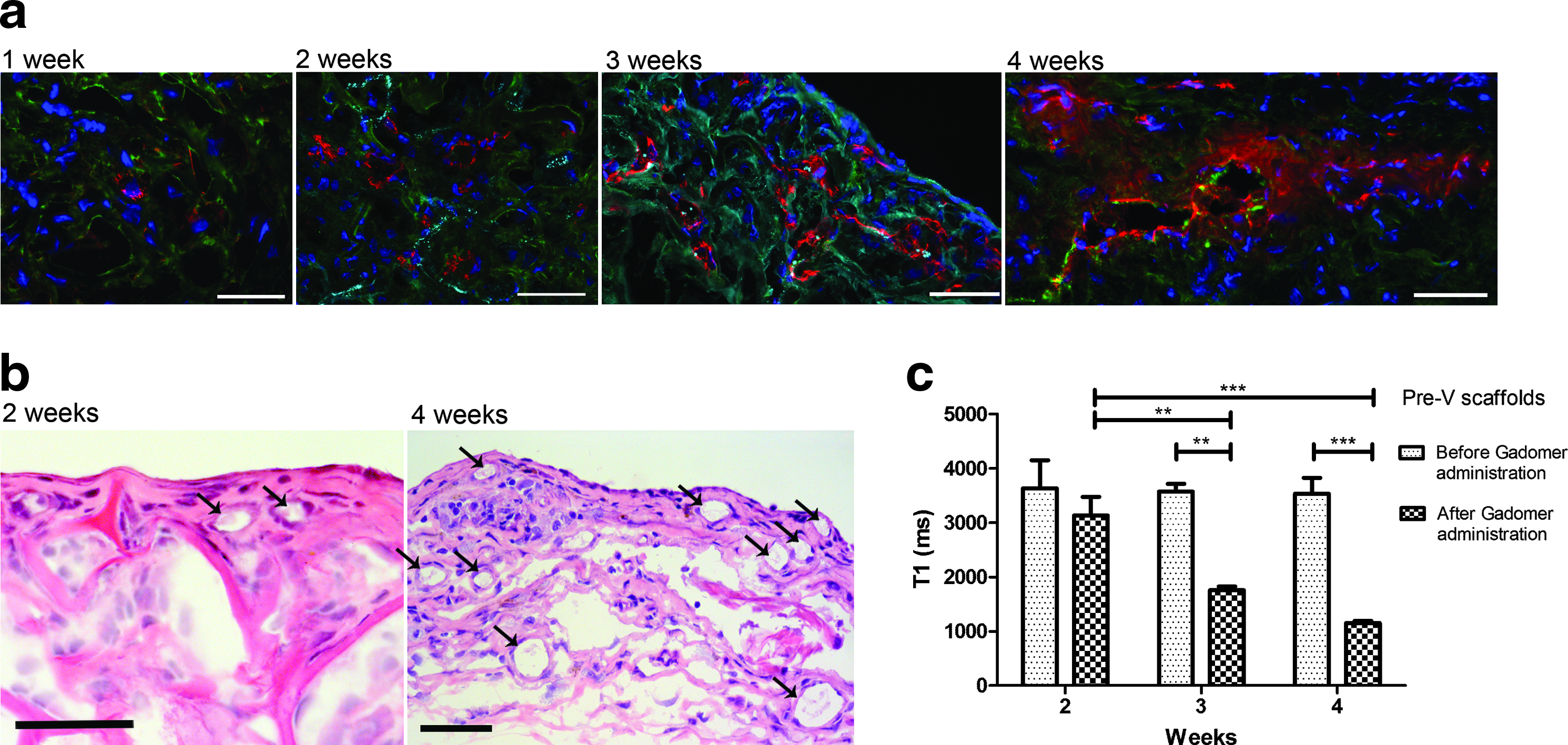
One week after implantation of pre-V templates, cells distributed equally throughout the template, well beyond the preimplant distribution. Small capillary-like structures (vWF+, pre-engineered, Fig. 3a, 1 week) containing exclusively human cells were present in the center of the templates. One week later, mouse neoangiogenesis became apparent as evidenced by the presence of mCD31+ mouse endothelial cells on the template surface in the vicinity of pre-engineered microvasculature (vWF+, Fig. 3a, 2 weeks). One to 2 weeks later the number of small capillaries had increased as well as the length of the capillaries was increased to 200 μm and the complete vasculature was of mouse origin (Fig. 3a; 3 and 4 weeks). Morphological analysis with HE (Fig. 3b) showed the presence of erythrocytes in the small capillaries 2 weeks after implantation.
Analysis of Pre-V templates in vivo.
The surface of the Acellular templates was covered with newly formed vessels (Fig. 4a) and mouse cells penetrated into the hybrid templates up to 200 μm 4 weeks after implantation. When hUCs were seeded on top of the hybrid templates and evaluated 4 weeks later, the surface of the templates was covered with a cell layer of Krt5+ cells (Fig. 4a) and newly formed vessels (mCD31+). In contrast, the mouse vasculature (mCD31+) penetrated deeply into Pre-V and Pre-V + UC templates (300–400 μm; Fig. 4a) and mature vascular structures were observed 4 weeks postimplantation (Fig. 4a).

Analysis of different templates 4 weeks postimplantation.
To evaluate the functionality and inosculation of the pre-engineered vasculature, MRI studies were performed. At 2 weeks postimplantation, the T1 of the Pre-V templates decreased due to Gadomer-17 application, but this was not significant. However, at 3 and 4 weeks, T1 was significantly decreased, which is a profound perfusion of the templates (p < 0.01, Fig. 3c).
MR analysis of the four templates showed that at 4 weeks the difference in T1 due to Gadomer-17 was significant for all (p < 0.001, Fig. 4b), although the difference of T1 was more prominent for the prevascularized templates (Pre-V and Pre-V + UC). The percentage decrease in T1 was 34%, 48%, 67%, and 66% for Acellular, UC, Pre-V, and Pre-V + UC templates, respectively (Fig. 4b).
The sequential images acquired in the dynamic contrast-enhanced MRI 4 weeks postimplantation showed clear SI changes (Fig. 4c). From the signal–time curves, the time to peak, highest SI, and the AUC from 0 to 2000 s were obtained. Both the highest SI and AUC were larger for the Pre-V and Pre-V + UC than for the other templates (UC and Acellular), indicating more Gadomer-17 uptake and therefore more extensive vascularization (Table 2). The time to peak varied among the templates, but was highest in the Pre-V, suggesting that Gadomer-17 was more rapidly taken up (Table 2).
AUC, area under curve; MRI, magnetic resonance imaging; SI, signal intensity.
A subset of dynamic contrast-enhanced MR images of the four different hybrid templates 4 weeks postimplantation is shown in Figure 5. Clearly the SI enhancement in Pre-V and Pre-V + UC was rapid and strong; SI enhancement in Acellular and UC increased as well, but the intensities were much lower than those observed in Pre-V and Pre-V + UC templates. As expected, the surrounding tissues also showed enhanced SI due to vascular extravasation and diffusion of the administered contrast agent. An animation of dynamic Gadomer-17 uptake in Pre-V template is presented in Supplementary Video 1.

Dynamic contrast-enhanced MR images of four different types of templates 4 weeks after implanting. Images were recorded for 42 min after Gadomer administration, only the first pass between 5 and 205 s is shown. The well-perfused areas became bright rapidly, which is most obvious in the Pre-V and Pre-V + UC templates (at 55 s); UC became bright during 55 to 105 s; the Acellular template stayed darker than the surrounding tissues. The different implants can be easily distinguished on the MR images. White arrows indicate the hybrid templates. Black dots mark ROI, that is, the hybrid template. ROI, region of interest.
Discussion
Functional and quantitative studies may aid in the analysis of engineered/newly formed tissues, but (in general) analysis is limited to morphological and histological analysis, which does not provide this information. Imaging tools enable a more objective analysis of tissue formation and provide functional information of biological responses. 12 Implanted templates are becoming analogous to the tissue they replace, and classical imaging strategies are poorly equipped to image these implants. Consequently, the fate and effect of the implants, including vascularization, cannot be followed adequately. In this study, the progress of vascular growth in hybrid templates was monitored with contrast-enhanced MRI. The initial tissue uptake of the intermediate-sized contrast agent, Gadomer-17, reflects tissue vasculature (blood volume and flow and vessel permeability).13,14
Although MRI is extensively used in the clinic and in (pre-) clinical research,6,15 it has been rarely used in studies addressing tissue engineering questions.13,16 Several imaging tools have been used to monitor degradation and vasculogenesis in scaffolds: near-infrared fluorescence microscopy, 17 fluorescence microscopy, 18 photoacoustic imaging,19,20 and intravital confocal imaging.21,22 These imaging tools provide high resolution up to cellular level, but the penetration depth is limited, in contrast to MRI, and anatomical information is lacking. In this study, we demonstrate that MRI is very well suited to monitor in vivo tissue engineering approaches, and to our knowledge, this is the first study to analyze functional vascularity in cell-seeded templates in vivo by MRI.
Red blood cells were already detected in the prevascularized templates 2 weeks postimplantation, but in our experiments, this did not yet result in a significant effect of the Gadomer-17 on T1 relaxation. In the third week postimplantation, the vasculature of prevascularized templates, as judged by this effect on the T1 relaxation time, significantly increased and in the fourth week even more. This suggests the progressive extension of a functional blood vessel network connecting the host and the in vitro pre-engineered templates. The histology confirmed that more and more mature vessels were present at 4 weeks: the extracellular matrix contained laminin and type IV collagen, indications of a mature vasculature, and formed a sleeve around endothelial tubes (mCD31+) reminiscent of a basement membrane (Supplementary Fig. S3). Clearly, once inosculation has occurred, contrast-enhanced MR adequately visualizes the functional vasculature in the templates.
Furthermore, AUC2000 values were higher in the prevascularized templates than in neovascularized templates. Contrast shortening of T1 relaxation times was observed for all templates 4 weeks postimplantation. The data suggest that in neovascularized templates, the vasculature was restricted to the surface, whereas in prevascularized templates, perfusion occurred throughout the template. Immunohistochemistry (IHC) analysis confirmed that the surface of Acellular and UC templates was covered by newly formed vessels, explaining the contrast effect on T1 relaxation time. Thus, the MR analysis correlated well with the histological observations and this shows that contrast-enhanced MR can be used to distinguish prevascularization from neovascularization. We conclude that contrast-enhanced MRI is an attractive tool for longitudinal follow-up to evaluate the vascular outcome without the need to sacrifice animals. Moreover, the functional information can be superimposed on the static histological information.
One limitation of MR is that it cannot distinguish host from donor cells. To study the degree of mouse endothelial cell involvement, IHC was performed, demonstrating that human cells (hMt+) were lining the prevascular structures in the hybrid templates 1 week after implantation. One week later, mouse endothelial cells (mCD31+) and human endothelial cells (vWF+) were present simultaneously in the template vasculature. Two weeks later, the template vasculature consisted exclusively of CD31+ mouse cells. This occupancy by mouse endothelial cells is most likely the consequence of the mouse blood flow that facilitated the process of mouse endothelial cell grafting and elimination of human cells.
For future studies, multimodality imaging, for example, MRI combined with PET or SPECT with antibody-conjugated probes might enable distinction of human and murine cells.23,24 Alternatively, human cells could be labeled with superparamagnetic iron oxide (SPIO) or ultrasmall SPIO (USPIO) in vitro 25 and be implanted in animals to permit MRI evaluations.
It is well established that VEGF plays a crucial role in angiogenesis, 26 and we envisioned that preseeded cells would release VEGF after template implantation because hypoxic conditions would prevail. 27 In general, Pre-V templates showed the lowest mVEGF-A level at all time points and mVEGF-A RNA levels decreased in time (Supplementary Fig. S4). It is likely that the lower VEGF expression levels are the consequence of vasculature in templates (i.e., a less hypoxic condition). This information again suggests changes in vascularity in the Pre-V templates, substantiating the MRI and histological observations.
Because epithelial cells (hUCs) seeded on top of prevascularized scaffolds may function as a barrier, they influence inosculation and neovascularization. By IHC analysis, no major differences between epithelial-covered and noncovered templates were observed. However, the MRI did show that the permeability of the prevascularized template was higher than the epithelial-covered vascularized template. Thus, the MRI suggests that epithelial cells functioned as a barrier, influencing the vascularity of the template, probably by blocking inosculation and/or preventing connection of neovasculature to the engineered vessels.
Studies have shown that inosculation of prevascularized fibrin- or collagen-based hydrogels was faster and the density of lumen was much higher than those achieved in our collagen/fibrin template.28–30 This slow progress might be explained by the fundamental difference between soft hydrogels where cellular outgrowth is not restrained from collagen sponges. This leads to a lower prevascular density in our templates and this diminishes the chances of inosculation. Moreover, the soft fibrin and collagen gels are more favorable materials for cell growth. Nevertheless, despite this slower inosculation, human endothelial cells survived for 2 weeks, demonstrating that these prevascularized materials can provide a temporary environment for cellular accommodation and might be useful for larger grafts that require (temporary) mechanical support.
Conclusion
MR is a valuable tool to monitor implanted materials in vivo in longitudinal studies. Moreover, with the use of contrast agent, it represents a powerful noninvasive imaging tool to follow vasculogenesis in these implanted materials.
Footnotes
Acknowledgments
The authors would like to acknowledge Gijs Rikken (technical support), Aleksandra Dudek-Madej (primer design), and Silvia Mihaila (discussion) at the Department of Urology. The technical support of Bianca Lemmers-van de Weem, Central Animal Laboratory and Andors Veltien (MRI), the Department of Radiology, is greatly acknowledged. The authors would like to thank the Microscopic Imaging Centre for use of facilities. The research leading to these results has received funding from the European Community's Seventh Framework Programme (MultiTERM, grant agreement nr 238551) and from NWO investment grant (nr 40-00506-98-06021).
Disclosure Statement
No competing financial interests exist.
References
Supplementary Material
Please find the following supplemental material available below.
For Open Access articles published under a Creative Commons License, all supplemental material carries the same license as the article it is associated with.
For non-Open Access articles published, all supplemental material carries a non-exclusive license, and permission requests for re-use of supplemental material or any part of supplemental material shall be sent directly to the copyright owner as specified in the copyright notice associated with the article.