




Abstract
Scaffold contraction is a common but underestimated problem in the field of tissue engineering. It becomes particularly problematic when creating anatomically complex shapes such as the ear. The aim of this study was to develop a contraction-free biocompatible scaffold construct for ear cartilage tissue engineering. To address this aim, we used three constructs: (i) a fibrin/hyaluronic acid (FB/HA) hydrogel, (ii) a FB/HA hydrogel combined with a collagen I/III scaffold, and (iii) a cage construct containing (ii) surrounded by a 3D-printed poly-ɛ-caprolactone mold. A wide range of different cell types were tested within these constructs, including chondrocytes, perichondrocytes, adipose-derived mesenchymal stem cells, and their combinations. After in vitro culturing for 1, 14, and 28 days, all constructs were analyzed. Macroscopic observation showed severe contraction of the cell-seeded hydrogel (i). This could be prevented, in part, by combining the hydrogel with the collagen scaffold (ii) and prevented in total using the 3D-printed cage construct (iii). (Immuno)histological analysis, multiphoton laser scanning microscopy, and biomechanical analysis showed extracellular matrix deposition and increased Young's modulus and thereby the feasibility of ear cartilage engineering. These results demonstrated that the 3D-printed cage construct is an adequate model for contraction-free ear cartilage engineering using a range of cell combinations.
Introduction
T
Hydrogels, which can be made from either synthetic or natural polymers, have been studied extensively in the context of cartilage tissue engineering.13,14 Hydrogels are particularly attractive since they are biocompatible,15,16 can be remodeled by cells, 17 and are permeable to oxygen, metabolic waste, and growth factors. 18 Often used hydrogel materials are (mixtures of) collagens, because of their excellent biocompatibility and negligible immunogenicity, commercial availability, and active use in clinical applications.19–22 Unfortunately, severe contractile remodeling of some hydrogels by cells, causing a decrease in volume to only 20–30% of the original volume, disrupts the shape of the engineered structure.23–25 This becomes particularly problematic when engineering anatomically complex shapes such as the ear. Therefore, most current auricular constructs are made from natural-based hydrogels with an interspersed lattice network of a synthetic polymer. Although these constructs seem feasible, 26 synthetic materials are very slowly degraded and may provoke a foreign body reaction that damages the implant. 27 In addition, insufficient long-term degradation studies have been undertaken to understand the behavior of these polymers.
In this study, we aim to construct rapidly producible and biomechanically stable scaffold structures with an optimized biochemical extracellular matrix (ECM) environment to generate and maintain proper ear cartilage. For this purpose, we evaluated combinations of (i) a novel cartilage-mimicking hydrogel, (ii) various cell types, and (iii) auxiliary scaffolds for creating lattice- and contraction-free scaffolds.
Materials and Methods
Cell isolation and culture
Mesenchymal stem cells
Goat mesenchymal stem cells (MSCs) were isolated from subcutaneous adipose tissue of skeletally mature Dutch milk goats (age 3–5 years) as described by Zuk et al.
28
The research protocol was approved by the scientific board and the animal ethics committee of the VU University, and is in accordance with national guidelines and regulations. In brief, adipose tissue was washed with phosphate buffered saline (PBS; Life Technologies) to remove blood and debris. The tissue was minced, washed three times with PBS, and enzymatically dissociated with 1U Liberase™ (Roche Diagnostics) per gram of tissue for 60 min at 37°C under gentle agitation. Digested tissue was filtered through a 100 μm mesh filter to remove residual ECM, pelleted using centrifugation at 600 g for 10 min, and washed several times with PBS. The resulting cell suspension was directly used for culture or repelleted, resuspended in cryoprotective medium (Recovery™ Freezing Medium, Life Technologies), and stored in liquid nitrogen. Cells were cultured as adherent cells in T175 flasks (Cellstar®; Greiner Bio-one) in GIBCO® Dulbecco's modified Eagle medium (DMEM; Life Technologies) supplemented with 10% fetal bovine serum (FBS; Hyclone–Thermo Scientific), 1% PSF (10,000 U/mL Penicillin, 10 mg/mL Streptomycin, 25 mg/mL Amphotericine B (Fungizone); Sigma Aldrich), and 50 μg/mL
Chondrocytes and perichondrocytes
Ear cartilage and perichondrium were obtained from the dorsal side of the ears of sacrificed skeletally mature Dutch milk goats (age 3–5 years) acquired from a local abattoir. Ears (n = 5) were transported to the laboratory on ice, thoroughly washed with soap to remove blood and debris, and shaved. Under sterile conditions, the perichondrium and cartilage were carefully dissected and isolated and immediately preserved in clear DMEM containing 2% PSF to prevent drying out. The cartilage and perichondrium were cut into 1 mm2 pieces and treated with 0.2% type II collagenase (Roche diagnostics) under gentle agitation at 37°C for 16 and 2 h, respectively. Digested tissue was filtered through a 100 μm mesh filter to remove residual ECM, pelleted using centrifugation at 600 g for 10 min, and washed several times with PBS. The resulting cell suspension was directly used for culture. For subsequent experiments, chondrocytes and perichondrocytes were cultured up to passage 2 under culture conditions similar to adipose-derived MSCs (adipose stem cells [ASCs]).
Scaffold terminology
Three constructs were tested in this study: (i) a proteoglycan-resembling hyaluronic acid moiety covalently coupled to a fibrin backbone (fibrin/hyaluronic acid [FB/HA] hydrogel), from now on referred to as the hydrogel, (ii) the combination of (i) and a collagen I/III scaffold (Optimaix®), from now on referred to as the scaffold, and (iii) the combination of (ii) and a surrounding 3D-printed PCL mold, from now on referred to as the cage construct. When referring to (ii) inside the cage construct, we use the term internal scaffold (Fig. 1).
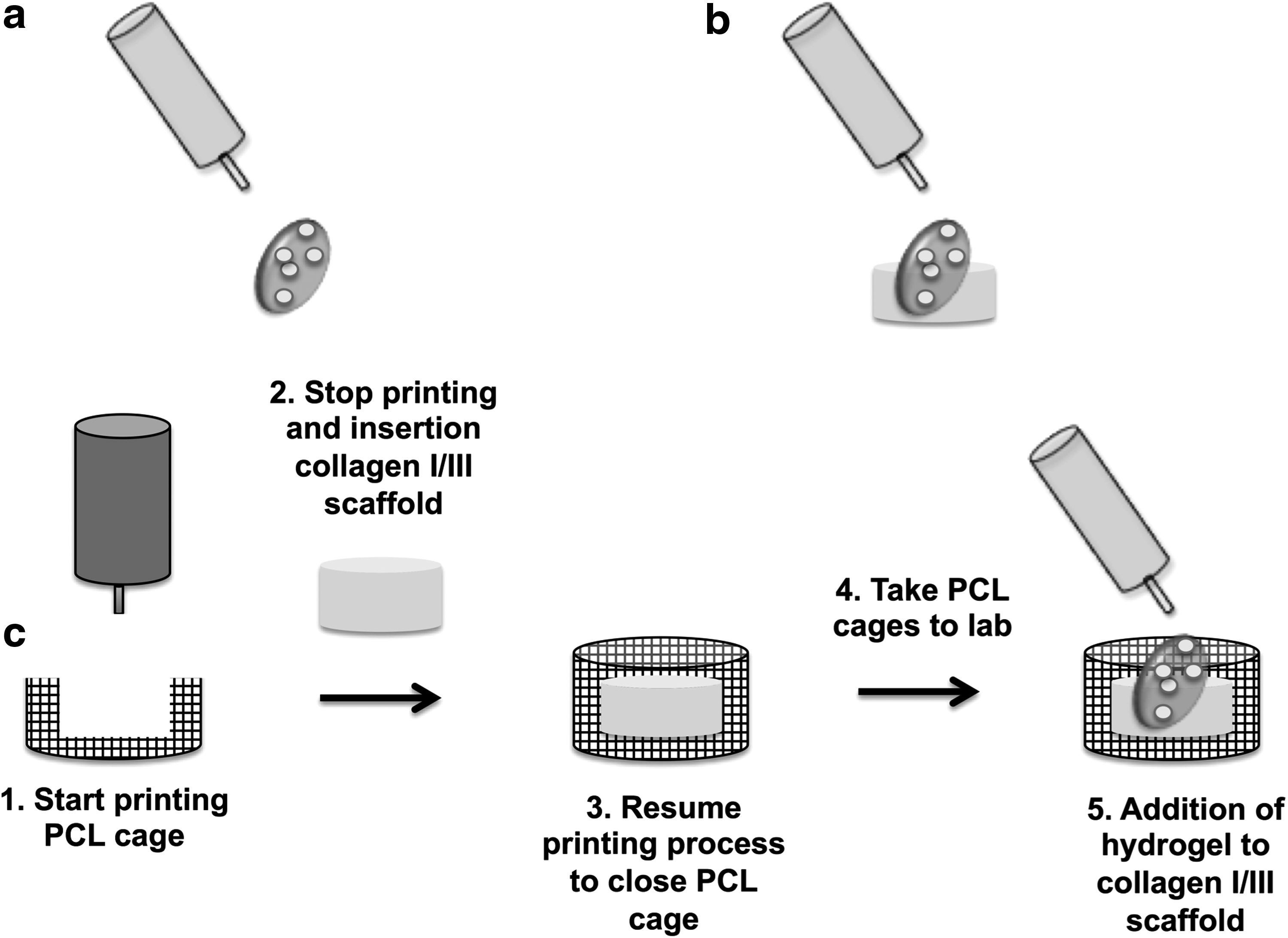
Schematic of the three cage constructs.
Preparation of the hydrogel (i)
After trypsinization, cells were counted using a Muse™ Cell Analyzer (Merck Millipore) and kept in plain DMEM until used. A FB/HA hydrogel, synthesized by the reaction of a buffered fibrinogen solution with a HA-active ester solution using HA with a molecular weight of 235 kDa, was obtained from ProCore Bio Med Ltd. The ratio of FB/HA was 17:4. Chondrocytes (C), ASCs, and a combination of chondrocytes–ASCs (CA, 20:80 ratio) were individually resuspended in a thrombin solution (3.2 U/mL) diluted with a 15 mM CaCl2 solution and added in a 1:2 ratio to the FB/HA hydrogel to cause gelation of the final cell/hydrogel mixture (Table 1). A total of 1 × 10⁶ cells were added to a final volume of 50 μL hydrogel and seeded into a six-well plate (Cellstar; Greiner Bio-one). The gels were cultured in 1.5% agarose (Invitrogen) cups in DMEM supplemented with 10% FBS, 1% PSF, and 50 μg/ml
Hydrogel.
Scaffold.
Internal scaffold.
ASC, adipose stem cells.
Preparation of the scaffold (ii)
The scaffold was prepared as described in the first steps of “Preparation of the Hydrogel (i)”. However, before gelation of the final cell/hydrogel mixture, a final volume of 50 μL hydrogel with a cell density of 1 × 10⁶ cells was seeded onto a 5 mm Ø ×3 mm height collagen I/III scaffold (Optimaix; Matricel GmbH) (Table1). The following cell combinations were tested in the scaffold: C, ASC, CA, perichondrocytes (P), perichondrocytes–ASCs (PA, 20:80 ratio), and CP (20:80 ratio). The scaffolds were cultured for 14 and 28 days as described in “Preparation of the Hydrogel (i)”.
Preparation of the cage construct (iii)
3D model designing
The 3D cages were designed using bioCAD® software version 1.0 (RegenHU). In the bioCAD software, construct outlines were drawn in layers. To create pores in the walls of the constructs, a double wall was drawn and alternated in thickness between 0.22 and 0.24 mm. Layers were subsequently stacked to obtain 3D shapes. Feed rate, indicating printing speed per layer, was continuously set to alternate between 3.2 and 10 mm/s depending on the complexity of the printed layer. Final shapes were generated as ISO files, containing a G-code that was imported into the 3D bioprinter (3DDiscovery®; RegenHU).
Preparation of 3D-printed cage constructs
A 3DDiscovery bioprinter (RegenHU) was used to print the constructs designed with bioCAD. All cage constructs were printed using the HM-300H thermo polymer extruder (RegenHU) at 35 revs/min, equipped with a needle with an inner diameter of 300 μm (designed and created by the Department of Physics and Medical Technology [FMT] at the VU University Medical Center [VUMC, Netherlands]). Printing was performed under semisterile conditions; the 3DDiscovery bioprinter and all materials were thoroughly cleaned with 70% ethanol using sterile gloves. Medical grade PCL (Purasorb; Purac Biomaterials) was melted at 85°C in the heating tank. The PCL was extruded through a preheated needle (75°C) at 0.3 MPa (3 barr), and the filament of PCL was plotted as layer-by-layer deposition on the lid of a sterile six-well plate (Cellstar; Greiner Bio-one). Printing parameters were optimized by changing the pressure and deposition speed accordingly, which allowed minimization of strand diameter and oozing of the material. During the printing process, time was allocated for insertion of the scaffold (ii) in the PCL cage (Fig. 1). This was done by reprogramming of the G-code before the start of the printing process.
Macroscopic size evaluation
The size of the hydrogel (i), scaffold (ii), and internal scaffold (in iii) was determined by macroscopic imaging using a Zeiss Axio Zoom V16 Stereo microscope (Carl Zeiss). The surface area of the hydrogel and scaffold was measured immediately after cell seeding, at day 14, and at day 28 of cell culture using Zeiss Efficient Navigation digital imaging software. The surface area was measured in square millimeters by drawing a region of interest along the parameters of the scaffolds.
Sample processing
After macroscopic size evaluation after 14 and 28 days, the scaffold (ii) and internal scaffold (iii) were processed for additional testing. Both the scaffolds (ii) and cage constructs (iii) were harvested and cut in half using a sterile surgical blade. After cutting the cage construct in half, the internal scaffold (iii) was carefully removed from the PCL cage using sterile tweezers. For both the scaffold (ii) and the internal scaffold (iii), one half was used for histological and immunohistological testing and the other half for second harmonic generation (SHG) analysis and subsequent mechanical testing.
The halves of the scaffolds (ii and iii) allocated for histology and immunohistochemistry were carefully washed in PBS twice, fixed in 4% phosphate buffered formalin (pH 7.2) for at least 5 h, and embedded in paraffin. Paraffin-embedded scaffolds were cut to 5 μm thick sections and mounted on glass slides. The halves (ii and iii) allocated for SHG and mechanical testing were washed in PBS and stabilized in a petri dish using 4% agar (Fig. 2A).
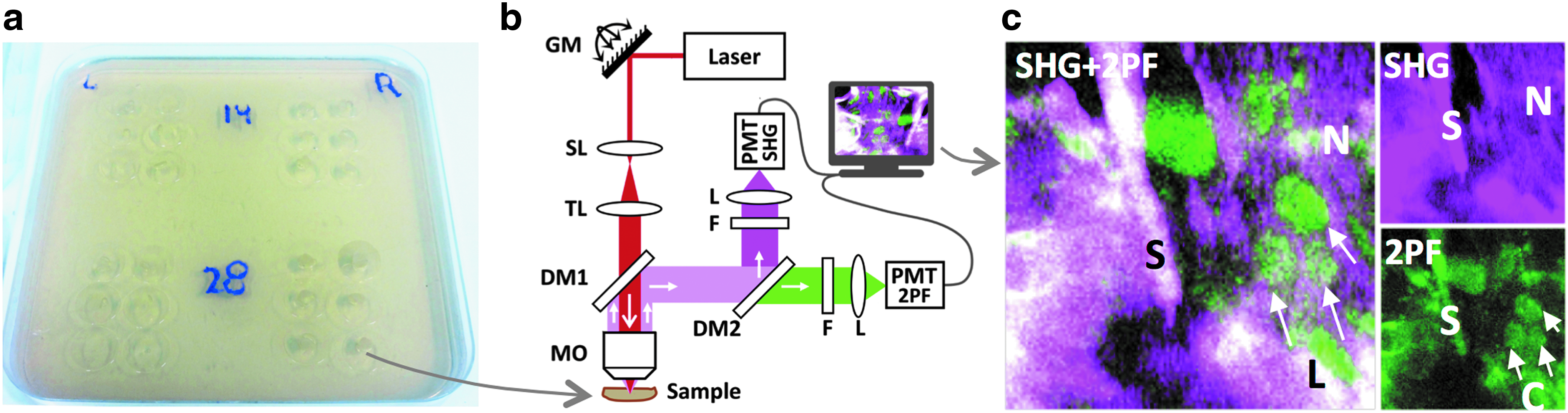
SHG/2PF imaging of collagen I/III scaffolds.
Histological evaluation and immunohistochemistry
Glass slides were systematically stained with hematoxylin and eosin (H&E) and Alcian Blue by the department of pathology (VU medical Center, Amsterdam) using standard histological techniques or treated with monoclonal antibodies to type II collagen (II-II6B3, Developmental Studies Hybridoma Bank).
For immunohistochemistry, sections were pretreated with 10 μg/mL proteinase K for 10 min at 37°C, rinsed thoroughly with PBS, and incubated at 4°C overnight with a 1:50 dilution of primary antibody. The next day, sections were incubated with rabbit antimouse secondary antibody (Zymed) for 30 min at room temperature. Positive staining was observed by Dako REAL™ EnVision™/HRP (Dako) with 3,3′-diaminobenzidine solution as substrate. The sections were counterstained in hematoxylin. Negative control was performed with mouse IgG under identical conditions.
Second harmonic imaging of collagen
SHG and two-photon excited autofluorescence (2PF) microscopy were used to observe collagen bundles and cellular material in the scaffold (ii), which has been done previously in.29–32 This technique provides a 3D observation of collagen bundles and cells without fixation, slicing, and labeling. The scaffolds (ii and iii) were covered with a 0.17 mm cover glass slides to generate flat interface (Fig. 2A). A commercial two-photon laser-scanning microscope (TrimScope I, Lavision BioTec GmbH) and a femtosecond Ti-sapphire laser source (Coherent Chameleon Ultra II) generating 200 fs pulses at 800 nm with linear polarization and repetition rate of 80 MHz were used for SHG/2PF imaging (Fig. 2B). The laser beam was focused on the samples by a high-numerical aperture water-dipping objective (25×/1.10, Nikon APO LWD), providing transverse resolution of 0.5 μm and axial resolution of 2 μm. Laser power was adjusted in the range of 5–50 mW to attain sufficient signal-to-noise ratio and avoid tissue photo damage. The laser beam was scanned transversely over the samples by galvo mirrors on the microscope objective through scan and tube lens. Depth scanning was accomplished by vertical translation of the objective with a stepper motor. SHG and 2PF photons were collected in the epi-detection geometry, filtered from the 800 nm excitation photons by the DM1 dichroic mirror (Chroma T695lpxrxt), split into SHG and 2PF channels by the dichroic mirror DM2 (Chroma 425lp), passed through interference filters (F) for SHG (Chroma Z400/10X) and 2PF (Chroma HQ500/140M-2P), and detected by high-sensitivity GaAsP photomultiplier tubes (Hamamatsu H7422-40). Data acquisition was performed with TriMScope I software (Imspector Pro). Image stacks were stored in 16-bit tiff-format and further processed and analyzed with ImageJ software (MacBioPhotonics).
Mechanical analysis
Mechanical analysis of the engineered cartilage was performed using a commercial microindenter (Piuma, Optics11) equipped with a built-in displacement control and a spherical indenter (Ø 78 μm) with a surface tension of 0.710 N/m. Cantilever-bending calibrations were performed on a rigid surface before analysis. Each scaffold was indented with a linear extension and retraction of 20 μm for 5 s, holding the probe for 2 s at peak extension. During analysis, the scaffolds were placed on a PBS-covered medical mesh to prevent dehydration and minimize adhesive forces between probe and tissue surface. To average out possible surface roughness from the surgical cut as well as inhomogeneity, each scaffold was indented on nine locations with 300 μm spacing to ensure independent measurements. To maximize contact area and minimize viscoelasticity, an extension rate of 4 μm/s and a retraction rate of 8 μm/s were used. Stress–strain curves were analyzed using the model for a spherical indenter (Oliver & Pharr) to determine the effective Young modulus.
Statistical analysis
All data were analyzed using GraphPad Prism® version 6.02. Data are represented as mean ± standard deviation. All experiments were performed in triplicate (n = 3) unless stated otherwise. Values of p < 0.05 were considered statistically significant. For scaffold contraction, individual paired t-tests were performed to compare difference in scaffold contraction between days 1, 14, and 28 of culturing.
Results
Gross observation of cage constructs and scaffold contraction
Six PCL cage constructs could be manufactured and printed in 2 h. Printing accuracy was excellent in all molds, with minor negligible oozing effects. The constructs showed adequate porosity on the lateral walls using the alternating layer thickness and double wall method (Fig. 3a). After 28 days of in vitro culturing at 37°C, all cage constructs showed no deformation or shrinkage. Culture medium was distributed evenly over the cage constructs during culturing as seen in the Optimaix scaffold (Fig. 3c).
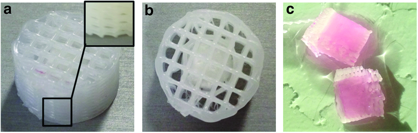
Gross morphology of the cage construct (iii).
Cell-seeded hydrogels (i) showed severe macroscopic contraction in vitro (Fig. 4a). After 28 days of culturing, the hydrogels contracted to 15–48% of their original volume depending on the cell type (p < 0.05). Less contraction was seen when combining the hydrogel with the Optimaix scaffold (scaffold ii). Scaffold (ii) showed an initial contraction of ∼20% after 14 days (p < 0.05, Fig. 4b) and additional 10% after 28 days of in vitro culturing (p < 0.05, Fig. 4b). Because of the clinical significance of CA and CP, we chose to further explore only these cell types in the cage construct (iii). No contraction occurred in the internal scaffold (Fig. 4c). As a control we tested acellular constructs (i) and (ii). These constructs did not contract (data not shown).

In vitro macroscopic contraction of
Cartilage-specific ECM production
Scaffold (ii) and internal scaffold (iii) were analyzed for ECM production after in vitro culturing using Alcian Blue staining for glycosaminoglycan and collagen type II for collagen production. All the scaffolds (ii) and internal scaffolds (iii) showed histological proof of glycosaminoglycan deposition, except for scaffolds with A and CA, which produced notably less matrix (Figs. 5 and 6). As shown in Figure 5, cells were distributed evenly throughout the scaffolds. There was evidence of collagen type II production in all scaffolds (ii). Chondrocytes and CP-seeded scaffolds (ii) produced most collagen. Higher magnification showed synthesis of collagen type II throughout the scaffolds, but less so in adipose stem cell- and PA-seeded scaffolds. An earlier study from our group 22 has shown that the collagen I/III scaffolds do not stain for type II collagen. Immunohistological analysis of the internal scaffold (iii) showed similar staining for CP compared with scaffolds (ii), but more intense staining for the CA group, which is probably explained by the increased cell density (Fig. 6).

Alcian Blue and immunohistological staining for type II collagen of scaffold (ii) after 28 days of in vitro culturing. Scaffolds were seeded with different cell types as shown in the figure. Black squares indicate magnified areas. Color images available online at
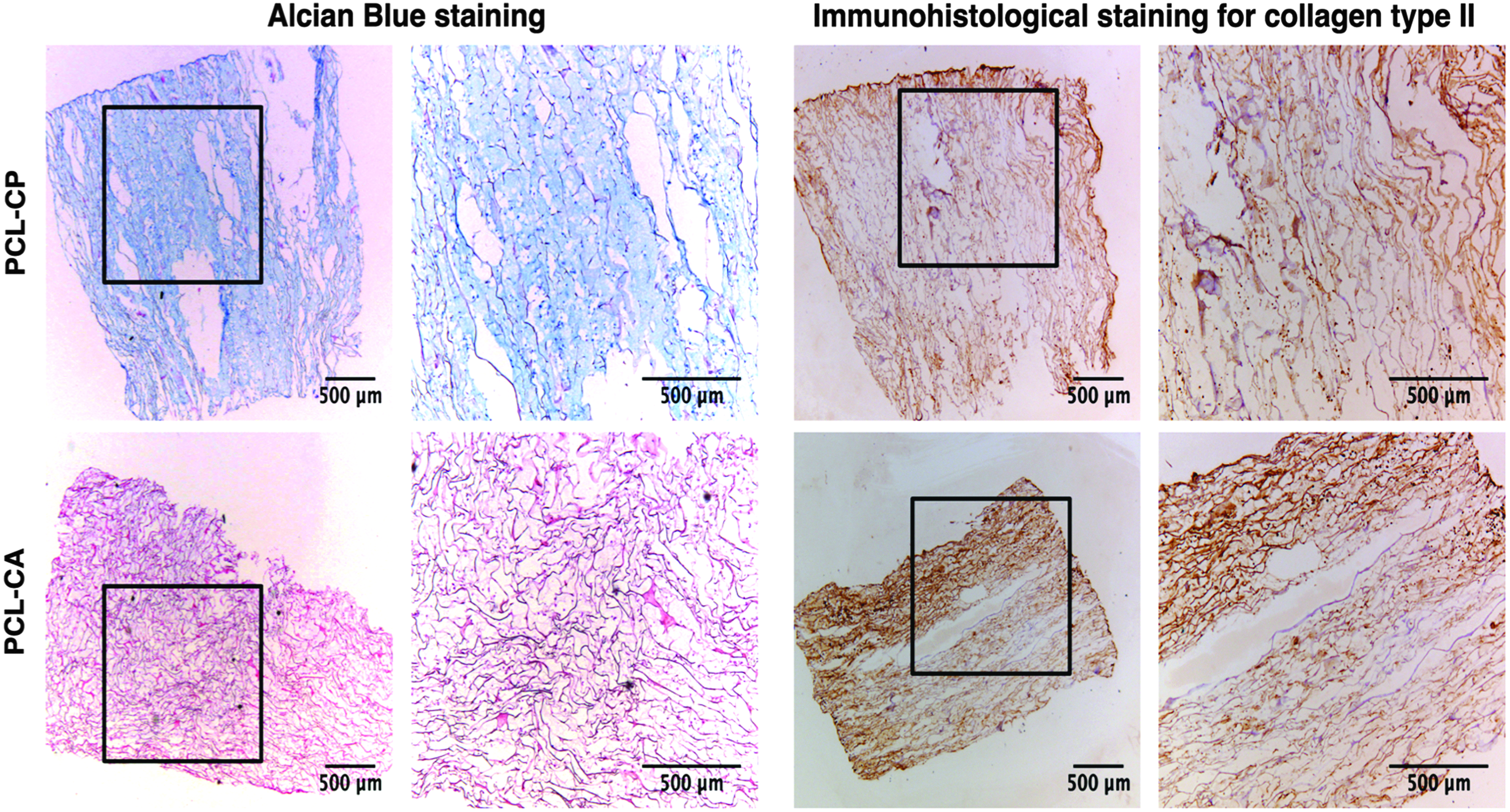
Alcian Blue and immunohistological staining for type II collagen of the internal scaffold (iii) after 28 days of in vitro culturing. CP, chondrocytes–perichondrocytes (20:80 ratio); CA, chondrocytes–adipose-derived mesenchymal stem cells (20:80 ratio). Black squares indicate magnified areas. Color images available online at
Second harmonic generation of collagen
SHG/2PF images of cartilage tissue correspond well with the “gold standard” H&E histology images and other stained sections commonly used to observe tissues33–37
and show collagen fibers and cells in detail. All scaffolds (ii) showed collagen production after 14 and 28 days of in vitro culturing. Detailed collagen bundles in the scaffold could be imaged by SGH and are shown in purple. White color indicates the backbone structure of the collagen I/III scaffold generating both SHG and 2PF, and cells with surrounding ECM proteins are shown in green because of two-photon excited autofluorescene (Fig. 7). After 14 days of in vitro culturing, all cell types and combinations show collagen production (Fig. 7). After 28 days of culturing, chondrocytes, perichondrocytes, and their combination (CP) seemed to produce most new collagen fibers (Fig. 7). Magnified regions of interest containing typical collagen (N), scaffold (S), and cells (L) (Fig. 2C) are indicated with yellow squares in Figure 7A–K and M–W and shown on corresponding Figure 7B–L and N–X. Depth scans of Figure 7A–X areas were performed with a 2 μm step. Figures 7G, 7I, 7S and 7U are shown in Supplementary Movies SG, SI, SS, and SU (Supplementary Data are available online at

SHG/2PF imaging of collagen fibers in the scaffold (ii). Images were taken at 14 and 28 days of in vitro culturing. Collagen is shown in purple. Green represents cellular material. White represents the backbone structure of collagen I/III scaffold (ii). When compared with day 14 of culturing, collagen fibers increased in all tissues after 28 days. Yellow squares indicate magnified areas. See Supplementary Movies for labels G, I, S and U. Color images available online at
Mechanical cartilage quality
Young's modulus was measured for both scaffold (ii) and internal scaffold (iii) after 14 and 28 days of in vitro culturing. Overall, Young's modulus ranged from 2 to 60 kPa. Young's modulus of all scaffolds (ii) and internal scaffolds (iii) increased with ∼50% after 28 days of in vitro culturing when compared with 14 days of culturing (Fig. 8). The internal scaffolds (iii) showed a significant increase in biomechanical properties after 28 days of in vitro culturing (p < 0.05).
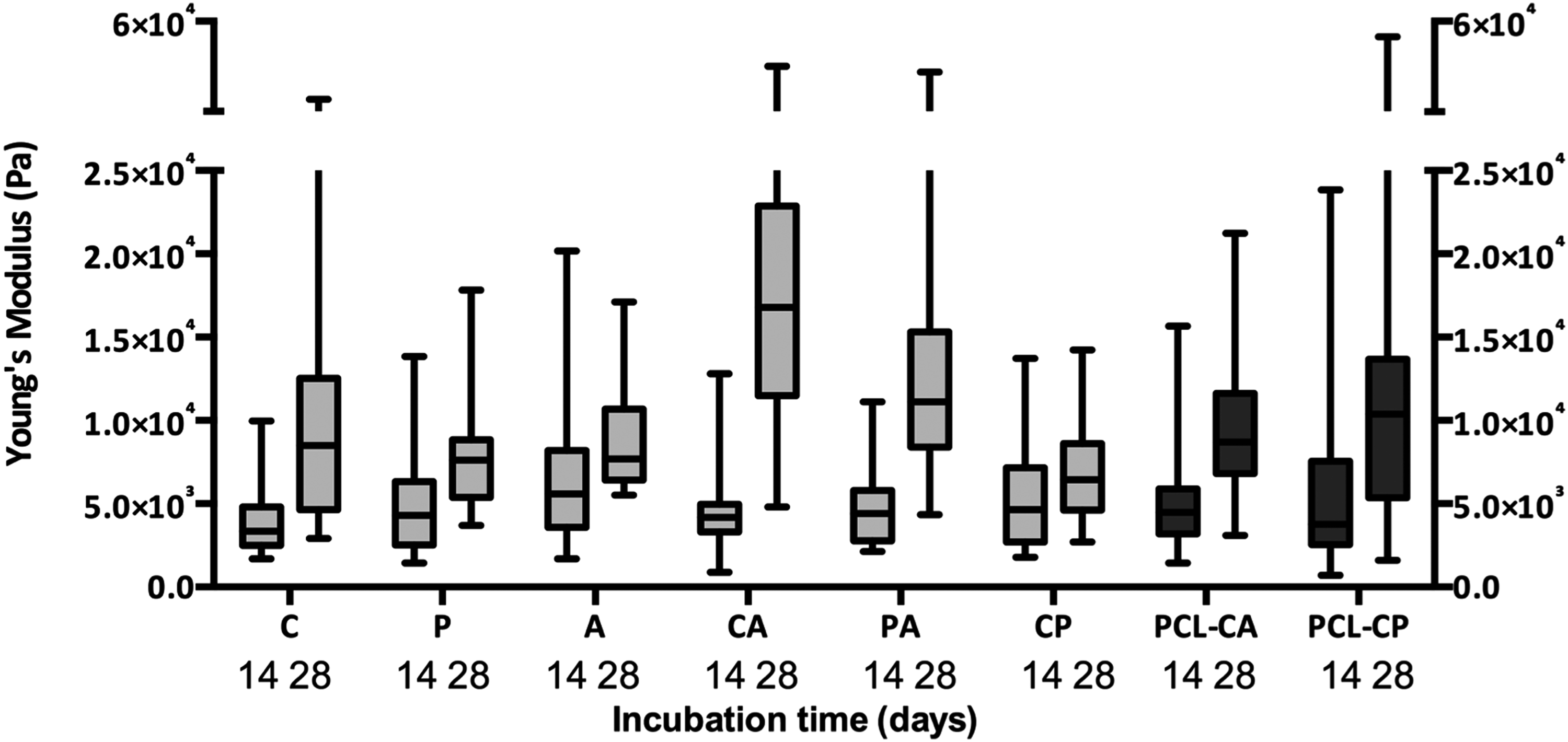
Box plot comparing biomechanical properties of cell-seeded scaffolds (ii) and internal scaffolds (iii). Analyses were performed after 14 and 28 days of in vitro culturing using a ferrule-top microindenter. C, chondrocytes; P, perichondrocytes; A, adipose stem cells; CA, chondrocytes–adipose stem cells (20:80 ratio); PA, perichondrocytes–adipose stem cells (20:80 ratio); CP, chondrocytes–perichondrocytes (20:80 ratio); PCL-CA, internal scaffolds seeded with CA; PCL-CP, internal scaffolds seeded with CP. Whiskers lower and upper limit were set to 5th and 95th percentile. Horizontal bars within the box represent the median value.
Discussion
Fundamental problems in the field of ear cartilage tissue engineering are failure of maintenance of implant shape because of cell-mediated scaffold contraction and a lack of clinically applicable and available cell sources that promote adequate neocartilage formation. These problems call for novel contraction-free biocompatible scaffolds and appropriate cell combinations to create adequately shaped, tissue-engineered cartilage implants. A wide range of cell types were tested to find the optimal cell combination for neocartilage formation without the need for additional chondrogenic stimuli. These were subsequently cultured in the cage construct resulting in cartilage-specific matrix deposition (collagen type II and GAG) throughout the scaffold. Mechanical analysis showed increased Young's moduli of the cartilaginous core within the constructs over time. These results demonstrate that a 3D-printed lattice-free cage construct can be used for ear cartilage tissue engineering and may provide a promising strategy for clinical translation of engineered ear-shaped cartilage implants.
Chondrocytes (C) and adipose-derived MSCs (A) are known for their contractile remodeling.4,38–40 However, no previous studies have reported on scaffold contraction using perichondrocytes (P), a potential candidate for ear cartilage tissue engineering.11,41 We found that the cell-seeded hydrogels contracted to less than 15–45% of their original volume independent of the cell type used, which has also been reported by others. 42 There are some contradictions in the literature with regard to whether cell-mediated tissue contraction should be inhibited. Cell-mediated contraction seems to play an important role in obtaining highly organized matrix deposition.43,44 However, scaffold contraction reduces pore size and decreases cell migration. 45 More important, contraction changes the size of tissue-engineered implants and causes difficulty in fitting a specific implant site. Therefore, inhibition of contraction is considered essential to obtain a highly accurate cartilage implant.
In a previous in vivo osteochondral defect model in goat articular cartilage, we employed Optimaix scaffolds, consisting of a highly porous collagen I/III matrix with aligned pores, which upon seeding with adipose stem cells showed strong hyaline cartilage-like ECM production in vivo after 4 months. 22 Since no evident shrinkage was observed in that study, we postulated that the porous collagen I/III scaffold could act as a backbone scaffold for cells encapsulated in hydrogel and decrease gel contraction. However, although decreased contraction was observed (60–70% of the original size was maintained), this still appeared insufficient to maintain shape.
Currently, much attention has been paid to 3D-printed biodegradable PCL scaffolds26,46,47 because of their biodegradability and low melting point (60°C).48,49 In addition, PCL promotes rapid cell attachment and cell proliferation.50–52 However, in contrast to currently published studies, we used a lattice-free construct with the hypothesis that long-term degradation products of an interspersed PCL lattice may affect cartilage quality. 27 In addition, only using an outer cage construct requires less material, which may promote biocompatibility. Strikingly, in our studies, PCL cages completely inhibited contraction of the internal scaffolds. This could be explained by an interaction of cells attached to the periphery of the inner scaffold and the surface of the PCL cage. The fact that increased levels of integrin activation are seen when cells are seeded onto a PCL scaffold 53 and decreased cell attachment is seen when integrin is blocked 54 supports this hypothesis. Because the rate of neocartilage formation will be substantially faster than the rate of PCL degradation,4,55 the cage can function as a temporary shape-stabilizing support that dissolves over time.
Apart from the maintenance of shape, it is obviously important that adequate ear cartilage formation is obtained within the construct. We tried to accomplish this by a dual approach: by using a novel chondroinductive natural-based FB/HA hydrogel, reported to promote neocartilage formation,15–22 in combination with the optimal (combination of) cell type(s) for chondrogenesis without the need for additional biochemical stimuli.
In our hydrogel-only experiments, we found that adipose-derived stem cells (A) and chondrocytes (C) combined with A (CA) showed less glycosaminoglycan production than other cell types. This is in contrast with other studies, which have shown ASCs to be capable of substantial cartilage remodeling, both in vitro 56 and in vivo.12,57,58 However, since we did not add any additional chondrogenic cues, ASC differentiation induction might have been insufficient. Nevertheless, the CA outcome was unexpected, since in earlier studies we showed that freshly isolated or cultured ASCs could promote the chondrocyte phenotype in articular chondrocyte–ASC coculture studies.59,60 It may be that the 20/80 ratio employed in those and the current study should be re-evaluated for ear chondrocytes. Alternatively, the major marker analyzed, the glycosaminoglycan content, may be more prominent in articular cartilage, whereas for ear cartilage, elastin may be a relevant and important matrix component. However, under the conditions and time frame tested, we were unable to observe elastin fibers with Elastica van Giesson staining.
Subsequent evaluation in the Optimaix and cage constructs showed staining for GAGs and collagen type II throughout the internal scaffold, although less intense than in other studies using pellet or micromass cultures. In this regard, it is very important to realize that in the latter situation where dense, contracted structures have formed, also “condensation” of staining will have occurred. In other words, less intense Alcian Blue and type II collagen staining in the hydrogel + collagen I/III scaffolds, either or not enveloped with the polymeric cage, is because of inhibition of contraction rather than aberrant matrix and collagen production. Nevertheless, the prevention of scaffold shrinkage concomitantly leads to lower cell densities, may have reduced interactions between cells, and subsequently may have caused delayed chondrogenic induction.
Interestingly, PA produced slightly more matrix than CA. Previous studies have shown that perichondrium cells can easily differentiate into chondrocytes11,61,62 and form cartilage that resembles native auricular cartilage both in vitro and in vivo.11,63–65 However, to our knowledge this is the first study that shows neocartilage formation by coseeding perichondrocytes with adipose stem cells. These results suggest that perichondrocytes have properties similar to chondrocytes and in combination with ASCs can deposit matrix to form neocartilage.
Finally, we sought to analyze whether matrix functionality was increased, that is, whether tissue was formed with an increased biomechanical Young's modulus and with not only merely increased but also well-organized matrix structure. The latter was evaluated using SHG/2PF microscopy. SHG/2PF, other than the “gold standard” histological techniques, is able to observe specific molecular orientations of materials once they assemble into fairly ordered, large noncentrosymmetric structures, for example, as collagen bundles. Moreover, SHG/2PF allows observation of these bundles three dimensionally, with greater detail than with simple histology. Collagen bundles were observed in all scaffold types using SHG/2PF, indicating structural reorganization toward neocartilage over a course of 28 days. Although our SHG imaging technique is not able to distinguish between different collagen types, our immunohistological stainings indicate that the bundles observed will primarily consist of collagen type II, the major collagen type in auricular cartilage. 66
Young's moduli from our engineered tissues were substantially lower than from native elastic cartilage, 67 in agreement with previous reports comparing in vitro and in vivo cartilage tissue engineering. 7 Nevertheless, Young's modulus of the scaffolds increased with longer culture times, which could explain differentiation toward neocartilage; an increase in Young's modulus may be linked to increased matrix deposition by the cells.68,69 Specifically, in our cage construct, more intense Alcian Blue staining correlated with higher Young's modulus.
Conclusion
In summary, this study demonstrated that a novel cage construct consisting of a combined hydrogel–collagen I/III scaffold surrounded by a 3D-printed synthetic PCL cage could prevent in vitro scaffold contraction, which is crucial for the production of adequately shaped tissue-engineered ear cartilage. Using this construct as implant model provides a promising strategy for clinical translation of engineered ear-shaped cartilage. Because complex ear-shaped PCL cages have already been produced in our laboratory, future studies should be aimed at in vivo testing of the presented scaffold construct in an anatomical ear shape.
Footnotes
Acknowledgments
The Dutch Burns Foundation financially supported the work presented in this article. The authors would like to thank Matricel GmbH for kind donation of Optimaix collagen scaffolds and Prof. Avner Yayon for the production and donation of FB/HA hydrogels. We thank Matthijs Barkman for his help with cell isolation.
Disclosure Statement
No competing financial interests exist.
References
Supplementary Material
Please find the following supplemental material available below.
For Open Access articles published under a Creative Commons License, all supplemental material carries the same license as the article it is associated with.
For non-Open Access articles published, all supplemental material carries a non-exclusive license, and permission requests for re-use of supplemental material or any part of supplemental material shall be sent directly to the copyright owner as specified in the copyright notice associated with the article.
