
Abstract
Micro- and nanotextured biomaterial surfaces have been widely studied for their capacity to drive the regeneration of organized tissues. Nanotopographical features in the shape of groove–ridge patterns aim at mimicking the extracellular matrix organization. However, to date, a wide array of groove and ridge sizes has been described. In this work, we therefore tested a device composed of a multipatterned array consisting of six patterns of radially arranged parallel nanogrooves, with a pitch ranging from 0 to 1000 nm and a depth ranging from 0 to 170 nm, to be used as a tool for the expeditious and simultaneous screening of surface topographies aiming the regeneration of anisotropically organized tissues such as ligament. The topographies were reproduced in (1) epoxy resin or (2) membranes produced by the crosslinking of platelet lysate (PL) with genipin (gPL). Both materials were seeded with periodontal ligament cells (PDLCs) and the proliferation, migration, as well as cell alignment were assessed. The effect of topography in PDLCs was only evident in terms of cell organization, resulting in a highly anisotropic organization of the cells for the 1000 and 600 nm patterns, and in an increased isotropic organization for shallower topographies. Overall, our results suggest that this multipatterned system can be a valuable diagnostic tool for biomaterials aiming at the regeneration of anisotropically organized tissues, such as periodontal ligament.
Introduction
T
The application of structurally organized scaffolds with aligned fibers or ridges/grooves has been proposed for the regeneration of anisotropically organized tissues.2–5 Recent advances in nanoengineering, such as photolithography, have allowed the development of biomimetic scaffolds that recapitulate the ECM of anisotropically organized tissues. Micro- and nanogrooves have been shown to modulate cell adhesion,6,7 orientation, 7 migration,7,8 and differentiation.6,8 However, such experiments were mainly performed on tissue culture plastic and therefore could not be clinically relevant. 3
The reproduction of such topographies into biomaterials, which can be used for tissue regenerative purposes, would allow for a possibility to fine-tune the performance of clinical applications. For example, the periodontal ligament (PDL) is a highly organized tissue composed mainly of collagen fibers connecting the tooth to the alveolar bone, whose anatomy and function can be affected by periodontitis. 9 The use of substrates with patterned grooves would be useful to promote fast and directional cellular migration for PDL regeneration. 4 However, topographies change cellular behavior often in a cell-type-specific manner.10,11 Also, interpatient heterogeneity12,13 might result in a diverse reaction to the same topography. Therefore, a suitable screening array could aid the selection of the tissue engineering construct with the most optimal topography.
In the current study, a concentric multipatterned culturing platform was used as a diagnostic device for screening of multiple submicron surface topographies for TE membranes used in periodontal regenerative therapy. The advantages of a device that features multiple topographies, instead of comparing individual samples, were already enunciated elsewhere 7 and can be summed up in three points: (1) reduced variation related with the material processing between substrates; (2) possibility to simultaneously compare cell behavior on multiple topographies, saving time and resources; and (3) reduction of statistical confounding effects. Besides the surface topography, also the biomaterial itself can be a variable.
Our previous research 14 evidences that platelet lysate (PL)-based membranes, fabricated by crosslinking of the PL proteins with genipin, and followed by solvent casting (gPL membrane), harbor several advantages as biomaterial for periodontal tissue regeneration. The biocompatible gPL membranes possess specific viscoelastic properties suitable for the regeneration of soft tissues that are subjected to dynamic forces. 14 Moreover, the gPL membranes were shown to be stable and do not retract as observed in other hemoderivatives, such as platelet-rich plasma and platelet-rich fibrin.15–17 Furthermore, PL is a natural source of growth factors and signaling molecules that are crucial for the wound healing process.18,19 We hypothesize that the multipatterned device can show differences in the response of periodontal ligament cells (PDLCs) to both the substrate, that is, gPL and a control polymer substrate, and the topography. Therefore, an optimum combination between substrate and topography could be found.
The ability of the gPL membrane to replicate the nanopatterns and maintain the topographic integrity after hydration was tested. Subsequently, the influence of parallel nanogrooves ranging from 150 to 1000 nm on the behavior of human periodontal ligament cells (hPDLCs) was assessed in terms of (1) cell proliferation, (2) cell migration, and (3) orientation after short (7 days) and long (14 days) culture periods.
Materials and Methods
Materials
Genipin for the crosslinking of PL-based membranes was purchased from Wako Pure Chemical Industries (Japan).
The basal medium for cell culture was composed of alpha-minimum essential culture medium purchased from Invitrogen, supplemented with 10% fetal bovine serum (FBS) (provided by Biochrom AG, Germany), and penicillin/streptomycin antibiotics (purchased from GIBCO®, Life Technologies, Bleiswijk, The Netherlands) at a final concentration of 0.25 and 0.60 μg/mL, respectively. Phosphate-buffered saline (PBS) 1× solution, pH 7.4, was purchased from GIBCO, Life Technologies.
Two-component epoxy resin Araldite/Aradur was purchased from Huntsman Corporation (Salt Lake City, UT). Polydimethylsiloxane (PDMS) Elastosil RT 601 was purchased from Wacker-Chemie (München, Germany). Liquinox™ critical cleaning liquid detergent was purchased from Alconox (White Plains, NY).
All the other reagents used in this work were research grade.
Platelet lysate
PL was prepared by freeze/thaw processing of platelet concentrate obtained by plasma apheresis, by adaptation of a previously reported method.18,19 The platelet concentrate batches, with an average platelet count of 106/μL, were provided by Instituto Português do Sangue (IPS, Porto, Portugal), under a previously established cooperation protocol and qualified according to the Portuguese legislation (Decreto-Lei No. 100/2011).
Briefly, batches from at least three different donors were pooled and subjected to two repeated freezing and melting cycles (frozen in liquid nitrogen at −196°C and thawed in a 37°C water bath). Then, the resulting lysis product was aliquoted (5 mL) in 15-mL falcon tubes and frozen at −20°C until further use. Just before use, the PL was defrosted in a 37°C water bath, completing the third freeze/thaw cycle. This process induces the platelets lysis and therefore the release of the protein content.18,19 The resulting cellular debris was removed by centrifugation at 400 g for 2 min.
Casting of multipatterned substrates by soft lithography
Multipatterned silicon wafers were produced by laser beam lithography by Lamers et al. 7 The Si wafers featured six different topographies, namely five parallel nanogrooved surfaces with the nominal pitch dimensions of 1000, 600, 300, 200, and 150 nm, and a depth of 170, 130, 50, 50, and 35 nm, respectively, and a smooth surface. The grooved patterns were radially organized as depictured in Figure 1A.
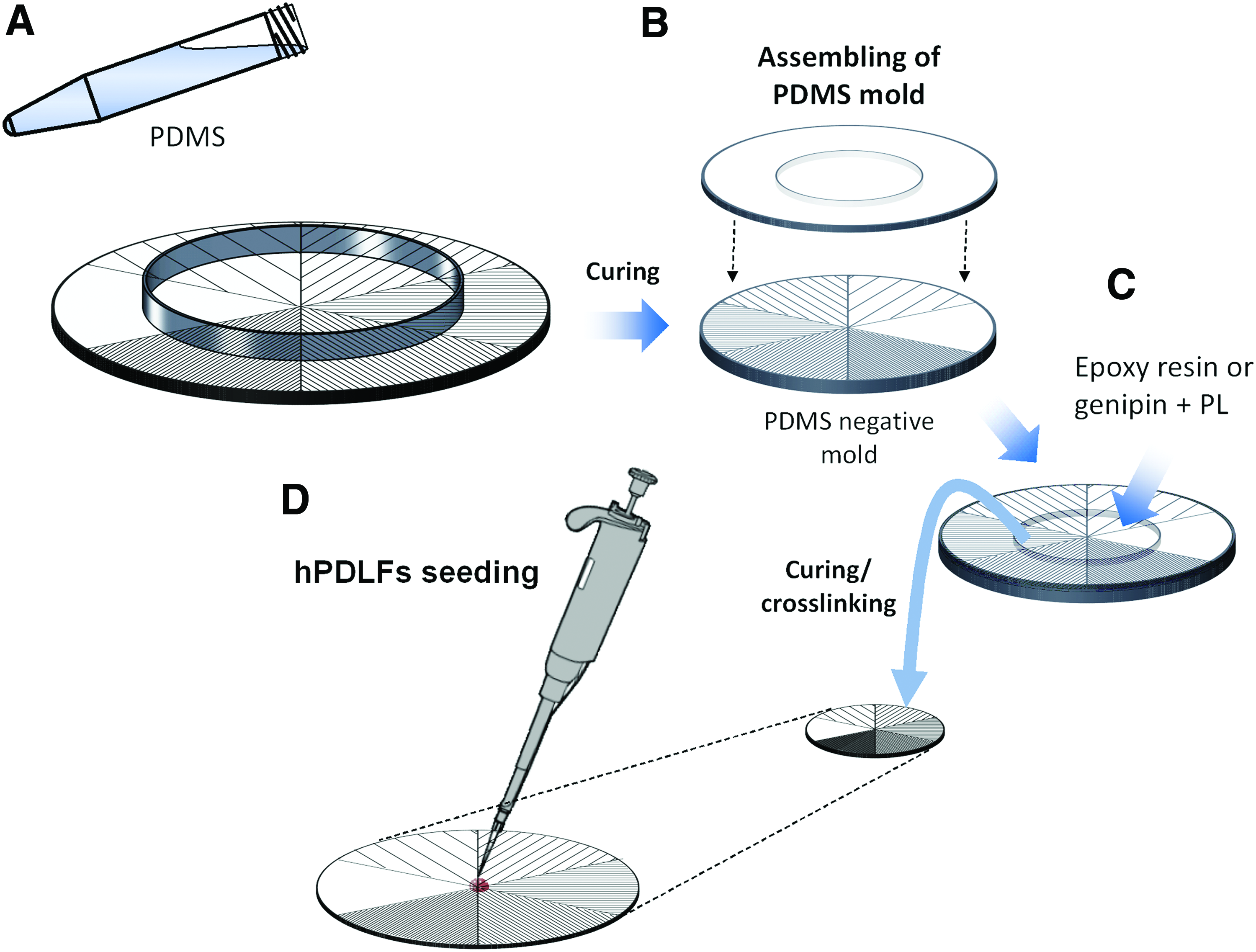
Scheme illustrating the production of multipatterned substrates and cell seeding.
PDMS replicates of the Si multipatterned wafer were produced as molds for the replication of the original patterns into the PL membranes. Therefore, premixed elastomer components were prepared according to the manufacturer's specifications and poured into a ring-shaped container that was placed over the multipatterned surface (as depicted in Fig. 1A). The viscous PDMS elastomer was evenly distributed and the air bubbles were removed inside a vacuum desiccator during a period of 10 min. After curing at 60°C for 3 h, the PDMS mold was washed with 10% Liquinox solution to remove the excess monomer for 10 min in an ultrasound bath. Then, the molds were thoroughly rinsed with tap water and left in MilliQ water for 10 min in the ultrasound bath. Also, 2-mm-thick PDMS rings with a central opening of 30 mm were created. Combined, the PDMS mold and the ring created a cylindrical container with a multipatterned bottom (Fig. 1B).
PL-based membranes with multipatterns were produced by genipin crosslinking of the PL proteins, followed by solvent casting, as described elsewhere. 14 In brief, genipin, which was prepared as described above, was added to PL to reach a final concentration of 0.25% w/v. After mixing, 1.5 mL of the transparent liquid PL solution was injected into the PDMS mold (Fig. 1C), and for sterilization, exposed to UV light for 15 min. The PL was allowed to crosslink and was dried for 4 days in a flow chamber, resulting in disk-shaped multipatterned membranes.
In addition, epoxy (Araldite; Huntsman Corporation) multipatterned substrates were created. In short, 10 mL of epoxy resin precursors was vigorously mixed with 2 mL of the hardener solution. Entrapped air bubbles were removed by centrifugation, and 250 μL of the final mixture was added to each PDMS mold, which were assembled as previously described (Fig. 1C). After 10 min under vacuum to remove any remaining air bubbles, the resin was cured at 60°C for 4 h. The epoxy resin disks were washed with isopropanol (IPA) under ultrasound for 30 min, followed by washing with tap water and MilliQ water. Before cell seeding, the resin disks were disinfected by incubation in 10% bleach for 30 min and rinsed thoroughly with sterile PBS.
Atomic force microscopy analysis
The presence of the nanopatterns on the surface of freshly produced or hydrated gPL substrates and epoxy substrates was confirmed by analysis with atomic force microscopy (AFM) (Catalyst; Bruker, Santa Barbara, CA).
For AFM analyses, freshly prepared dried samples of multipatterned gPL or epoxy substrates were mounted on a glass slide. To evaluate the preservation of the nanotopography after hydration, a sample of a multipatterned gPL substrate previously incubated in PBS overnight, at 37°C, was thoroughly rinsed with MilliQ water and dehydrated by incubation in increasing ethanol gradient series. After air drying, the membranes were mounted on a glass slide for AFM analysis.
The measurements were performed in AFM tapping mode in ambient air at 50% humidity with high-aspect ratio NW-AR5T-NHCR cantilevers (NanoWorld AG, Wetzlar, Germany) with average nominal spring constants of 30 Nm−1. The analyzed field was scanned at a rate of 1.0 Hz and 512 scanning lines.
Harvesting and culture of human periodontal ligament fibroblasts
The human periodontal ligament fibroblasts (hPDLFs) were harvested and isolated from extracted third molars (one male adult human donor). The isolation was performed by adaptation of a procedure previously described by Brunette et al. 20 Briefly, after extraction, the tooth was washed three times (with 10 min of incubation between washes) in PBS containing 1000 units/mL of penicillin and streptomycin. The PDL tissue was scraped with a scalpel from the middle third of the roots, thereby avoiding contamination with epithelial or pulpal cells. The tissue debris was smeared inside of a 25-cm2 tissue culture plate, which was incubated in an inverted position for 2 h at 37°C with 5% CO2. Then, the basal medium was added slowly to the cells, avoiding the resuspension of the tissue debris. The medium was refreshed every 2–3 days.
Cellular outgrowth originating from the tissue debris was consistently seen between 8 and 10 days after the start of culture. On subconfluency, cells were trypsinized (0.25% w/v trypsin, 0.02% EDTA; Sigma-Aldrich, Taukirchen, Germany) and subcultured for two passages before use in the experiment.
Assessment of topography effect on human periodontal ligament fibroblasts phenotype and physiology
Cell seeding on multipatterned substrates and culture
The effect of topography on the behavior of hPDLFs was assessed in both the epoxy resin and gPL multipatterned substrates. In brief, resin and gPL disks were distributed in six-well plates and hydrated by incubation in PBS for 3 h. The PBS was removed and a drop of 10 μL containing 2 × 10⁴ cells was placed in the intersection of the different topography sextants, as depicted in Figure 1D. After 2 h of incubation, 2 mL of culture medium was carefully added to each well to completely cover the disks. The cells were cultured at 37°C in a 5% CO2 humidified atmosphere.
Cell proliferation
After 7 or 14 days in culture, the samples were washed twice in PBS, sliced into six sextants corresponding to the topographies with a surgical blade, and immersed into 1 mL MilliQ water in a 1.5-mL Eppendorf tube. The samples were frozen at −20°C. The DNA present in the membranes was exposed during the freeze/thaw process and quantified using a PicoGreen dsDNA quantification kit, according to the manufacturer's specifications (Life Technologies). Cell proliferation was quantified as a function of amount of double-strand DNA (in ng) in the cell lysate.
Evaluation of cell migration distance and orientation
After 7 and 14 days in culture, the cells seeded on the resin or gPL multipatterned surfaces were fixed in 10% formalin overnight at 4°C. Then, the cells were washed thoroughly with PBS to remove the excess of formalin and permeabilized by incubation in a 1× PBS solution containing 0.5% triton-X100 and 10% of FBS for 15 min at room temperature.
The nuclei were stained with DAPI (Roche) and the cytoskeleton with Phalloidin-Alexa 568 (Molecular Probes). Cell distribution in the multitopography substrates was analyzed by fluorescence microscopy (Axio Imager Z1m; Zeiss, Sliedrecht, The Netherlands). Preferential hPDLC migration in the multipatterned substrates was measured as the distance between the seeding site (intersection of the different topography sextants) and the cell migration front for each topography generally located in the midline of the sextant.
To investigate the directional orientation of hPDLCs on the multipatterned substrates, Fourier component analysis for directionality was performed on representative micrographs of cell sheets growing in each topography, after 7 and 14 days of culture using the ImageJ plug-ins “Directionality” created by Tinevez 21 and “OrientationJ” created by Sage 22 following their respective instructions. The actin filaments have a polar orientation toward the cell migration orientation 23 ; thus, the micrographs were preprocessed before analysis to enhance the actin cytoskeleton signal intensity. The regions near sextant contact areas were avoided to prevent any possible confounders.
From the main fiber direction it was possible to calculate the divergence between the cell orientation and the pattern orientation. Coherency gives a measure of the anisotropy of the system and is bounded between 0 and 1, with 1 indicating highly oriented structures and 0 indicating isotropic areas. 24
Statistical analyses
All the experiments were performed with at least three replicates. Results are expressed as mean ± standard error of mean. Statistical analyses were performed with GraphPad Prism 5 Demo software (GraphPad Software, Inc., San Diego, CA) using one-way analyses of variance (ANOVA) with a Bonferroni's multiple comparison post-test to compare the differences between all the variables tested for each time point. Differences between the groups with p < 0.05 were considered to be statistically significant.
Results
Nanopatterned substrates
The AFM analyses of the epoxy resin and gPL membrane substrates produced in the PDMS counter-molds of the silicon multipatterned wafer confirmed that it is possible to cast parallel nanogrooves on these substrates (Fig. 2A, C). The quality of groove pattern reproduction was very consistent for epoxy resin substrates. The gPL membranes tended to shrink during the replication process, resulting in a groove pitch smaller than the nominal pitch, as well as a reduced groove depth (Fig. 2B). The gPL substrates submitted to a hydration step showed that the nanogrooves smaller than 600 nm were not stable on hydration. Only gPL membranes casted with nanopatterns with pitches of 600 and 1000 nm maintained a repetitive uniform grooved pattern after the second dehydration step.
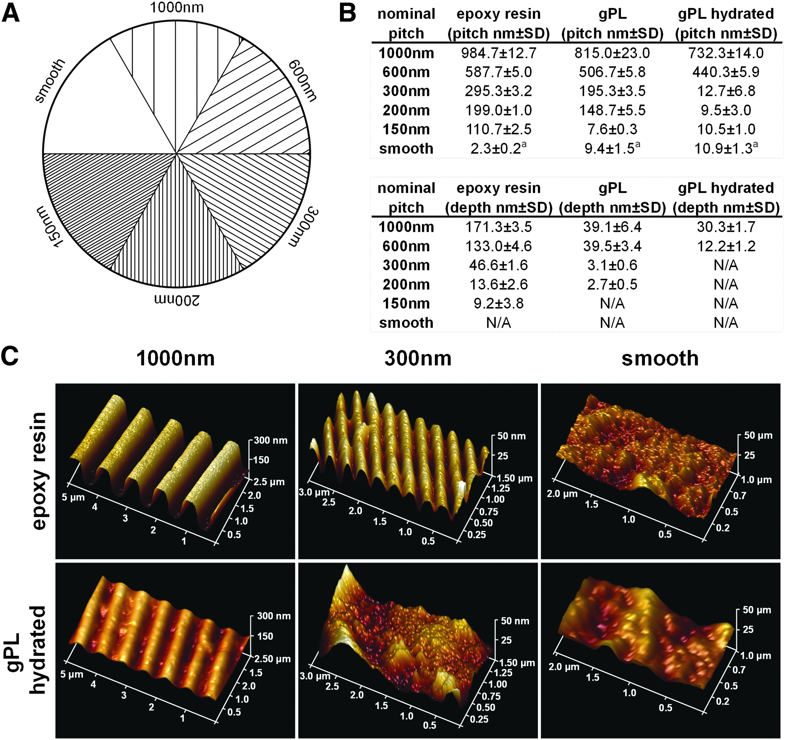
Casting of nanopatterns in gPL and epoxy substrates.
Effect of topography on cell proliferation
The analysis of dsDNA content on both the epoxy (Fig. 3A) and gPL substrates (Fig. 3B) after 7 and 14 days in culture demonstrated that the hPDLCs were proliferating in both the substrates. Nevertheless, the topography had no significant influence on cell proliferation.
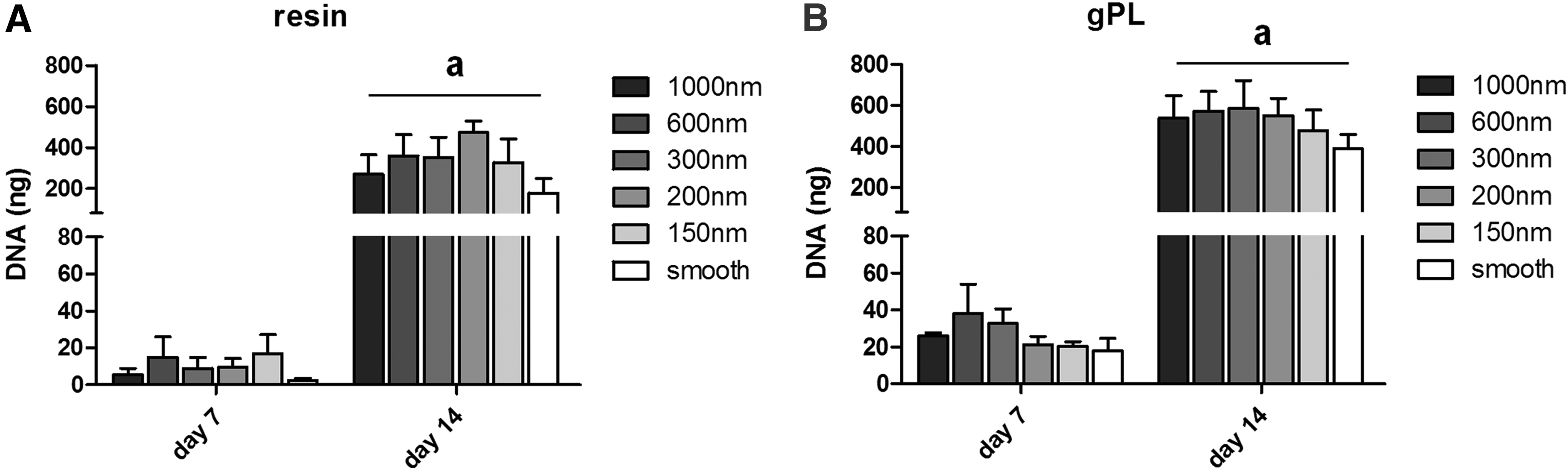
hPDLC proliferation evaluated as a function of dsDNA content of cells growing on
Directional cell migration
Figure 4 represents the outward migration distance covered by hPDLCs on epoxy and gPL multipatterned substrates after 7 and 14 days in culture. The cells migrated at an average speed of 1 mm/day and reached the circumference of the disk radius (15 mm) on both the substrates, regardless the topography (Fig. 4B, D). It was not possible to detect significant differences in outward migration related with the topography.

Outward migration distance between the cell seeding site (intersection of the sextants) and the cell migration front on
Cell organization on the multipatterned substrates
Figure 5 shows the organization of the hPDLCs on both the epoxy resin and gPL nanopatterned substrates. The alignment of the hPDLCs along the axes formed by the nanogrooves with higher nominal pitch (1000 and 600 nm) was observed on both the epoxy (Fig. 5A) and gPL substrates. It was also evident in the micrographs presented in Figure 5A that the hPDLCs tended to a more random and disperse orientation with the narrowing of the groove pitch. Furthermore, the analysis of the cell orientation using the OrientationJ software, and represented in the graphics depicted in Figure 5B and C, supports this observation. The orientation of the hPDLCs was roughly parallel to the 1000 and 600 nm nanogrooves. For lower groove pitch, the cells tended to distribute more randomly on the substrate surface, in some cases aligning preferentially with the sextant walls (at −30° and +30°).
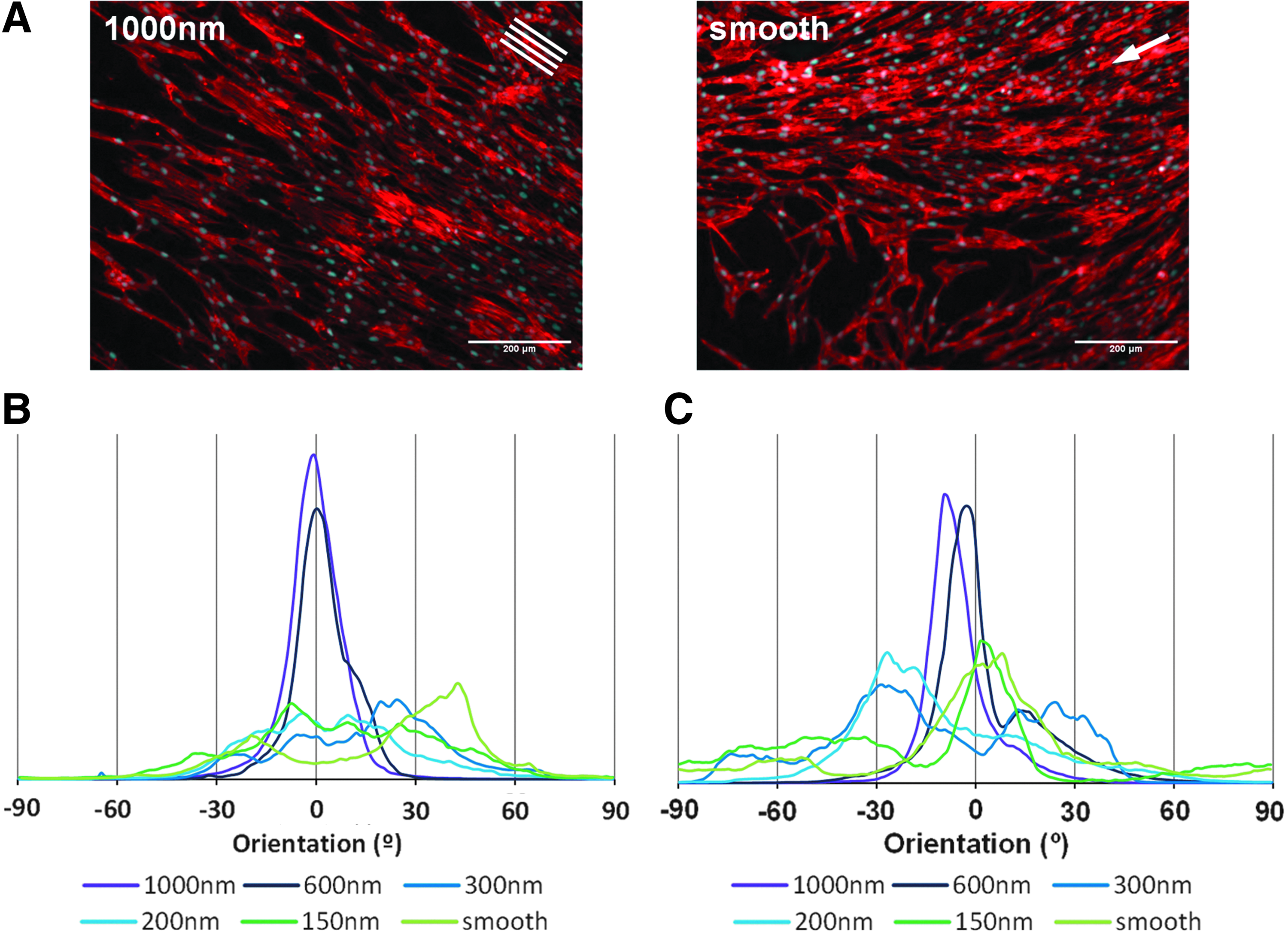
hPDLC orientation on the multipatterned substrates after 14 days in culture.
The average of main cell orientation angle, analyzed in representative regions of the cells sheets of PDLCs after 7 and 14 days in culture, is represented in Figure 6. The alignment consistency with groove orientation regardless the hPDLC donor or substrate for the 1000 and 600 nm topographies was evident. On the contrary, while the orientation of the hPDLCs seeded on the epoxy resin substrate after 7 days in culture was still influenced by the topographies with narrower pitch (300–150 nm) (Fig. 6A), organization was partially lost for the same topographies after 14 days in culture.

Characteristic organization of hPDLCs on the multipatterned substrates. Distribution of hPDLCs main orientation angle, relative to the orientation of nanogrooves in
Regarding the cells growing on gPL multipatterned substrate, for nominal pitch lower than 300 nm, the main cell orientation is random, similarly with the smooth surface, throughout the duration of the assay (Fig. 6B).
Likewise, the general organization of the cells was also influenced. The coherency of cellular orientation, that is, the percentage of anisotropy, was generally higher for the higher nanopattern pitches and reduced with the narrowing of the grooves, regardless the substrate (Fig. 6C, D).
Discussion
The casting of micropatterns on the surface of scaffold materials for the regeneration of periodontal tissue has shown to be an effective strategy to improve their efficacy.4,25 The submicron 500 nm-pitched parallel grooves have been shown to promote PDLC elongation parallel to groove axes, while did not affect the migration speed when compared to smooth control. 26 Nevertheless, the effect of topographical features in enhancing contact guidance and promoting the fast repopulation of root surface with PDLCs is poorly understood. 26 Therefore, in this work, we tested the suitability of an array of submicrometer-pitch grooves to be used as an in vitro screening platform for the determination of the most potent cell-dependent topography for periodontal tissue regeneration.
When looking at the setup of our study, we selected two markedly different materials. The membrane produced by the crosslinking of PL proteins with genipin (gPL) was previously proposed as a potential material for periodontal tissue engineering. 14 The gPL membrane possesses viscoelasticity with an E′ of ∼0.55 MPa, 14 which is within the range of magnitude reported for natural PDL. 27 Furthermore, the platelet-rich hemoderivatives have been widely proposed for periodontal tissue regeneration as a provisional matrix for new tissue ingrowth and source or growth factors.28–30 In addition, an epoxy resin (araldite) was chosen as an example of a stiff material (elastic storage modulus [E′] in the GPa magnitude) devoid of biochemical cues, but capable of supporting cellular adhesion and proliferation. 31 Moreover, this resin was previously shown to enable an excellent reproduction of various topographical features. 32
Although the introduction of a “pure control,” generally a polystyrene substrate, would be desirable, in the scope of this study, the radially organized system was hypothesized as a tool for the screening of different biomaterials for biomedical applications; therefore, it was decided to choose for nonconventional materials, whose application has not been already heavily studied in the literature.
Both substrate materials were casted with nanogrooves featuring pitches, that is, distance between two consecutive groove ridges, ranging from 1000 to 0 nm (smooth surface). The presence of a smooth surface among the array of patterns was envisioned as an internal control to the substrates. The epoxy resin substrate was able to reproduce the multipatterns with an adequate reproducibility. A qualitative reproduction into gPL was only possible for the 600 and 1000 nm pitch topographies. The reduction of groove pitch size observed in the gPL specimens is correlated with shrinkage of the gPL during the casting process. For narrower groove dimensions, the mold will act as a sieve, hindering the penetration of the template grooves with the high-molecular-weight PL proteins, which hampers the stabilization of the crosslinked matrix. The matrix will therefore be easily dissolved, erasing the casted physical features.
The effect of the different nanopatterned surfaces on hPDLCs was assessed in terms of cell proliferation, outward migration, and cell alignment. Previous work demonstrated that the casting of nanogrooves with ∼500 nm increases the initial adhesion of PDLCs, 26 but has no significant influence on cell proliferation after 4 days of culture. 33 In the present study, the parallel nanogrooves were not found to have a significant influence on cell proliferation. The cells proliferated similar to the smooth control on all tested topographies after the short (7 days) and long (14 days) culture periods, regardless the substrate. The same result was also observed for the outward cell migration, which was constant for the different topographies and substrate compositions. The cell proliferation and migration results reported were supported in duplication studies using hPDLCs derived from different donors (data not shown).
As mentioned before, the effects of micro- and nanotopographic cues on cell proliferation and migration are poorly understood. Recent studies have shown that the mechanisms that control both proliferation and migration as response to topographical cues are connected and dependent on the reorganization of cytoskeleton and cell adhesions mediated by the Rho-dependent signaling pathway.34,35 Nevertheless, the influence of topography on cell proliferation seems to vary with cell type and pattern. For example, Lamers et al. 36 reported a faster and slower directional migration of osteoblast-like cells on, respectively, 600 and 150 nm pitch grooves, which was in an inverse correlation with the adhesion of the cells to the substrate. On the contrary, Hamilton et al. described equal migration velocities for PDLCs spreading on smooth and nanogrooved (500 nm pitch) substrates. 26
Regarding the effect of the patterns, Klymov et al. showed that cells are able to select their favorite substrate in terms of submicron- and nanopillars, pits, ridges, or grooves. 37 Therefore, to develop biomaterials that promote a faster colonization of periodontal wound with PDLCs, future studies should focus on the response of PDLCs to different nanopatterns.
In contrast, the topography had a strong influence on hPDLC organization. Parallel grooves with a dimension closer to the micrometer scale, 1000 and 600 nm nominal pitch, effectively promoted the hPDLC anisotropic organization, independent of the substrate composition. On the epoxy substrates, the hPDLCs aligned consistently along the parallel grooves with a pitch as small as 300 nm with a coherency higher than 0.5 (0 being highly isotropic and 1 highly anisotropic). However, in the gPL substrates, grooves smaller than 600 nm could not be reproduced. Therefore, topography-related differences in cell alignment could not be expected to occur for these dimensions in gPL substrates. In literature, the anisotropic micro- and submicron topographies have been described to induce the alignment of a broad spectrum of cell types in vitro.3,35,36,38,39
These dimensions recapitulate the collagen fiber bundles of connective tissue, which in PDL range from 3–10 to 10–20 μm in diameter, near cementum and alveolar bone, respectively, 40 and 1–8 μm in diameter in the oriented collagen meshwork that forms the PDL core. 41 In the PDL, the PDLCs organize alongside and within the dense collagen fiber bundles that connect the alveolar bone and the cementum. 42 Evidently, the 600 and 1000 nm pitch grooves are more appropriate to recapitulate the microenvironment of the PDL. In fact, the range of groove dimensions that effectively promote the cell anisotropic organization seems to be transversal to a wide variety of tissues in mammal species.
The anisotropic organization of some tissues, including bone, muscles, and tendons/ligaments, is supported by the aligned microstructure of the ECM. 1 Smaller nanogroove dimensions are compliant with the dimensions of collagen bundles in normal PDL, which have a unimodal distribution around 45 nm. 40 Therefore, smaller nanogroove dimensions may be involved in cell adhesion 7 and ECM remodeling 43 rather that in tissue functionality.
From a technical point of view, the system as proposed in the current study allows for a simple and fast screening of the most efficient pattern that could be casted in constructs as used for tissue engineering applications. Considering gPL, this material was proven to be valuable for tissue engineering and regenerative medicine applications targeting anisotropic tissues, but not limited to periodontal applications. The limitation in reproducing grooves with pitches lower than 300 nm was shown to be negligible, given the importance of the micron and submicron grooves in driving cell orientation. The effectiveness of the micro- and submicro-grooved gPL membranes to promote (periodontal) ligament regeneration should be further investigated in vivo.
Conclusions
In this work, the suitability of a radially organized multipatterned device for the screening of the best topographies to be casted into biomaterials, envisioning the regeneration of anisotropically organized tissues, was tested. The multipatterned device was shown to be able to cast materials with different origins, processing methods, and mechanical properties. Parallel nanogrooves have no significant effect on PDLC proliferation and directional migration. Nevertheless, the 600 and 1000 nm pitches were shown to induce an anisotropic organization in PDLCs. Therefore, this multipatterned system could be a valuable diagnostic device for the screening of topographies on tissue engineering scaffolds aiming the regeneration of anisotropically organized tissues.
Footnotes
Acknowledgments
P.S.B. acknowledges Fundação para a Ciência e Tecnologia (FCT) for the PhD Grant SFRH/BD/73403/2010. We thank the RIMLS Microscopic Imaging Center for the use of their facilities. J.t.R. is supported by a Dutch NWO Veni grant (680-47-421). This research received funding from an NWO Medium Sized Investment (NOW-ZonMV 91110007).
Disclosure Statement
No competing financial interest exists.