
Abstract
Decellularized scaffolds composed of extracellular matrix (ECM) hold promise for repair and regeneration of the meniscus, given the potential for ECM-based biomaterials to aid in stem cell recruitment, infiltration, and differentiation. The objectives of this study were to decellularize canine menisci to fabricate a micronized, ECM-derived scaffold and to determine the cytocompatibility and repair potential of the scaffold ex vivo. Menisci were decellularized with a combination of physical agitation and chemical treatments. For scaffold fabrication, decellularized menisci were cryoground into a powder and the size and morphology of the ECM particles were evaluated using scanning electron microscopy. Histologic and biochemical analyses of the scaffold confirmed effective decellularization with loss of proteoglycan from the tissue but no significant reduction in collagen content. When washed effectively, the decellularized scaffold was cytocompatible to meniscal fibrochondrocytes, synoviocytes, and whole meniscal tissue based on the resazurin reduction assay and histologic evaluation. In an ex vivo model for meniscal repair, radial tears were augmented with the scaffold delivered with platelet-rich plasma as a carrier, and compared to nonaugmented (standard-of-care) suture techniques. Histologically, there was no evidence of cellular migration or proliferation noted in any of the untreated or standard-of-care treatment groups after 40 days of culture. Conversely, cellular infiltration and proliferation were noted in scaffold-augmented repairs. These data suggest the potential for the scaffold to promote cellular survival, migration, and proliferation ex vivo. Further investigations are necessary to examine the potential for the scaffold to induce cellular differentiation and functional meniscal fibrochondrogenesis.
Introduction
A
There are a number of biological approaches reported in the literature that aim to enhance the healing response following meniscal repair. These include delivery of growth factors, mechanical stimulation, or surgical techniques such as trephination, cellular conduit placement, and synovial abrasion and flaps; as well as fibrin clot suturing used alone or in conjunction with meniscal repair.9–23 Small intestinal submucosa grafts have also been reported for the treatment of large meniscal defects by enhancing meniscal tissue regeneration in a canine model.24–26 The majority of these approaches provide a scaffold for repair tissue production, and/or delivery of blood, cells, and other bioactive factors that are essential for healing.27–31 However, functional healing with preservation of meniscal size, geometry, and composition does not consistently occur with any of the available treatment options for avascular meniscal defects, often leading to meniscal resection and a joint doomed to OA. 32 Accordingly, improving the healing response for avascular meniscal tears is an ongoing challenge for clinicians and researchers.
To address this common and costly orthopedic problem, we propose that a scaffold derived from particulate meniscal extracellular matrix (ECM) could promote biological augmentation of meniscal repair, or stimulate meniscal regeneration in avascular meniscal defects.33,34 The optimal scaffold for an engineered tissue should mimic the in vivo functions of the native ECM. 35 Such functions include structural support as well as regulation of cell survival, proliferation, morphogenesis, and differentiation. 36 Biological scaffolds composed of ECM derived from decellularized tissues have been reported for a number of tissue engineering applications, including urinary bladder, heart valve, dermis, blood vessels, nerves, tendon and ligament, meniscus, and articular cartilage.37–48 In addition, previous studies have examined the regenerative potential of scaffolds derived from particulate ECM for articular cartilage repair and skin and soft tissue reconstruction.46,49–59 Thus, there is a growing body of evidence to support the potential utility of ECM-derived scaffolds for use in meniscal tissue regeneration and repair. 60
Our overarching goal is to develop an arthroscopically deliverable, orthobiologic composed of micronized meniscus ECM, combined with readily available, point-of-care biologics as a carrier, such as platelet-rich plasma (PRP) or bone marrow aspirate concentrate. Toward this goal, the primary objectives of the present study were to decellularize canine menisci to fabricate a micronized, ECM-derived scaffold, and to determine the cytocompatibility and repair potential of the scaffold ex vivo. Decellularization was based on a series of preliminary experiments using a modification of previously reported decellularization and tissue washing techniques.61–63 It was hypothesized that decellularized canine meniscal tissue could be processed into a cytocompatible, ECM-derived scaffold, and that radial tears augmented with the scaffold delivered with PRP would demonstrate superior production of repair tissue compared to menisci repaired with standard-of-care suture techniques.
Materials and Methods
Tissue procurement and harvest
All procedures were performed with Institutional Animal Care and Use Committee approval for the use of cadaveric tissues in research and for blood collection and processing from canine blood donors (IACUC #8235 and #8285). Grossly normal menisci (N = 18) were harvested from the stifle joints (knees) of adult dogs euthanatized for reasons unrelated to this research. Menisci were collected from purpose-bred research dogs and/or canine donors from several different licensed animal control shelter facilities within 2 h postmortem. Canine tissue donors were apparently healthy adults (skeletal maturity, >20 kg) with grossly normal knees and menisci.
Decellularization and scaffold fabrication
Decellularization was performed based on a modification of procedures previously described.61–63 Scaffold fabrication was performed as demonstrated in the flow chart in Figure 1. All menisci were processed individually in 4–6 mL of the appropriate solution, based on the mass of tissue determined at the time of harvest (5:1 liquid to tissue ratio). Following harvest, the menisci were aseptically minced into 1–3 mm2 pieces, rinsed by pipetting in wash buffer (0.9% NaCl +10% antibiotic–antimycotic solution), and then washed with agitation on a microplate shaker at 200 rpm for a minimum of 12 h at room temperature (RT) in diH2O. All reagents were purchased from Sigma-Aldrich (St. Louis, MO) unless otherwise noted. All steps of decellularization and washing were carried out under agitation (200 rpm) at RT unless otherwise noted.

Schematic of canine meniscus processing for fabrication of a micronized (cryoground) meniscus extracellular matrix scaffold.
First, cellular membranes were disrupted with a solution of deoxycholic acid (2% v/v), Triton-X 100 (2% v/v), and ethylenediaminetetracetic acid (EDTA) dissolved in diH2O (0.1% v/v; Thermo Fisher Scientific, Waltham, MA) brought to pH of 8.0. Next, the decellularization chemicals were removed after 24 h and the tissue was washed with agitation in diH2O for a minimum of 12 h to rinse the tissue in hypotonic solution and further disrupt cellular membranes. The tissue was then treated with a 48-h cycle of sodium dodecyl sulfate (SDS, 2% v/v) and Tris(hydroxymethyl)aminomethane reagent (10 mM) at 4°C with agitation to further solubilize and remove cells. Next, the decellularization chemicals were removed and the tissues were again washed in diH2O for a minimum of 12 h. Last, the tissue was treated with peracetic acid (0.1% v/v) for 4 h, followed by PBS overnight, to further remove residual nucleic acids.
After decellularization, the tissues underwent final washing in PBS with centrifugation to further remove any residual cell and nuclear material and to completely remove decellularization chemicals. To determine the optimal endpoint for washing of the scaffold after decellularization, two wash protocols were compared. For wash protocol 1, decellularized menisci were washed at least three times in PBS with centrifugation (3000 g for 20 min). For wash protocol 2, menisci were washed and centrifuged (3000 g for 20 min), and the supernatant was saved for further analysis. Briefly, washing and centrifugation were repeated until SDS was no longer detected using a spectrophotometric assay developed in our laboratory using 1–9-dimethylmethylene blue (DMMB) as the indicator for SDS levels in the rinse water. 64
After washing, decellularized menisci were flash frozen in liquid nitrogen and cryoground into a powder with a Micro-Mill grinder (BEL-ART, Wayne, NJ). The powder was lyophilized for a minimum of 12 h and packaged for sterilization by exposure to UV light irradiation for 48–72 h. 65
Scanning electron microscopy
The size and morphology of the powdered meniscal ECM scaffold particles were observed in representative samples from two dogs using environmental scanning electron microscopy at the institutional electron microscopy core (ESEM; FEI Quanta 600F; Thermo Fisher Scientific). Before imaging, the scaffolds were mounted using double-sided carbon adhesive tape and imaged in low-vacuum mode with an accelerating voltage of 10 kV.
Biochemical content
The biochemical and DNA content of the menisci was assessed before and after decellularization to compare control (untreated) versus treated tissue. The glycosaminoglycan (GAG) content of each sample was determined using the DMMB spectrophotometric assay. 66 Total collagen content was determined using a hydroxyproline (HP) colorimetric assay. 67 DNA content (ng/mg dry weight) was determined using the Quant-iT PicoGreen dsDNA Assay Kit (Thermo Fisher Scientific) according to the manufacturer's protocol. GAG and HP content was normalized to dry weight (μg/mg) of each sample for data analysis.
Histology
Histologic evaluation of the menisci was performed before and after decellularization to compare cellularity and matrix composition in control versus treated tissue. Briefly, samples were fixed in 10% neutral buffered formalin fixative overnight, placed in 10% EDTA solution for 10 days to soften tissue, and then paraffin embedded, sectioned, and stained with hematoxylin and eosin (H&E), toluidine blue for proteoglycans, or picrosirius red for collagen content. Tissue samples were evaluated by light microscopy for cell morphology and matrix composition by a veterinary pathologist blinded to treatment.
Cell isolation and culture
Synovium was freshly harvested (<2 h postmortem) from the knees of healthy dogs (N = 2) euthanized for reasons unrelated to this study. Briefly, the synovial intima was sharply excised, minced, and rinsed with wash buffer. The synovium from each dog was pooled and placed in a flask containing 10 mL of Dulbecco's Modified Eagle's Medium (DMEM) (Gibco, Life Technologies, Waltham, MA) and clostridial collagenase (Sigma; 0.5 mg/mL) at 37°C with 5% CO2 until the tissue was completely digested (12–18 h). The suspension was centrifuged (1000 rpm for 10 min), the supernatant discarded, and then, the cells resuspended in DMEM and 10% fetal bovine serum (FBS; Sigma). Synoviocytes were plated in 25-cm2 (T25) tissue culture flasks and incubated in standard conditions (37°C with 5% CO2 and 95% humidity). Culture medium was changed every 2–3 days. At near confluence, the adherent cells were detached by trypsinization, plated in T75 culture flasks, and then collected after the third passage.
Canine dermis and menisci were aseptically collected, minced, digested, and the cells were expanded and isolated as previously described for synoviocytes. At near confluence, the adherent cells were detached by trypsinization and plated in T75 culture flasks. Meniscal cells were collected after the second passage and dermal fibroblasts (DFB) were collected after the fourth passage.
In vitro cytotoxicity
To determine if the powdered scaffold leached any cytotoxic chemicals after treatment, canine dermal fibroblasts (seeding density, 1.6 × 10⁶ cells) were seeded onto 12-well plates for 24 h, and the scaffold (wash protocol 1, N = 8; wash protocol 2, N = 12) was placed into 8.0-μm filter well inserts that did not allow contact between the scaffold and the cells. Cell viability was assessed using the resazurin reduction assay after 3 days of culture in standard conditions. Second, to determine if direct contact with the washed scaffold was cytotoxic, meniscal fibrochondrocytes (MFC, seeding density, 1.3 × 10⁶ cells) and synoviocytes (SYN, seeding density, 4.4 × 10⁵ cells) were seeded directly on the ECM scaffold with or without prior PRP treatment (ACP; Arthrex, Inc., Naples, FL). Pellet cultures of MFC (N = 3) and SYN (N = 3) were used as culture controls. Cell viability was assessed after 8 days of culture in standard conditions using the resazurin reduction assay.
In vitro cytocompatibility
To evaluate cytocompatibility of the scaffold with cells and meniscal tissue in vitro, grossly normal menisci (N = 4) were harvested from the knee of an adult dog (recipient) euthanized for reasons unrelated to this research. Scaffold fabrication and analysis were performed as previously described with menisci obtained from a single adult, canine donor and washed until SDS levels were no longer detected in the rinse water.
For the recipient menisci, full-thickness cylindrical defects were created in the cranial (anterior) and caudal (posterior) portions of the inner 2/3 using a 2 mm biopsy punch. Menisci were bisected into two portions (N = 8), which were then randomly assigned for treatment and packed with either an ex vivo fibrin clot as the standard-of-care biological scaffold 10 or with the meniscal ECM scaffold combined with PRP into a paste. Venous blood was collected from a donor research hound and PRP was prepared using the Angel system (Arthrex). One-half of the bisected menisci were randomly seeded with canine MFC (seeding density, 1 × 10⁶ cells). After cell seeding, menisci were incubated at 37°C for 1 h before adding complete, serum-free culture medium to promote cellular adherence and diffusion into the defects. Menisci were cultured in standard conditions for 42 days and then collected for histologic evaluation and processed as previously described. After histologic processing, the menisci were stained with H&E. All sections were evaluated by light microscopy by a veterinary pathologist blinded to treatment. Samples were assessed for cellularity in and around the defects, for evidence of repair tissue production, and for normal meniscus architecture and cellularity.
Ex vivo repair model
Scaffold fabrication and PRP collection were performed as previously described from single canine donors. For the culture model, grossly normal menisci (N = 12) were harvested from three adult, canine donors euthanized for reasons unrelated to this research. With a No. 11 scalpel blade, a 5 mm radial tear was created in the midbody of medial and lateral menisci, including both the red-white and white-white zones. Menisci were randomly assigned for treatment in one of the following groups: scaffold-augmented repair (scaffold+PRP+suture), standard-of-care repair (suture only), no repair, or intact menisci (tissue control). All repairs were performed with two horizontal mattress sutures using 4-0 FiberWire (Arthrex). Samples were cultured in complete, serum-free growth medium in standard conditions for 40 days before assessment of tissue cell viability and histology.
Determination of tissue cell viability of the cultured menisci was performed using the LIVE/DEAD Viability/Cytotoxicity kit according to the manufacturer's protocol (Thermo Fisher Scientific). Briefly, 1 mm full-thickness tissue slices were obtained from each sample (minimum three sections) and stained with calcein acetoxymethyl ester to detect live cells and ethidium homodimer-1 to detect dead cells.
Menisci were collected for histologic evaluation, processed as previously described, and stained with H&E. All sections were evaluated by light microscopy by a blinded veterinary pathologist. Samples were subjectively assessed for cellularity in and around the defects, and for evidence of repair tissue production, as well as for normal meniscus architecture and cellularity.
Statistical analysis
Data were assessed for normality with the Shapiro–Wilk test using a computer software program (SigmaPlot 13.0; Systat Software, Inc., San Jose, CA). Continuous data (mean ± SD) were compared for statistically significant differences between groups using a t-test with p < 0.05 considered statistically significant. Nonparametric data (median ± SEM) were compared with the Mann–Whitney rank sum test when appropriate.
Results
Scaffold biochemical content, cellularity, and matrix composition
DNA content was significantly reduced in decellularized menisci compared to native (15.43 ± 8.635 vs. 26.26 ± 8.01 ng/mg dry wt; p < 0.001) (Fig. 2A). Histological examination of decellularized menisci revealed a marked decrease in cell content and nuclear material after treatment, associated with a decreased amount of proteoglycans and variable thickness and packing of collagen fibers (Fig. 3). Tissue GAG content significantly (p < 0.001) decreased and tissue HP content significantly (p = 0.009) increased in the decellularized menisci compared to native control tissue (Fig. 2B, C). Furthermore, the GAG:HP ratio was significantly (p < 0.001) lower in the decellularized menisci compared to the native control tissue (Fig. 2D).
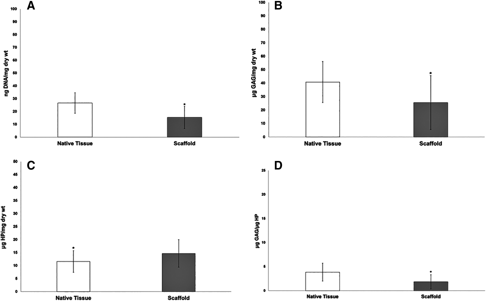
Tissue DNA content

Representative photomicrographs depicting histologic differences in cellularity
Scanning electron microscopy
SEM imaging revealed that the ECM particles were heterogeneous in morphology and size. A representative particle of the ECM scaffold is shown in Figure 4.
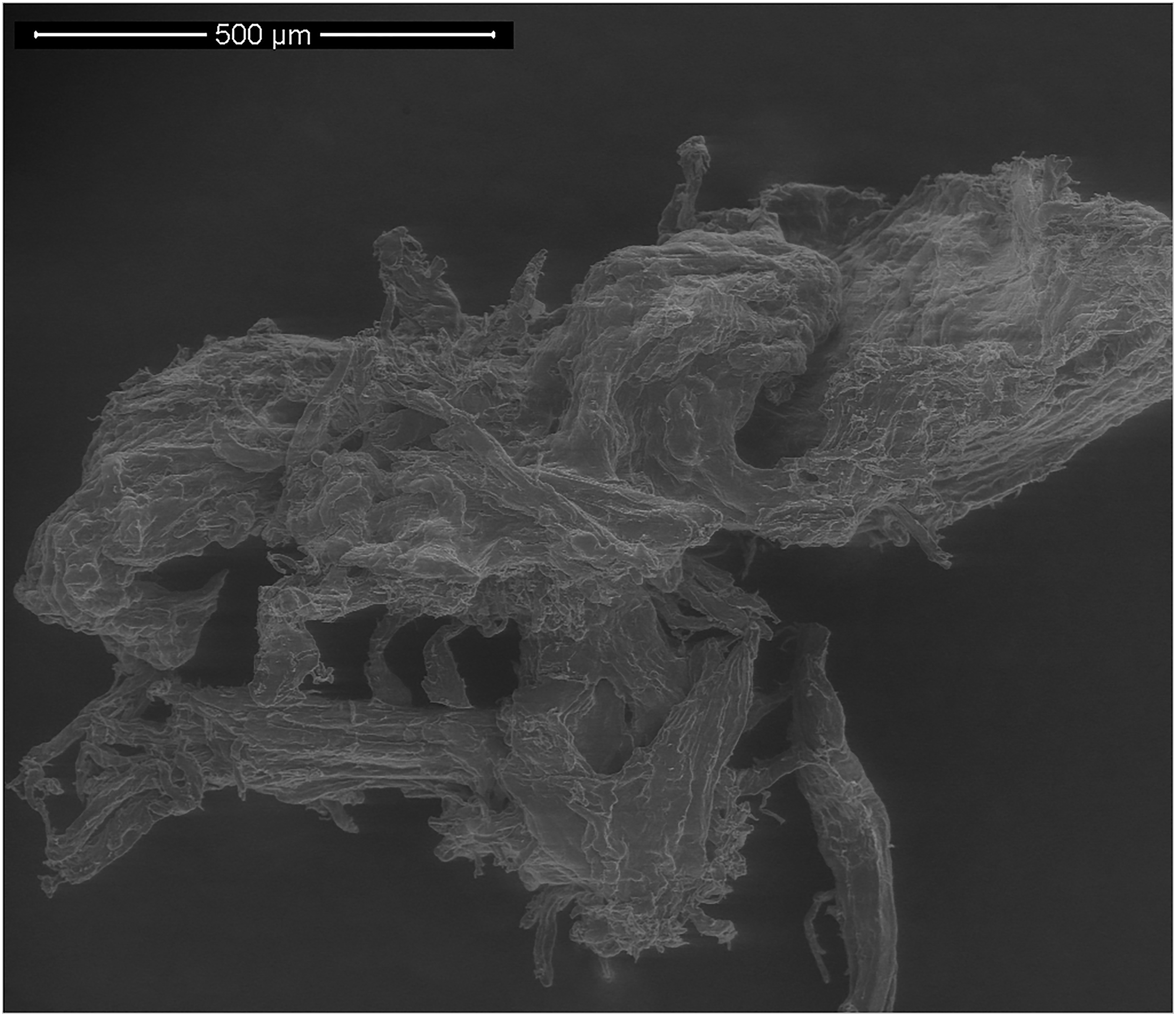
Representative SEM image of micronized (cryoground) meniscus extracellular matrix. Accelerating voltage 10.00 kV. Magnification 200 × .
In vitro cytotoxicity and cytocompatibility
Scaffolds washed with a defined protocol (protocol 1) were cytotoxic to DFBs in monolayer culture (Fig. 5A). However, scaffolds washed until SDS levels were no longer detected in the rinse water (protocol 2) were not cytotoxic to DFBs in monolayer culture (Fig. 5A). Furthermore, the washed ECM scaffold, with or without the addition of PRP, was not cytotoxic to MFC and SYN cells (Fig. 5B).
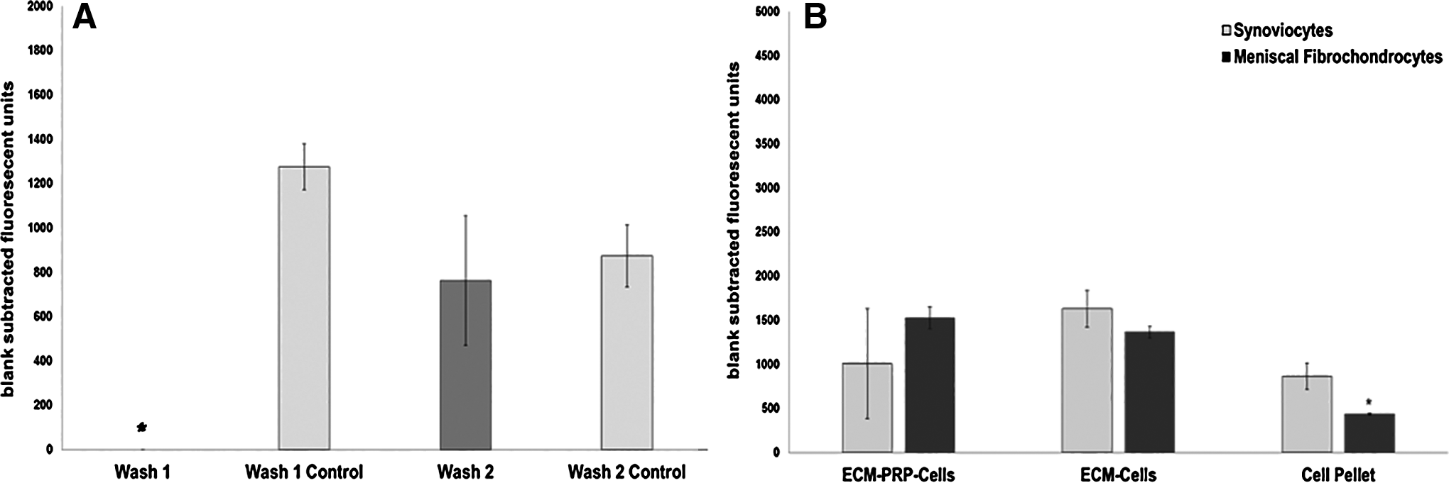
Cytotoxicity analysis:
After 42 days of culture in 3 mm meniscal defects, histological evaluation showed normal tissue architecture and cellularity of both scaffold and fibrin clot-treated menisci, with the presence of spindle cells within the defects of both seeded and noncell seeded samples (Fig. 6).

Representative photomicrographs of canine menisci following treatment of full-thickness defects with an ECM-derived scaffold
Ex vivo repair model
Scaffold DNA content was significantly reduced compared to control tissue (17.09 ± 3.339 vs. 37.482 ± 6.11 ng/mg dry wt; p = 0.001). When standardized to the dry weight of the tissue, tissue HP content significantly (p = 0.029) decreased. There was no significant difference in tissue GAG content between the scaffolds and controls. While the GAG:HP ratio was lower in the scaffold compared to control tissue, this difference was not significant (p = 0.050).
Following 40 days of culture, subjective evaluation of tissue cell viability of the repaired menisci was excellent for all groups (Fig. 7). Histologically, there was no evidence of cellular migration or proliferation detected in any of the untreated or standard-of-care treatment groups (Fig. 8A, B). Conversely, cellular infiltration and proliferation were discovered in scaffold-augmented repairs, as demonstrated by the presence of spindle cells within the defects. The extent of cellular proliferation was variable in scaffold-augmented repairs and ranged from a few visible cells to moderate quantities of spindle cells arranged in loose streams and supported by amorphous matrix within the defect (Fig. 8C, D).
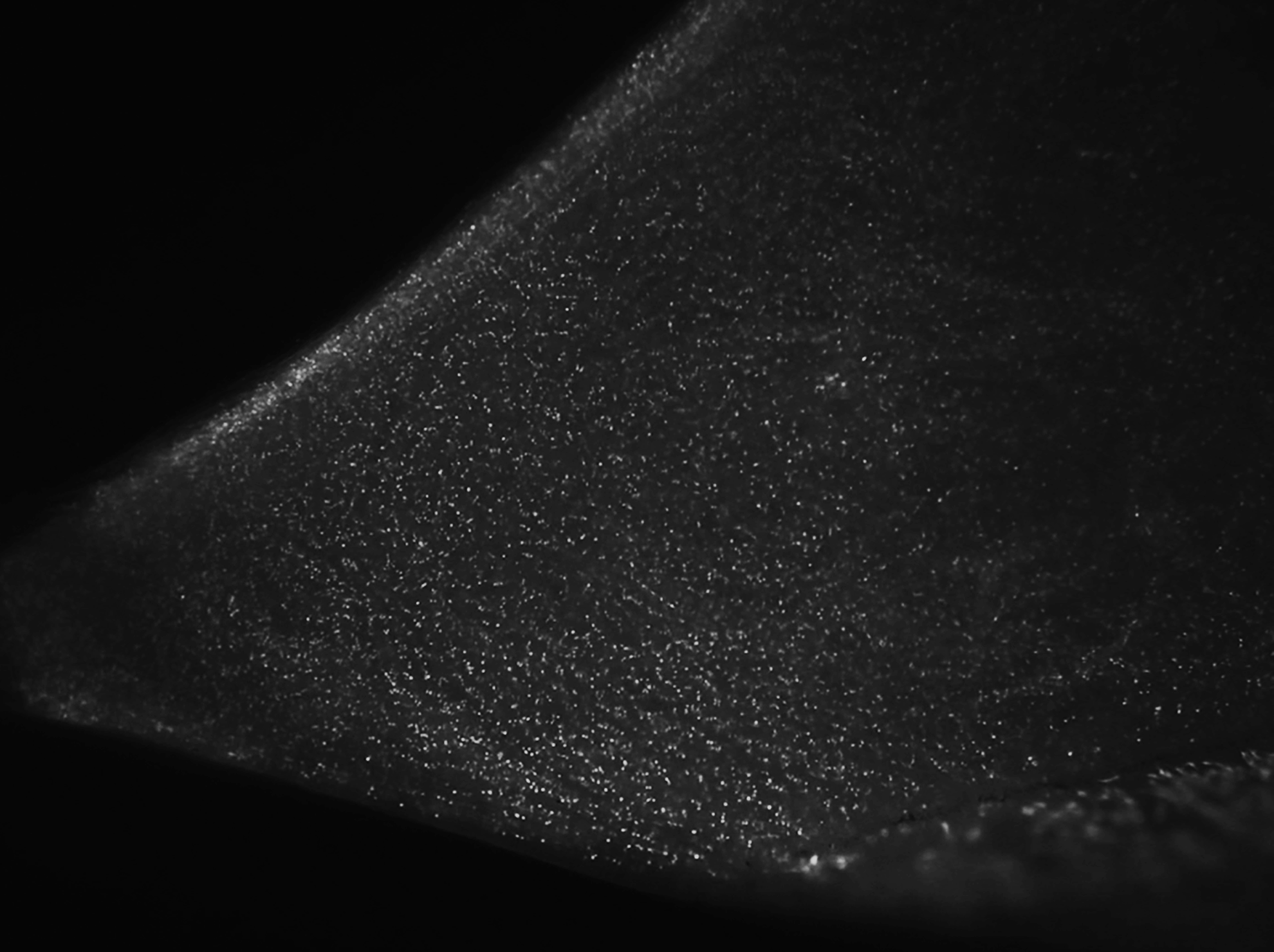
Representative photomicrograph of fluorescent cell viability of canine menisci following 40 days of culture (4 × image).

Representative day 40 histologic photomicrographs of canine menisci following creation of a 5-mm radial defect with
Discussion
Our study results indicate that canine menisci were effectively decellularized and processed into an ECM-derived scaffold that was cytocompatible to the meniscus and to key cell types for normal meniscal healing. On histology, spindle cells were noted within noncell seeded, ECM+PRP-treated menisci, suggesting cellular survival, migration, and proliferation within the scaffold. Furthermore, cellular infiltration and proliferation were discovered in scaffold-augmented repairs of radial meniscal defects ex vivo after 40 days of culture.
Our long-term objective is to develop a clinically applicable orthobiologic composed of micronized meniscus ECM to biologically enhance meniscal tissue healing and repair. For the present study, menisci were cryoground into a powder to broaden the potential clinical application in the future—from repair augmentation to possible treatment of full-thickness meniscal defects. Similar products that are commercially available for tissues other than meniscus include BioCartilage (Arthrex) combined with PRP as an adjunct to microfracture for enhanced repair of articular cartilage defects,49–52 and acellular dermal matrices, including AlloDerm Regenerative Tissue Matrix (LifeCell Corporation, Bridgewater, NJ), for skin and soft tissue reconstruction.52,53 Accordingly, decellularized scaffolds composed of particulate ECM show promise for use in tissue engineering, given the critical role of the ECM for normal tissue regeneration.35,54–59
There is a growing body of research in regenerative orthopedics evaluating the effects of PRP and stem cell therapy to promote healing of musculoskeletal tissues.68–75 For the present study, PRP was used as a biological carrier for delivery of the ECM scaffold to full-thickness and radial meniscal defects. PRP is a readily available, point-of-care biologic, which could promote meniscal healing and facilitate application of the scaffold to meniscal defects, with immediate regulatory approval and clinical translation.68,73,76 Furthermore, release of blood and bone marrow contents into the joint space is the theoretical basis for improved meniscus healing and outcomes following meniscal repair with concurrent anterior ligament reconstruction.12,77,78 While the optimal PRP for treatment of meniscal injuries has not been determined, previous evaluation of the bioactive molecules in PRP has documented growth factor concentrations that promote tissue healing. 79
Cells, scaffolds, and signals are the key components of tissue engineering. Scaffolds provide the structural support for cell attachment and new tissue formation, and are critical for recapitulating the native tissue's microenvironment. Decellularized scaffolds composed of ECM have been investigated for the meniscus and other orthopedic tissue engineering applications.45,46,80,81 However, challenges in the decellularization of dense, collagenous tissues include incomplete decellularization and poor cellular infiltration in vitro and in vivo.45,46,81,82 Therefore, finding an optimal method for decellularization of meniscal tissue was a necessary first step in the development of a biocompatible, meniscus ECM-derived scaffold for use in meniscal tissue engineering and repair. Additional challenges in the initial development of the scaffold included finding an effective method for micronizing the meniscus into a powder, which can also be attributed to its dense and fibrous composition.
Our study exposed canine menisci to a combination of chemical and physical treatments based on a series of preliminary experiments designed with modification of protocols previously reported.61–63 Early experiments were performed to determine the optimal decellularization protocol for canine meniscal tissue as defined by a significant reduction in dsDNA content between native and treated tissue, and with a reduction in cells and visible nuclear material in histologic tissue sections stained with H&E. 81 These objectives were achieved in the present study based on evaluation of DNA content in decellularized meniscal tissue and confirmed with histological evaluation. After decellularization, tissue GAG content decreased and tissue HP content increased compared to fresh (native) meniscal tissue. However, the GAG:HP ratio was lower in decellularized menisci compared to control tissue, indicating the inevitable loss of some GAG from the tissue during decellularization and scaffold creation. 81 These matrix composition alterations were also observed histologically, in that decellularized menisci had reduced proteoglycan staining compared to native tissue. However, there were no significant alterations in the collagen content of decellularized tissue based on HP content and collagen staining.
Detergents (SDS, Triton X-100, deoxycholic acid) were combined to solubilize cell membranes and to dissociate DNA from proteins, and to help remove cell residues. 81 EDTA is a chelating agent that helps facilitate cell dissociation from ECM proteins by sequestering metal ions, and peracetic acid is a disinfection agent that also removes residual nucleic acids with minimal effect on the ECM. 81 Hypotonic and isotonic rinses were alternated to cause cell lysis with minimal changes in matrix molecules and to help rinse cell residue from the tissue. 81
Following decellularization, SDS levels were measured in the rinse water as a general marker for detecting residual chemicals and detergents, given the inherent challenges involved in completely removing SDS from decellularized tissues. This was primarily due to the relatively high concentration of SDS used in the present study, as well as previously reported data suggesting that residual detergents are cytotoxic and can potentially contribute to reduced cellular ingrowth and proliferation within decellularized tissues.82–84 Our study results suggest that decellularized meniscus, when washed effectively, can be processed into a cytocompatible scaffold for clinically relevant cell types and whole meniscus tissue.
The scaffold was sterilized by UV light given the feasibility for in vitro studies, and due to the unknown potential for matrix disruption and/or residual cytotoxic chemicals left on the scaffold following sterilization with other methods, including steam autoclave, plasma gas sterilization, or ethylene oxide sterilization.65,85 In addition, previous work in our laboratory has shown UV light to be an effective method for complete sterilization of the ECM scaffold based on aerobic culture and preliminary in vitro cell culture experiments. Subsequent studies are aimed at determining a safe and effective method for aseptic preparation of the scaffold with the least disruption in scaffold biocompatibility and matrix composition.
For the ex vivo repair model, there was excellent tissue cell viability in all control and repaired menisci after 6 weeks of culture. Maintaining viable recipient tissue is critical for ex vivo studies evaluating the repair potential of a tissue-engineered meniscal scaffold. On histological evaluation, there was no evidence of cellular migration or proliferation noted in any of the untreated or standard-of-care treatment groups. However, cellular infiltration and proliferation were revealed in scaffold-augmented repairs. These data suggest the potential for the ECM+PRP to support cellular survival, proliferation, and to potentially provide the appropriate chemotactic and mitogenic factors necessary for cellular differentiation and matrix synthesis. Importantly, these data provide initial evidence for validation of the described meniscal repair model for subsequent in vitro and in vivo studies.
There is limited evidence for the use of in vitro models for meniscal repair reported in the literature, most of which do not evaluate radial tear healing as described in the present study.86,87 Shimomura et al. evaluated a cell-seeded, electrospun scaffold using an in vitro radial meniscal tear model and saw partial repair histologically and improved mechanical strength, providing critical data for how much radial tear healing may be expected in vitro. 87 Radial tears result in disruption of the circumferential fibers of the meniscus and are among the most difficult types of meniscal tears to heal due to both biological and biomechanical factors.2,87 Therefore, the majority of meniscal radial tears are often treated by partial meniscectomy. 88 This often leads to complete or near complete functional meniscectomy if the radial tear extends to the red-red zone. Since the amount of meniscal tissue resected directly affects the degree of joint dysfunction and subsequent progression of osteoarthritis, surgeons should attempt to repair and preserve functional meniscal tissue whenever possible.5,6,89–93 Therefore, improved healing of meniscal tears ex vivo could have profound ramifications for clinical patients in the future, as it is expected that the native environment would be more conducive for meniscal tissue repair and regeneration.
We used canine menisci for the present study to aid in preparation for future studies, in which the dog would serve as a translational model for meniscal repair and replacement.94–96 In addition, the canine model is a commonly used and accepted preclinical model for appropriate safety and efficacy testing, regulatory pathways, and evaluation of therapeutic strategies in human orthopedics. These translational data would likewise be of benefit to veterinary patients, given the need for basic science and clinical investigations that provide more evidence, outcomes, and treatment guidelines for canine meniscal injuries. 97
Limitations of the present study should be considered when interpreting the data. Primarily, there was a relatively small sample size in both the in vitro and ex vivo studies, which may have led to type II errors for continuous data that were not considered statistically significant. However, statistically significant differences were noted between groups for scaffold fabrication, and the primary outcome measures for the repair study were subjective in nature. Therefore, the conclusions drawn regarding significant differences between groups appear to be valid.
There are also inherent limitations for in vitro and ex vivo studies, particularly those that evaluate meniscal healing and repair. As previously stated, the meniscus is a tissue with already limited intrinsic healing potential, so failure of successful repair tissue production in vitro could be the result of tissue removal from the natural environment, which effectively eliminates the surrounding tissues, synovial fluid, blood supply, and other biological and biomechanical stimuli necessary for normal tissue healing and repair. However, our study results are encouraging in that there was histological evidence of biological activity at the repair site in scaffold-augmented repairs, without any supplemental cells or growth factors in the culture medium. The origin of these cells was not determined from this study, but they were thought to arise from the adjacent meniscal tissue and synovial membrane.10,98 Conversely, there was no evidence of cellular migration or proliferation noted in any of the untreated or standard-of-care treatment groups. Future studies are necessary to further elucidate the scaffold's potential to guide meniscal fibrochondrogenesis and promote integrative tissue repair.
Interestingly, there was a similar cellular response observed between full-thickness meniscal defects that were treated with either an ex vivo fibrin blood clot as a scaffold for tissue repair, or with the ECM scaffold combined with PRP. These data suggest the potential for the ECM scaffold to provide an appropriate scaffold for cellular migration and proliferation, which are necessary for early wound healing. In the seminal study by Arnoczky and colleagues, the ability of a fibrin clot to stimulate and support a reparative response in the avascular portion of the meniscus was evaluated in a translational canine study. 10 Over the 6-month study period, the fibrin clot appeared to act as a chemotactic and mitogenic stimulus for production of fibrocartilage and to promote tissue repair. However, these histologic findings were not immediately observed, which corroborates our ex vivo data over a shorter culture period. Future studies are necessary to determine how much healing in vitro would translate to synthesis of functional meniscal repair and replacement tissue in vivo.
In conclusion, we report a safe and effective method for decellularization and micronization of the canine meniscus for fabrication of a novel, ECM-derived scaffold for meniscal repair and tissue engineering. Forthcoming studies are aimed at further model development and evaluation of repair tissue composition and material properties.
Footnotes
Disclosure Statement
Parts of this study were funded by two grants from a University of Missouri Phi Zeta Award received by the first author. Authors JLC and SLS received consulting fees, grant support, and speakers' bureau fees from Arthrex, Inc., which is the maker of some of the devices and medical hardware used in this study; the company supplied no support to this study. No other authors have a potential financial interest. This research has been presented at the 2016 Orthopedic Research Society's Annual Conference in Orlando, FL, and also at the 2016 International Cartilage Repair Society's Annual Conference in Sorrento, Italy.