
Abstract
Low back pain (LBP) is an increasing global health problem associated with intervertebral disc (IVD) trauma and degeneration. Current treatment options include surgical interventions with partial unsatisfactory outcomes reported such as failure to relieve LBP, nonunions, nerve injuries, or adjacent segment disease. Cell-based therapy and tissue engineered IVD constructs supplemented with transfected disc cells that incorporate factors enhancing matrix synthesis represent an appealing approach to regenerate the IVD. Gene delivery approaches using transient nonviral gene therapy by electroporation are of a high clinical translational value since the incorporated DNA is lost after few cell generations, leaving the host's genome unmodified. Human primary cells isolated from clinically relevant samples were generally found very hard to transfect compared to cell lines. In this study, we present a range of parameters (voltage pulse, number, and duration) from the Neon® Transfection System for efficient transfection of human and bovine IVD cells. To demonstrate efficiency, these primary cells were exemplarily transfected with the commercially available plasmid pCMV6-AC-GFP tagged with copepod turbo green fluorescent protein. Flow cytometry was subsequently applied to quantify transfection efficiency. Our results showed that two pulses of 1400 V for 20 ms revealed good and reproducible results for both human and bovine IVD cells with efficiencies ≥47%. The presented parameters allow for successful human and bovine IVD cell transfection and provide an opportunity for subsequent regenerative medicine application.
Introduction
L
The IVD is formed by the inner nucleus pulposus (NP) surrounded by the annulus fibrosus (AF). Each disc is sealed by a superior and inferior cartilaginous end plate. 5 The disc has a relatively low cell density compared to other organs6,7 with an abundant extracellular matrix (ECM) rich in proteoglycans and collagens. 8 When IVDs degenerate, the ECM degrades resulting in a change in the biochemical and biomechanical properties of the disc.
Today's gold standard treatment for spinal disorders is spinal fusion, removing damaged disc tissue and bridging the vertebrae with an autograft or synthetic bone biomaterial to achieve the spinal segment stability. 9 However, apart from surgical therapies and conservative treatments such as medications, there are currently no reliable biological options to treat degenerated IVD. Postoperative complications of spinal fusion include failure to relieve LBP, spinal nonunions, nerve injuries, and adjacent segment disease. Therefore, novel therapeutic regimes to enhance IVD regeneration based on the molecular level are urgently needed. 10 Cell-based therapy and tissue engineered IVD constructs supplemented with transfected AF and NP cells that incorporate growth factors enhancing matrix synthesis represent an appealing approach to regenerate the IVD.
IVDs show only very limited self-repair capability; therefore, patient's autologous IVD cells or stem cells, with the ability to differentiate into IVD phenotype, supported by biological manipulations could be the optimal solution for designing future treatment strategies. 11 Regenerative medicine for disc degeneration could be enhanced by direct cellular targeting for specific protein synthesis or introducing natural growth factors to upregulate ECM production. The transfection of potentially therapeutic genes for in vitro and in vivo applications has been shown previously to increase proteoglycan synthesis and restore disc height.12,13
One potential target gene is the growth and differentiation factor 6 (GDF6, syn. BMP13), forming a subgroup with GDF 5 and 7 in the transforming growth factor beta (TGF-β) family. GDF6 has been proposed as a major growth factor important in the differentiation of mesenchymal stem cells (MSC) toward the NP phenotype or for the maintenance of IVD cells promoting the synthesis of the disc ECM like collagen type II and aggrecan.14,15 GDF5 and GDF6 seem to be involved in chondrocyte differentiation and cartilage formation.14,16
Gene delivery can be achieved by viral and nonviral transfection strategies. 17 Viral therapy has turned out to be a delicate treatment for human patients, with many disadvantages, shown by the negative example of Jesse Gelsinger, with little acceptance in public.18,19 The introduction of a viral vector in patients could lead to disruption of tumor suppressor genes or activation of oncogenes. 20
On the contrary, nonviral gene delivery, such as electroporation, represents a promising cell-based therapy decreasing immunogenicity and insertional mutagenesis. However, nonviral gene delivery is associated with generally low transfection efficiency in primary cells and shorter periods of transgene expression. 21 Electroporation has many beneficial aspects in gene therapy and handling is easier compared to viral transduction and more reproducible. 22 More recently, a “ménage à trois” among nucleic acids, materials, and the biological environment was proposed to optimize nonviral therapy.23,24 In this study, mainly cationic polymers and cationic lipids were discussed; however, these might also have their drawbacks for clinical usage. 23
Transfection for primary IVD cells is important for future gene therapies and subsequent cell-based therapy and tissue engineering applications, although at present, there is a lack of established protocols for in vitro transfection of IVD cells. 25 Previous studies reported feasibility of nonviral gene delivery for bovine and rabbit IVD cells with gene gun-mediated gene therapy.26,27 The electroporation system available by Lonza (Visp, Switzerland) offers standard protocol with few preset parameters, which cannot be easily modified. Parameters of transfection such as the number and duration of the impulse, as well as the voltage, are sensitive settings and vary largely depending on cell type and, therefore, require optimization.
The aim of this methodological study was to establish a reproducible electroporation protocol for successful in vitro transfection of IVD cells by the Neon® Transfection System, where parameters, such as voltage, duration, and number of electrical pulse, can be freely selected. The open system allows the optimization of transfection protocols. As an applied example of IVD transfection, polymerase chain reaction (PCR) data of transfected bovine NP cells (bNPC), human NP cells (hNPC), and human AF cells (hAFC) with a GDF6 overexpression plasmid are presented.
Materials and Methods
IVD donor material
Human IVD tissues were obtained from patients undergoing spinal surgery. The four donors for the transfections with pCMV6-AC-GFP (to investigate the transfection efficiency) were male with experienced trauma discs and ranging from 19–50 years of age (Table 1, donors 1–4), 34.0 ± 7.2 years old (mean ± standard error of mean [SEM]). For the two example transfections with pGDF6, donor 5 and 6 were used (Table 1). Patient's written consent was obtained, and the procedure was approved by the Ethics Committee of the Canton of Bern.
All intervertebral disc cells were obtained with written consent from patients undergoing spine surgery. For the transfection efficiency, experiments (cells transfected with pCMV6-AC-GFP and analyzed by FACS) were used on four male trauma patients (mean age ± SEM: 34.0 ± 7.2 years) undergoing spine surgery. Patients 5 and 6 were used for the pGDF6 (qPCR) transfection.
FACS, fluorescence-activated cell sorting; L, lumbar vertebral body; D, degenerative; qPCR, quantitative polymerase chain reaction; SEM, standard error of the mean; T, trauma and nondegenerative.
Immediately after harvesting the IVDs, an experienced surgeon divided the tissues into AF and NP, and the tissues were stored in sterile Ringer solution and sterile gaze at 4°C prior digestion. Subsequently the tissues were minced in the laboratory within 24 h after surgery under aseptic conditions and washed with sterile phosphate-buffered saline (PBS). Bovine IVDs (10–14 months in age) were obtained from a local abattoir within 5 h postmortem, and the discs were separated into AF and NP and minced into small tissue fragments.
IVD cell isolation
Human and bovine disc cells were isolated from their native ECM by sequential digestion of the tissues with 1.9 mg/mL pronase (Roche, Basel, Switzerland) for 1 h and 64 U/mL for NP and 129 U/mL for AF collagenase type 2 (Worthington, London, United Kingdom) on a plate shaker at 37°C overnight. The remaining undigested tissue fragments were removed by filtration through a 100 μm cell strainer (Falcon; Becton Dickinson, Allschwil, Switzerland), and subsequently, the IVD cells' viability was determined by trypan blue exclusion. The human and bovine NP cells (hNPC and bNPC) and AF cells (hAFC and bAFC) were separately seeded within T150 flasks (BD Falcon, Basel, Switzerland) and expanded in the proliferation medium [low-glucose (1 g/L) Dulbecco's modified Eagle's medium (LG-DMEM; Gibco; Life Technologies, Zug, Switzerland), supplemented with 10% fetal bovine serum (FBS) and penicillin/streptomycin (P/S, 100 μg/mL and 100 U/mL, respectively; Merck, Darmstadt, Germany)]. The medium was changed thrice a week up to passage two before transfection experiments.
Turbo green fluorescent protein plasmid (pCMV6-AC-GFP)
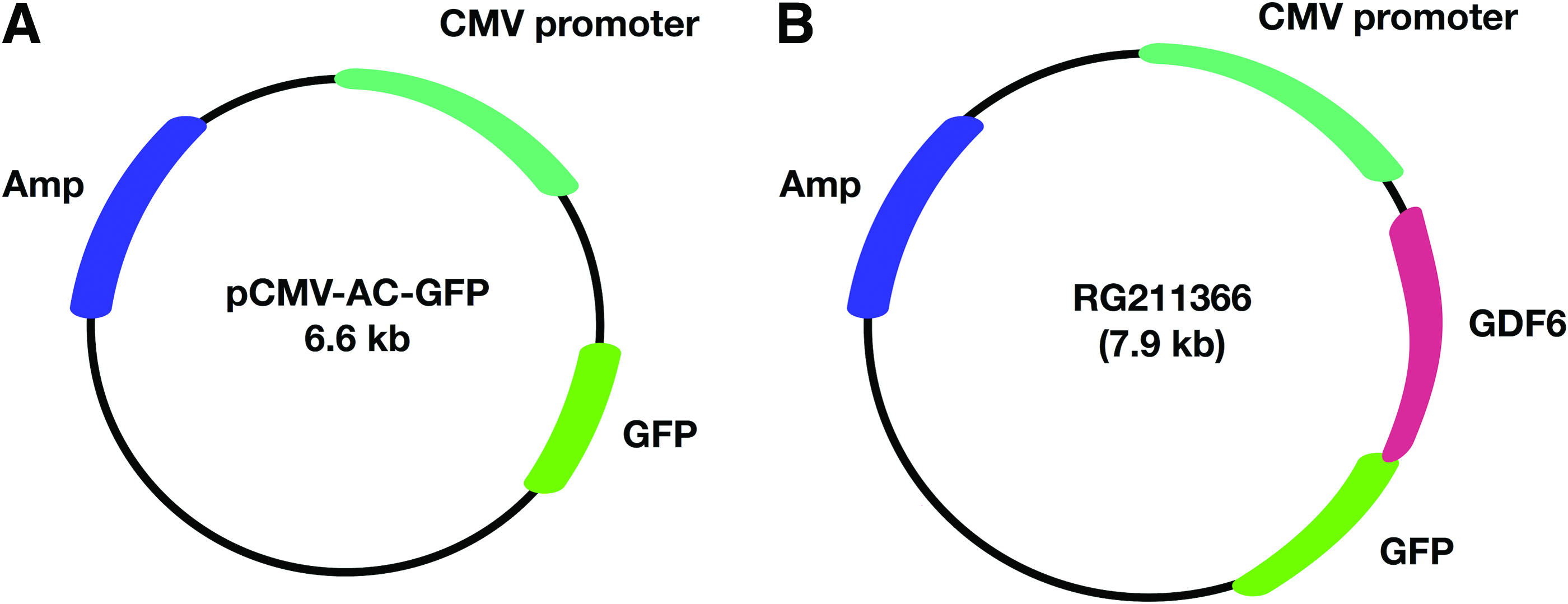
The mammalian cell vector pCMV6-AC-GFP (cat no. PS100010; OriGene Technologies, Inc., Rockville) was selected for visualization and quantification of the transfection efficiency (Fig. 1A). This plasmid contains the turbo green fluorescent protein (tGFP) sequence at the site of the ORF, which is an enhanced variant of the “CopGFP” cloned from copepod Pontellina plumata (Dana, 1849) (Crustacea: Pontellidae). The ORF is preceded with a cytomegalovirus (CMV) promoter, which overexpresses the protein of interest (Fig. 1A). The plasmid was clonally amplified overnight using heat-shock transformation of competent DH5α-E. coli. Positive clones were selected by addition of 100 μg/mL ampicillin (Sigma-Aldrich, St. Louis) and purified using the DNA purification kit, NucleoBond Miniprep (Macherey-Nagel, Oensingen, Switzerland).
Transfection with pCMV6-AC-GFP
The IVD cells were harvested at nearly 80% confluency. Cells were released from the plates by Trypsin/EDTA solution (Sigma) for 10 min at 37°C and washed once with PBS. The transfection of primary IVD cells was achieved by an implemented electroporation device system according to manufacturer's instructions (Neon Transfection System; Invitrogen, Basel, Switzerland). The Neon Transfection System 10 μL kit was used for the transfection of human and bovine IVD cells. Per transfection, 140,000 cells were taken up in 10 μL of resuspension buffer containing 0.8 μg of the plasmid with the 10 μL Neon tips using the Neon Transfection System pipette (Invitrogen). We used unpublished parameters for the required transfection procedure, that is, pulse voltage (V), pulse width (ms), and pulse number. First we optimized for both, human and bovine IVD cells, the parameters using a specific set of parameters shown in Table 2 and tested on human and bovine AFC and NPC, respectively.
The transfection efficiencies were determined using different settings, and the transfected cells were quantified by microscopy. + = 0–10%, ++ = 11–20%, +++ = 21–30%, ++++ = 31–60% transfected of total cell numbers.
Bold values emphasize parameter set B.
Finally, for the main experiment, electroporation was performed applying two pulses with 1400 V for 20 ms. Nontransfected cells and transfected cells without plasmid served as controls. After the electroporation, the cells were seeded in monolayer cultures in standard 6-well plates (BD Falcon, Basel, Switzerland) by adding 3 mL of medium containing LG-DMEM supplemented with 10% FBS without antibiotic supplements. The cells were cultured for 48 h and subsequently proceeded for analysis of transfection efficiency by flow cytometry.
Transfection with pGDF6
For GDF6 treatment transfections, the pCMV6-AC-GFP vector containing cDNA for the human GDF6 was used (Fig. 1B), further defined in this study as “pGDF6” (RG211366; OriGene Technologies, Inc.). The C-terminus for the ORF is further tagged with tGFP, producing a chimeric protein. The cloning of the plasmid was performed, as explained in the previous section for pCMV6-AC-GFP. The flanking T7 primer sequences on the vector map were used to check the sequence of the GDF6 ORF (performed by the sequencing service of Microsynth, Inc., Buchs, Switzerland). Prior to transfection, previously cultured bNPC and bAFC were harvested at 80% confluency and washed once with PBS. Due to the high amount of required cells, the Neon 100 μL kit was used. For transfection, the cells (450,000) were resuspended in 100 μL of resuspension buffer containing 9 μg plasmid DNA (pGDF6 or pCMV6-AC-GFP) and taken up with the 100 μL Neon Transfection System tip by the Neon Transfection System Pipette (Invitrogen). The human transfections were performed similar to the bovine transfections, except for the amount of DNA, which was lowered to 6 μg, to sustain viability of cells after transfection. We used three different parameter sets, to investigate their impact on gene expression: parameter set A: 1400 V, 10 ms, two pulses, B: 1400 V, 20 ms, two pulses, and C: 1100 V, 40 ms, one pulse. Nontransfected and transfected cells without plasmid served as controls.
After the electroporation, the cells were seeded in monolayer cultures into standard 6-well plates (Becton & Dickinson, Inc., Falcon, Brussels, Belgium) by adding 3 mL of medium containing LG-DMEM supplemented with 10% FBS without antibiotic supplements. After 48 h, the medium was replaced by LG-DMEM supplemented with 10% FCS, 100 μg/mL penicillin, and 100 U/mL streptomycin (Gibco®; Life Technologies Corporation, Basel, Switzerland). The cells were cultured for 14 days, and gene expression of different marker genes (Table 3) for the IVD phenotype and GDF6 28 was determined on day 7 and 14.
A two-step cycling with 95°C for 15 s and 57°C (bovine) or 61°C (human) for 30 s was used with 45 repeats.
qPCR, quantitative polymerase chain reaction.
Fluorescence microscopy
Efficiency of transfections and health status of the IVD cells were visualized on an inverse microscope (Leica, DM IL; filters: I3 S 450–490 nm and N2.1 S 515–560 nm).
Flow cytometry
To quantify the transfection efficiency, flow cytometry was applied. The electroporated and control cells (nontransfected and transfected control cells) from human and bovine IVD samples were trypsinized with 0.5% EDTA-Trypsin (Gibco; Life Technologies, Basel, Switzerland). The cells were resuspended in 500 μL of fluorescence-activated cell sorting buffer (PBS+2% FBS). Dead cells were excluded by addition of propidium iodide (PI, 1 μg/mL; Sigma-Aldrich). To compensate the GFP and PI channels, single stains were performed. The fluorescence signals were detected using 488 nm blue laser and the filters 525/50 GFP and 585/15 PI on a LSR II flow cytometry system (Becton & Dickinson), and the data were analyzed using FlowJo Software (LLC, Ashland, OR, United States, version 10.1 for MacOS X).
Quantitative polymerase chain reaction
IVD cells were lysed in TRI Reagent (Molecular Research Center, Cincinnati), and total RNA was extracted using a modified TRI spin method as reported previously.29,30 Briefly, RNA was mixed with polyacryl carrier (Molecular Research Center), and organic 1-bromo-3-chloropropane (BCP; Sigma Aldrich) was added and mixed vigorously. Phase-separation of total RNA from the DNA/protein fraction was performed by centrifugation at 800 g for 15 min at 4°C. Subsequently, the clear RNA supernatant fraction was loaded at a final concentration of 70% ethanol onto the GenElute™ Miniprep Kit (Sigma). After elution of the RNA, any possibly remaining genomic DNA was digested with recombinant DNase I (AMP-D1; Sigma Aldrich) for 15 min. RNA integrity and purity was then detected on selected samples by Experion™ Automated Electrophoresis System (Bio-Rad, Reinach, Switzerland).
For reverse-transcription, the iScript™ Kit was used according to manufacturer's instructions using ∼500 ng of total RNA per 20 μL reaction (Bio-Rad). cDNA was then diluted 1:5; prior to quantitative polymerase chain reaction (qPCR), genomic template and negative controls were run to exclude DNA contamination. Real-time PCR was performed in duplicates using SYBR Green PCR Master Mix on a CFX96 Touch RT-qPCR system (all from Bio-Rad). The following genes relevant for the IVD phenotype were monitored: growth and differentiation factor 6 (GDF6 syn. CDPM2 or BMP13), green fluorescent protein (GFP), collagen type 1 and 2 (COL1A2 and COL2A1), and aggrecan (ACAN) (Table 3). 31 The 18S ribosomal RNA gene was chosen as reference gene, which is highly expressed in IVD cells and proven independent from these experimental conditions. 32
Primers were synthesized by Microsynth, Inc. (Balgach, Switzerland) and were all tested for efficiency. The qPCR was run in a two-step protocol with an annealing temperature of 57°C for bovine or 61°C for human DNA (Table 3). Melting curve analysis was run as a control for the specificity of the amplicons. Relative gene expression was quantified using the CFX96 cycler software (Bio-Rad); the number of PCR cycles needed for each sample to reach that threshold level was recorded as the Cq value. 33 ΔΔCq values at end point were estimated relative to day 0 and transformed into relative mRNA values using the formula 2−ΔΔCq. 34
Statistics
Statistical difference in the transfection efficiencies between IVD cell types and between species was performed by the Student's t-test. qPCR data were evaluated by two-way ANOVA (i.e., parameters versus genes in a donor-matched design) and Tukey's correction of multiple comparisons. For statistical analysis Prism 6.0h for Mac OS X (GraphPad, La Jolla) was used. Values are given as mean ± SEM. A p value <0.05 was considered significant.
Results
Transfection with pCMV6-AC-GFP
The transfection efficiency of bovine and human IVD cells was quantitatively determined by flow cytometry. Representative transfections of human and bovine NPC and AFC with pCMV6-AC-GFP after 48 h were monitored by fluorescent microscopy (Fig. 2). The highest transfection efficiency was detected in bAFC (59.6% ± 5.0%) (Fig. 3), compared to bNPC and human IVD samples. In bNPC the efficiency was 52% ± 8%. hNPC showed an average transfection efficiency of 46.7% ± 1.4% and hAFC an average of 47.1% ± 2.4%.
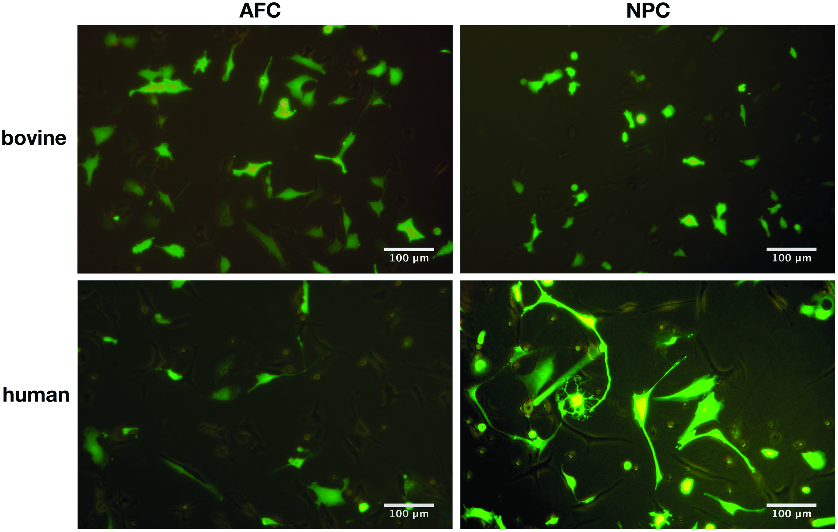
GFP-positive human and bovine annulus fibrosus cells (AFC), and nucleus pulposus cells (NPC) after 48 h of transfection with pCMV6-AC-GFP were detected under a light microscope. Color images available online at
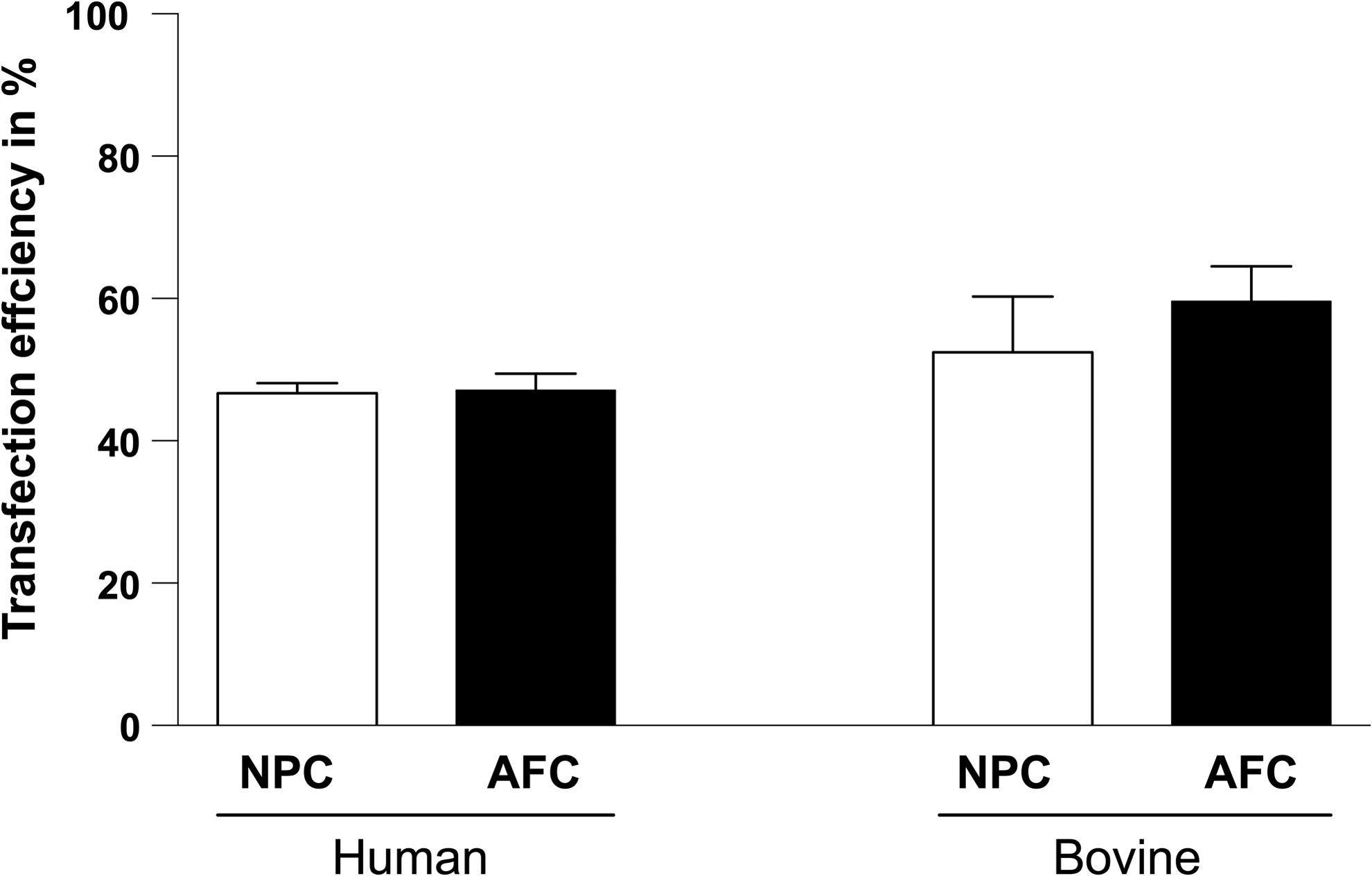
Percentage of transfection efficiency of human (N = 4) and bovine (N = 5) NPC and AFC as quantified by flow cytometry. The percentages of transfection efficiencies are (mean ± SEM): hNPC 46.7% ± 1.4%, hAFC 47.1% ± 2.4%, bNPC 52.44% ± 7.9%, and bAFC 59.6% ± 5.0%. AFC, annulus fibrosus cells; NPC, nucleus pulposus cells; SEM, standard error of the mean.
The two control groups, nontransfected and cells transfected without plasmid, showed lack of GFP expression. In particular, dead cells were also detected to be GFP positive. The rates of GFP+ and PI+ cells were 10.4% ± 3.5% in bNPC and 12.4% ± 2.3% in bAFC. A higher number of dead cells were detected in human cells (17.2% ± 1.3% in NP and 19.2% ± 2.3% in AF). There was no significant difference in efficiencies between hNPC and bNPC (p = 0.545) and between the two AFC populations, respectively (p = 0.076). Furthermore, there was no significant difference between hAFC and hNPC (p = 0.918) and between bAFC and bNPC (p = 0.542).
Transfection with pGDF6
Seven and 14 days after transfection, qPCR was performed to analyze the gene expression of GDF6, GFP, and ECM proteins (ACAN, COL1, and COL2) (Fig. 4). Seven days after transfection, an upregulation of GDF6 in bNPC, hNPC, and hAFC, transfected with pGDF6, was detected (Fig. 4 and Supplementary Fig. S1; Supplementary Data are available online at
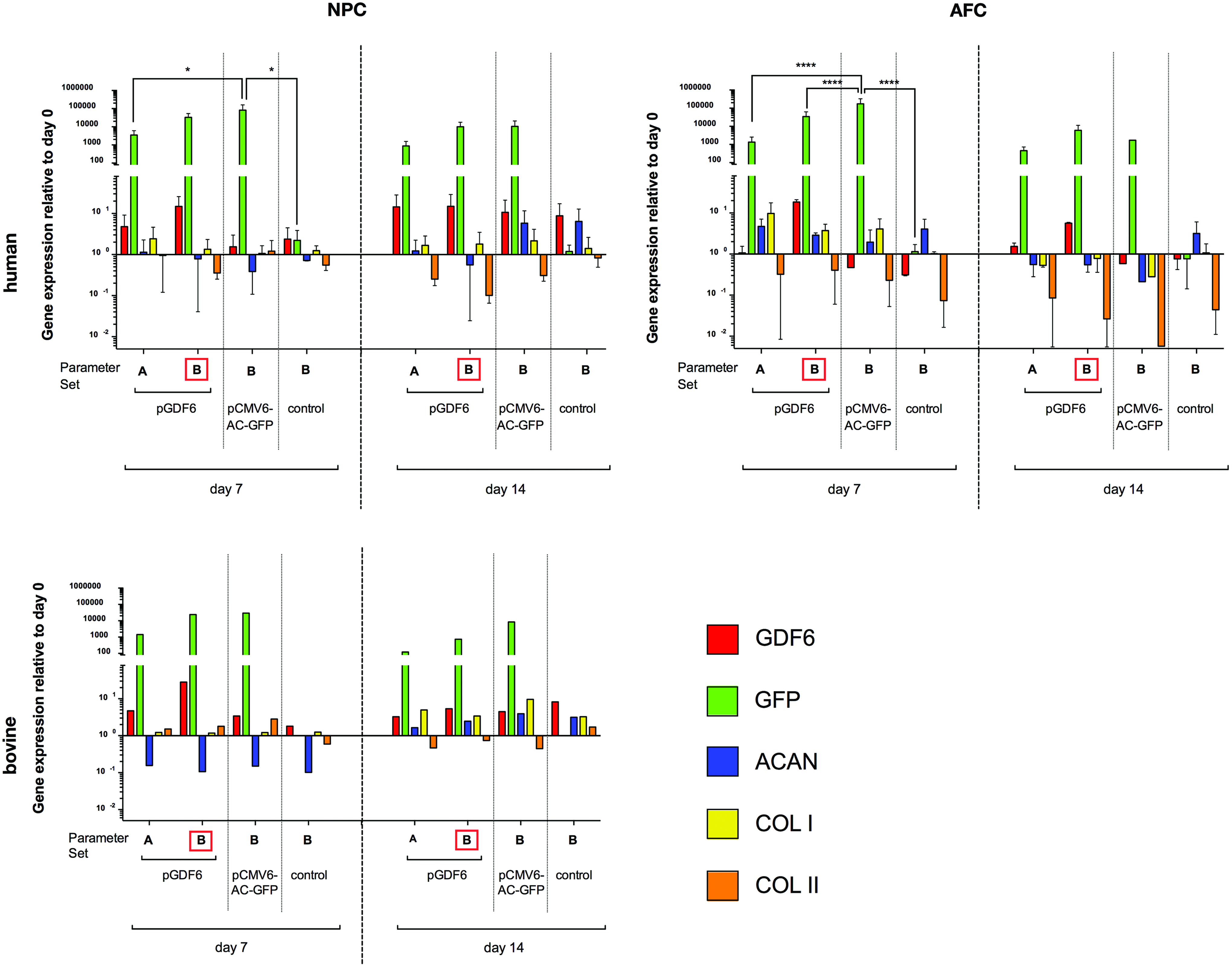
Relative gene expression of GDF6, GFP and major ECM genes in primary hNPC and hAFC (N = 2) and bNPC after transfection with pGDF6, pCMV6-AC-GFP, or as a control with no plasmid, after 7 and 14 days. pGDF6 was transfected with two different parameter sets: Set A: 1400 V, 10 ms, two pulses, Set B: 1400 V, 20 ms, two pulses. p Values (two-way ANOVA) *<0.05; **** <0.0001. AFC, annulus fibrosus cells; b, bovine; h, human; NPC, nucleus pulposus cells. Color images available online at
GFP showed in all transfected cells (pGDF6 and pCMV6-AC-GFP) a strong expression. However, the highest GFP expression level reached cells transfected with parameter B (Fig. 4). In hNPC, GFP in cells transfected with pCMV6-AC-GFP and parameter set B was significantly upregulated compared to the control cells (p = 0.0221). In hAFC transfected with pCMV6-AC-GFP and parameter set B, a significant upregulation of GFP expression compared to the control cells was detected (p value <0.00001). However, surprisingly no upregulation of ECM proteins such as ACAN and COL2A1 could be determined. After 14 days GFP was still present, but the expression of GDF6 was decreased.
Discussion
In this study, we aimed to determine parameters for efficient and reproducible transfection of human and bovine IVD cells with the Neon Transfection System. Successful transfection of pCMV6-AC-GFP could be confirmed in human and bovine AFC and NPC with 1400 V for 20 ms in two pulses (Figs. 3 and 4 and Supplementary Fig. S1).
In both species, human and bovine, AFC showed higher transfection efficiency than NPC (Fig. 3). This variance could arise from the different phenotypes of these cells, as NPC are described to be chondrocyte like and AFC are more similar to fibroblasts.35,36 Dickens et al. 37 could detect up to 46% transfection efficiency in human fibroblast by nucleofection. The present study showed transfection efficiency in the same range for hAFC (47.1% ± 2.4% for N = 4).
In comparison of human and bovine samples, we detected 10% higher transfection efficiency in bovine than human IVD cells. The bovine cells seemed to regenerate faster from the transfection procedure, as they adhered rapidly after seeding and started proliferating. Bovine IVD cells showed in general a higher viability than human cells as detected by microscopy and flow cytometry analysis. One possible explanation is the origin of disc material, whereas bovine discs were obtained from healthy individuals and human discs were donated from trauma patients. In addition, bovine and human discs differed in age, as animals reached only 10–14 months before slaughtering.
In this study, we provide an example of gene therapy using bNPC, hNPC, and hAFC that were transfected with the pGDF6 and pCMV6-AC-GFP with different parameter sets. A significant upregulation of GDF6 gene expression in pGDF6 with parameter set B transfected cells was detected. In addition, control groups showed a slight upregulation of GDF6 compared to the day 0 group, which indicates a native GDF6 expression in hIVD cells. It could be shown that hIVD transfected with pCMV6-AC-GFP and parameter B showed a significant upregulation of GFP expression, compared to the control groups, what again represent the strength of parameter set B.
In this experiment, we also observed major IVD ECM proteins to investigate the effect of GDF6 on the IVD cells. Clarke et al. 14 showed a shift from GDF6 stimulated adipose-derived MSC toward a NP-like phenotype, which resulted in more proteoglycan-rich matrix. Wei et al. 38 also demonstrated that the direct injection of GDF6 after disc injury promotes ECM proteins like aggrecan or collagen type 2 in a large animal ovine model. However, in our study, the overexpression plasmid for GDF6 did not upregulate aggrecan nor collagen type 2 in IVD transfected cells (Fig. 4 and Supplementary Fig. S1). A possible reason could be that the investigated cell population was not homogenous for the desired GDF6 overexpression and so there was no stimulation of ECM proteins. Another explanation is that GFP-tagged GDF6 fusion protein is not biologically functional. Thus, these parameters would need to be investigated in more detail using untagged overexpression vectors or vectors with two independent promoters.
Nonviral transfection is a promising approach for gene therapy as it fulfills the translational safety aspects of transiency and lacks the harmful effects compared to viral transduction and is of high interest for tissue engineering and regenerative medicine applications. High-voltage electric pulses to introduce DNA into cells can be used with most cell types and is gaining popularity as it can be applied for stable and transient transfection.22,39,40 The discovery of parameters, which led to highly efficient transfected cells, will positively influence future gene therapies in IVD cells. The possibility to manipulate primary bovine IVD cells is of high importance in the IVD research community as bovine coccygeal discs have advanced to a translational model system in spine and IVD research.41,42
Conclusion
In this study, parameters are presented for optimization of transfection efficiency and reproducibility in IVD cells. The investigation of a successful nonviral transfection method for IVD cells can serve as possible gene therapy for tissue engineered IVD constructs using incorporated growth factors to promote IVD tissue regeneration.
Footnotes
Acknowledgments
The authors acknowledge Eva Roth for technical assistance. The project was supported by the Lindenhof Foundation “Teaching & Research” project #15-05F and by direct funds from Hansjörg Wyss and the Hansjörg Wyss Medical, US, foundation.
Disclosure Statement
No competing financial interests exist.
References
Supplementary Material
Please find the following supplemental material available below.
For Open Access articles published under a Creative Commons License, all supplemental material carries the same license as the article it is associated with.
For non-Open Access articles published, all supplemental material carries a non-exclusive license, and permission requests for re-use of supplemental material or any part of supplemental material shall be sent directly to the copyright owner as specified in the copyright notice associated with the article.