
Abstract
Synthetic polymers such as polyglycolic acid (PGA) fibers are the traditional tissue engineering scaffolds that are widely used for engineering a variety of soft tissues. However, the major disadvantage of this polymer material is its released acidic degradation products that trigger inflammatory response and fibrotic process, which affects the biocompatibility and the quality of the engineered tissues. In this study, the effect of hyaluronic acid (HA) coating on improving PGA biocompatibility was explored. The results showed that 1% HA solution could better coat PGA fibers than other tested concentrations of HA, and coated PGA exhibited less inflammatory reaction upon in vivo subcutaneous implantation. In vitro characterization demonstrated that HA coating could enhance cell adhesion to the scaffold and reduce gene expression of IL-1, IL-6, IL-8, and α-SMA. It also decreased the acidity of degradation products in vitro. Furthermore, coated PGA could engineer better cartilages in vitro with higher content of total collagen and glycosaminoglycan, as well as higher gene expression levels of collagen II, aggrecan, and Sox9. Collectively, the data indicate that HA coating can significantly enhance the biocompatibility of this traditional scaffold material, which also enhances the quality of engineered tissues.
Introduction
B
Among them, poly (α-hydroxy acid) fibrous scaffold is a typical type of synthetic polymer-based scaffold. Naturally, hydrolysis of polyglycolic acid (PGA) causes its bulk degradation to glycolic acid, followed by further metabolism in the tricarboxylic acid cycle, which results in carbon dioxide and water. 3 This polymer type includes PGA, polylactic acid (PLA), or (poly(lactic-co-glycolic acid) (PLGA).1,4 At early stage of tissue engineering, PGA nonwoven fibers were almost the exclusive scaffold materials for proof-of-concept researches in engineered cartilage 5 and tendon. 6 Later, this scaffold has been widely used for engineering various types of soft tissues, including cartilage, 7 tendon, 8 blood vessel, 4 peripheral nerve, 9 and skin, 10 and succeeded in engineered tissue formation and repair. Despite this, the released acidic degradative products of the scaffolds are the disadvantage that could affect their biocompatibility to seeded cells and host tissues once they are implanted in vivo, particularly in large immunocompetent animals.
To improve the biocompatibility of polymer-based synthetic materials, surface modification either with physical treatment or with chemical treatment, such as plasma surface modification or pulsed plasma deposition, was applied. In addition, Ko et al. 11 utilized ozone oxidation of PEG [poly (ethylene glycol)] to enhance its hydrophilic properties.
The other important way is the surface coating of polymer with natural extracellular matrix molecules such as collagen and proteoglycans. 12 Hyaluronic acid (HA), a naturally occurring polysaccharide composed of N-acetyl-d-glucosamine and d-glucuronic acid, is known as an important component of the extracellular matrices in connective tissues, particularly in cartilage tissue. 13 HA has also been used for surface modification of poly(α-hydroxy acid) based polymer. 14
Based on our previous experience of using PGA fibers as scaffold materials,1,15 this study further explored the possibility of improving PGA biocompatibility with HA surface coating and observed its effect on PGA degradation, acidic degradation product release, and inflammatory response in vivo and its effect on engineered tissue fabrication using cartilage as an example.
Materials and Methods
General experimental design
In this study, unmodified nonwoven PGA scaffolds served as control group, whereas the test groups included the nonwoven PGA scaffolds modified with HA solution coating at various concentrations as described below. Coated and noncoated nonwoven PGA scaffolds were, respectively, tested for their degradation rates and acidic product release, induction of inflammatory gene expression in vitro, host inflammatory reaction in vivo, and capability of engineering cartilage in vitro and in vivo along with related characterization. The schematic flow chart is showed in Figure 1. The scaffold coating procedure was performed under asepsis, and no further sterilization was applied.
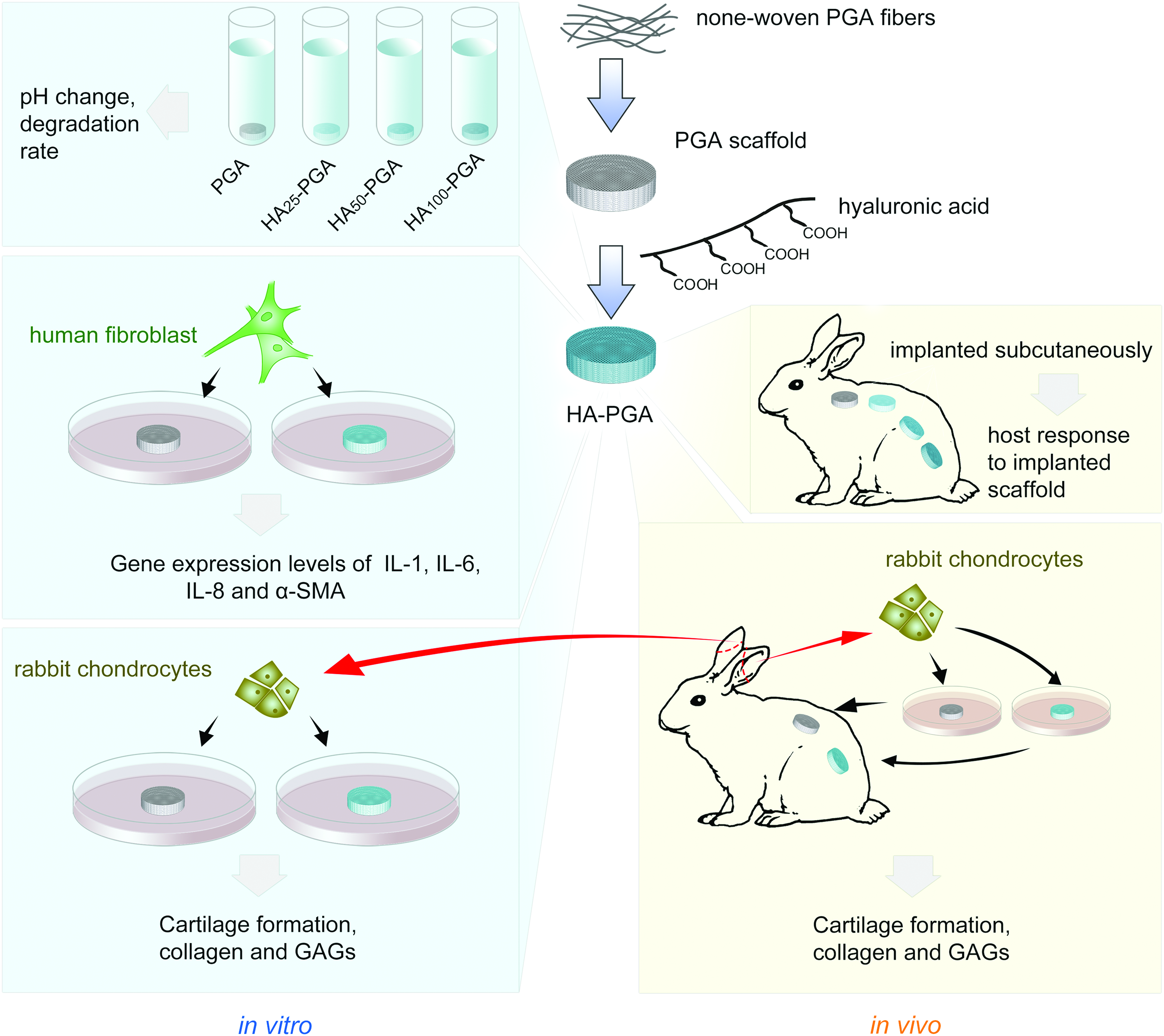
The schematic flowchart of experimental design. Blue areas show the experiments performed in vitro, and yellow areas show the in vivo experiments. Color images available online at
Animals
Total eight New Zealand white rabbits 2–3 month old were used in this research. The mean weight was 2.10 ± 0.12 kg. All animal experimental protocols were approved by Animal Care and Experiment Committee of Shanghai Jiao Tong University School of Medicine. The rabbits were used to test host inflammatory response to implanted nonwoven PGA scaffolds with or without HA coating and to test the effect of HA coating on engineered cartilage formation in vivo.
Preparation of noncoated and HA-coated PGA scaffolds
PGA fibers (Shanghai Ju Rui Biomaterials Co. Ltd. Shanghai, China) were used as previously described. 16 Briefly, nonwoven PGA scaffolds were pressed into a cylindrical shape and then disinfected with 75% ethanol solution for 60 min followed by two washes with phosphate-buffered saline (PBS). Then, 25, 50, or 100 mg HA powder (molecular weight of 2.5–2.6 million Da, Shandong Freda Biochem, Shandong, China) was, respectively, dissolved in 10 mL PBS (g/mL) at the final concentrations of 0.25%, 0.5%, and 1% (w/v). They were, respectively, labelled as HA25-PGA, HA50-PGA, and HA100-PGA. Briefly, to generate surface modification with HA, one PGA scaffold was put into one syringe with the same radius for all prepared samples, and the same amount of HA solution was added to each of the scaffold contained syringes. As a control, the same amount of PBS was added to each syringe. The immobilization reaction was carried out for 12 h with gentle stirring and vacuum dried.
Scanning electron microscopic examination
After vacuum drying, the scaffold constructs were prepared for scanning electron microscope (SEM) examination as previously described. 17 Briefly, samples were vacuum dried. Then the samples were sputter-coated with gold (BAL-TEC, Philips, Eindhoven, Netherlands) and examined finally with SEM (JCM-5000, NeoScope, Japan). Three samples for each group were examined.
In vitro assay of scaffold degradation and pH value of degradation products
To analyze pH value change during in vitro degradation process, 12 scaffold pieces of each group were included. Briefly, each sample was made of 50 mg nonwoven PGA scaffolds that were pressed into a cylindrical shape with 10 mm in diameter and 3 mm in thickness, then put into 40 mL of PBS in a separate tube for total 4 weeks in an incubator (37°C) without stirring and refreshing, and pH value was measured every 3 days (n = 3) using a pH meter (KEDIDA, CT-6023, China). At the end of each week, three pieces of scaffolds from each group were harvested, vacuum dried, and weighted to collect data of weight change.
Host response to implanted cell-free scaffold in vivo
Rabbits were anesthetized with intramuscular injection of ketamine (10 mg/kg) and Lumianning (5 mg/kg) and laid in prone position. As described above, a total of 50 mg nonwoven PGA fibers were used to prepare each scaffold into a cylindrical shape (10 mm in diameter and 3 mm in thickness). Four incisions with a length of 1.5 cm were made on the back at two sides of the spine. Subcutaneous tissue was separated to form four pockets, and cell-free PGA, HA25-PGA, HA50-PGA, and HA100-PGA scaffolds were, respectively, inserted into four pockets followed by wound closure with suture. Three samples of each group were tested.
Isolation and culture of human dermal fibroblasts
Protocols for the handling of human tissue and cells were approved by the Ethics Committee of Shanghai Jiao Tong University School of Medicine. Human foreskins were obtained with written informed consent during circumcisions. The cells were derived from the foreskin of nine patients, aged ranging 1–10 years old. Cells derived from three different patients were mixed as a combined cell source, and thus, total three cell samples were generated for the following experiments. Briefly, foreskin tissues were harvested under aseptic conditions and rinsed in 2.5% chloramphenicol solution thrice, then in PBS for 5 min twice. The tissues were sectioned into 2 × 4 mm pieces and treated with 0.2% dispase (Roche Diagnostics, Indianapolis, IN, dissolved in Dulbecco's modified Eagle's medium [DMEM] containing 10% fetal bovine serum [FBS]) overnight at 4°C. Then the dermis was mechanically separated from the epidermis and cut into smaller fragments followed by enzyme digestion in 0.2% collagenase (SERVA, GER) dissolved in DMEM containing 10% FBS for 2 h at 37°C on a rotator. Later, the cell suspension was centrifuged at 1500 rpm for 5 min. The cell pellet was resuspended in DMEM containing 10% FBS, penicillin (100 U/mL), and streptomycin (0.1 mg/mL) and seeded onto 10 cm culture dishes (BD FALCON, Franklin Lakes, NJ) at a density of 1.5 × 10⁶ per dish and cultured at 37°C, 5% CO2. When reaching confluence, cells were detached with 0.25% trypsin-EDTA (Gibco, Grand Island, NY) and subcultured at the same density. Passage 3 fibroblasts were used for following assays.
Isolation and culture of chondrocytes
Cartilage tissue slices with a size of 2.0 × 2.0 cm were obtained from harvested auricular cartilage of rabbits and minced into pieces sized about 1.0 mm3. The cartilage pieces were washed with PBS and digested with 0.25% collagenase type II (Worthington Biochemical Corp., Freehold, NJ) for 6 h to isolate chondrocytes as previously described. 18 Then, the cells were harvested, cultured, and expanded according to reported methods. 18 The primary cultured chondrocytes were used for in vivo cartilage engineering, while passage 2 cells were used for in vitro cartilage engineering.
Cell-scaffold adhesion assay
The assay was used to observe the effect of HA coating (at concentration of 1%, HA100-PGA) on cell adhesion to the scaffold. Briefly, 100 μL of cell suspension of dermal fibroblasts at a density of 3 × 10⁷ cells/mL was loaded onto the upper side of one of control or HA-coated scaffold (100 mg in weight and in a cylindrical shape with a diameter of 25 mm and a thickness of 1.5 mm), and another 100 μL cell suspension with the same concentration was injected into the scaffolds and incubated in 10 cm culture dishes (BD FALCON, Franklin Lakes, NJ) at 37°C in a humidified, 5% CO2 atmosphere for 4 h. Then the cell-loaded scaffolds were gently transferred into a new dish. The remaining cells left in the original dishes were collected and counted. The cell seeding efficiency on the scaffolds was calculated based on the formula: (Total loading cell number−Remaining cell number)/Total loading cell number × 100%. The assay was repeated in three samples for each group.
Culture of dermal fibroblast seeded scaffolds
As described above (last section), after the loading of 100 μL cell suspension and intra-scaffold injection of 100 μL cell suspension, the cell-seeded scaffolds were, respectively, cultured on cell culture dishes at 37°C in an incubator for 4 h. Then the cell-seeded scaffolds were cultured with 20 mL of DMEM containing 10% FBS, penicillin (100 U/mL), and streptomycin (0.1 mg/mL) per dish and cultured at 37°C in a humidified 5% CO2 atmosphere. The medium was replaced at the third day. At the sixth day, the constructs were transferred to new dishes, incubated in serum-free DMEM for 24 h, and then harvested at the seventh day for cytokine/growth factor expression analysis.
RNA extraction and real-time quantitative polymerase chain reaction
The cultured cell-seeded constructs were harvested at different time points (1 week for fibroblast-PGA scaffold as described in last section and 4 and 8 weeks for in vitro engineered cartilages). Total RNA was extracted from these constructs or engineered tissues using TRIzol Reagent (Invitrogen, Carlsbad, CA) without cell detachment. The complementary DNA (cDNA) was synthesized from 2 μg total RNA per sample with the use of AMV reverse transcriptase (Promega) in a 20 μL reaction solution consisting of 4 μL 5 × buffer, 2 μL dNTP, 1 μL oligo-(dT), 0.5 μL RNase inhibitor, and 0.5 μL AMV reverse transcriptase, using ddH2O to meet the final volume. The mixture was then incubated at 30°C for 10 min, 42°C for 60 min, 95°C for 5 min, and 5°C for 5 min. cDNA was then amplified using a Power SYBR Green PCR master mix (2 × ) (Applied Biosystems, Foster City, CA) in a real-time thermal cycler (Stratagene Mx3000PTM QPCR System, La Jolla, CA). The optimized primers for qPCR analysis are listed in Table 2. Glyceraldehyde 3-phosphate dehydrogenase gene was used as an internal control. The experiment was performed in triplicate and repeated in three samples for each group.
In vitro cartilage engineering using PGA scaffold with or without HA coating
For in vitro cartilage engineering, 10 mg PGA fibers were prepared for each piece of scaffold (cylindrical shaped scaffold 5 mm in diameter and 1 mm in thickness), then 20 μL of cell suspension of passage 2 chondrocytes at a density of 6 × 10⁷ cells/mL were seeded onto each scaffold (PGA or HA100-PGA). A total of six pieces of scaffolds were included in each group, and the cell-seeded constructs were put into 6-well plates (one piece per well) (BD FALCON, Franklin Lakes, NJ) filled with 8 mL/well chondrogenic medium of DMEM containing 10 ng/mL TGF-β1, 100 ng/mL IGF-1, 1% ITS, 40 ng/mL dexamethasone, 50 μg/mL vitamin C, 100 U/mL penicillin, and 0.1 mg/mL streptomycin. The cell-seeded scaffolds were then placed in an incubator at 37°C and cultivated for 4 and 8 weeks, respectively. The medium was changed every 3 days. And 100 μL of 1% HA was added to the HA-PGA group at days 7, 14, and 21. Three samples were included in each group.
In vivo cartilage engineering and surgical procedure
For in vivo cartilage engineering, the autologous chondrocytes harvested from the rabbit auricular cartilage were resuspended in DMEM containing 10% FBS to a final concentration of 1.0 × 10⁷ cells/mL, and 60 μL of cell suspension was seeded evenly onto each scaffold (PGA or HA100-PGA scaffold, prepared as described above). The chondrocyte-scaffold constructs were cultured in DMEM containing 10% FBS, penicillin (100 U/mL), and streptomycin (0.1 mg/mL) for 24 h at 37°C in a humidified, 5% CO2 atmosphere. Afterward, they were in vivo implanted into rabbit subcutaneously.
For surgical implantation, the rabbit was anesthetized with intramuscular injection of ketamine (5 mg/kg) and xylazine (0.05–1 mg/kg). Six incisions with a length of 1.5 cm were made on the back at both sides of the spine followed by subcutaneous tissue separation to form six tissue pockets. Afterward, three chondrocyte-PGA constructs and three chondrocyte-HA100-PGA constructs were inserted into the six pockets, respectively, followed by wound closure with suture. The experiment was repeated in four rabbits. The animals were euthanized for multiple analyses at the end of 2 months.
Histological analysis
In vitro engineered cartilages were harvested after 4 and 8 weeks of culture, and in vivo engineered cartilages were harvested after 8 weeks of implantation. In addition, cell-free PGA scaffolds were also, respectively, harvested at 2 and 4 weeks postimplantation. The harvested tissues were fixed in 4% paraformaldehyde for 24 h followed by three washes in PBS, dehydration through graded alcohols, and finally embedded in paraffin for tissue section at 8 μm thickness for hematoxylin and eosin (H&E) staining and Toluidine Blue staining as previously described. 19 Ten sections were observed for each sample.
Biochemical analyses
The sulfated glycosaminoglycan (GAG) content of both in vitro and in vivo engineered cartilages was quantified by Alcian Blue assay as previously described. 20 In addition, total collagen content was also quantified by orthohydroxyproline assay as previously described. 21 The assay was performed in three samples for each group.
Statistical analysis
All the experimental data are presented as mean ± standard deviation and statistically analyzed with software SPSS (version 19.0, SPSS, Inc., Chicago, IL). Due to non-normal distribution of the data, Kruskal–Wallis test was used to analyze the data of Figure 3B among different groups at a particular time point, and Dunnett's test was used to analyze the difference between paired control and experiment groups. As for the rest of the data, after normal distribution test and homogeneity of variance test, ANOVA and Student's t-test were applied. A p-value less than 0.05 was considered statistically significant.
Results
The influence of HA concentration on coating efficiency of the scaffolds
Scanning electron microscopic examination demonstrated that 1% HA solution could coat the nonwoven PGA scaffolds better with even distribution, compared to 0.25% and 0.5% concentrations, which could only partially coat the PGA fibers as shown in Figure 2. The weight change of HA-PGA scaffolds before and after HA coating was listed in Table 1. There is significant difference in weight increase between noncoated and HA50-PGA and between noncoated PGA and HA100-PGA,

Scanning electron microscopic (SEM) images of noncoated scaffold (PGA) and hyaluronic acid (HA) coated scaffold at the concentrations of 0.25% (HA25-PGA), 0.5% (HA50-PGA), and 1% (HA100-PGA), Bar = 100 μm. The red arrows indicate the presence of coated HA on PGA fibers. PGA, polyglycolic acid. Color images available online at
Significant difference in weight change before and post-HA coating. (p < 0.05).
HA, hyaluronic acid; PGA, polyglycolic acid.
HA-coating attenuated the acidity of degradation products
The time-dependent pH course was examined during in vitro scaffold degradation assay in PBS over a 28-day period, which showed that HA coating helped to reduce the acidity of released degradation products. As shown in Figure 3A, pH value sharply dropped in the first 3 days and then entered into a plateau phase between day 3 and 18 followed by another sharp drop of pH value in the following 10 days. In general, all these HA-coated groups exhibited similar pH values at most of the time points (p > 0.05), except for days 3 and 6 (p < 0.05). By contrast, noncoated PGA group exhibited relatively sharper drop of pH value. Statistical analysis showed significant difference among four groups at all the time points (p < 0.05) except for day 0. When performing a paired analysis, the pH value of control PGA was significantly lower than those of other three groups, respectively, at days 9, 12, and 18 (p < 0.05). And significant difference in pH value was found between PGA and HA50-PGA at days 3 and 6 (p < 0.05), between PGA and HA50-PGA, as well as between PGA and HA100-HA at days 15 and 21 (p < 0.05). Among the three HA-coated groups, significant difference in pH value was found between HA25 and HA50-HA at days 3 and 6 (p < 0.05) and HA50 and HA100-HA at day 3 (p < 0.05).
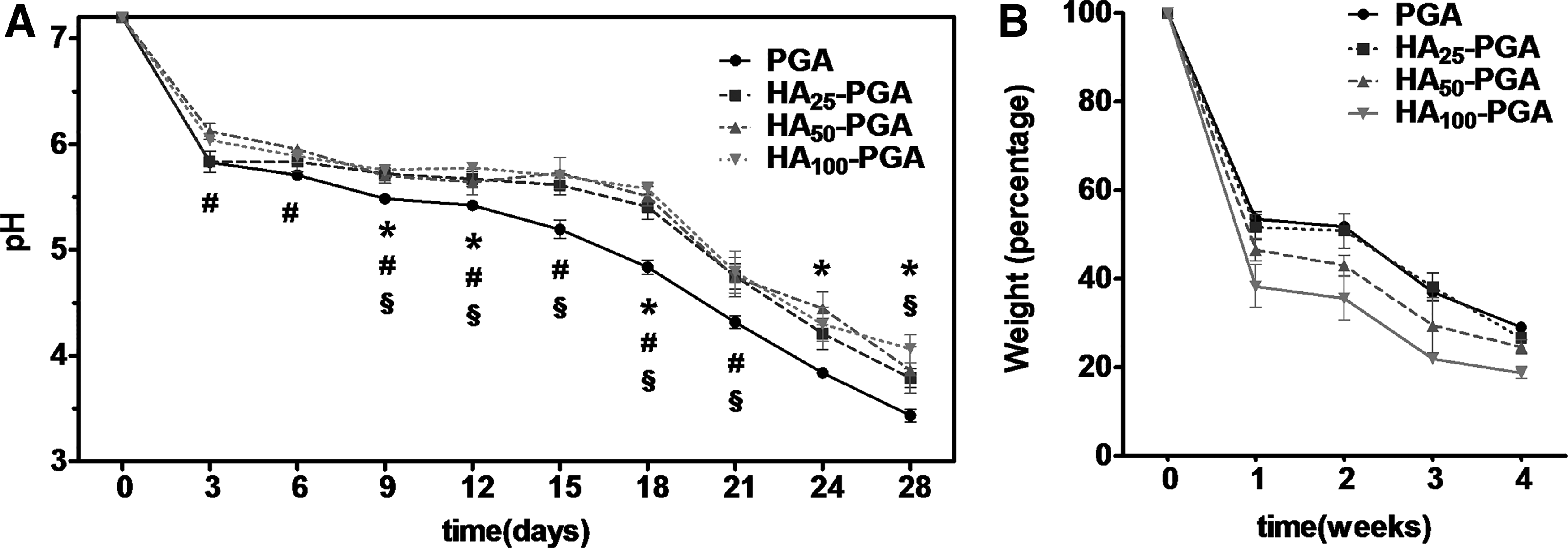
In vitro scaffold degradation assay.
The effect of HA coating on scaffold degradation
As shown in Figure 3B, all HA coating and noncoating scaffold showed a drop of scaffold weight as time went by. Statistic analysis showed only significant difference in scaffold weights among 4 groups at the third week (p < 0.05). When performing a paired comparison, significant difference was not found between any two groups (p > 0.05).
In vivo host response to implanted scaffold with or without HA coating
After 2 weeks of in vivo implantation of cell-free scaffolds, three rabbits were sacrificed and three specimens of each group were harvested and investigated for early inflammatory response. As shown in Figure 4, at the 2-week time point, PGA coated with higher concentrations of HA (HA50-PGA and HA100-PGA) exhibited less extent of acute inflammatory response with fewer cell infiltration (represented by round shaped nuclear cells) at the peripheral area of implanted scaffold, compared to noncoated scaffold or the scaffold coated with lower concentration of HA. In addition, significantly more vascular structures were observed at the peripheral area of control PGA scaffold (Fig. 4E, white arrow) compared with HA-coated scaffold with high concentration (Fig. 4H). At this time point, no obvious cell penetration into the inner part or obvious scaffold degradation was observed as shown in Figure 4M–T.
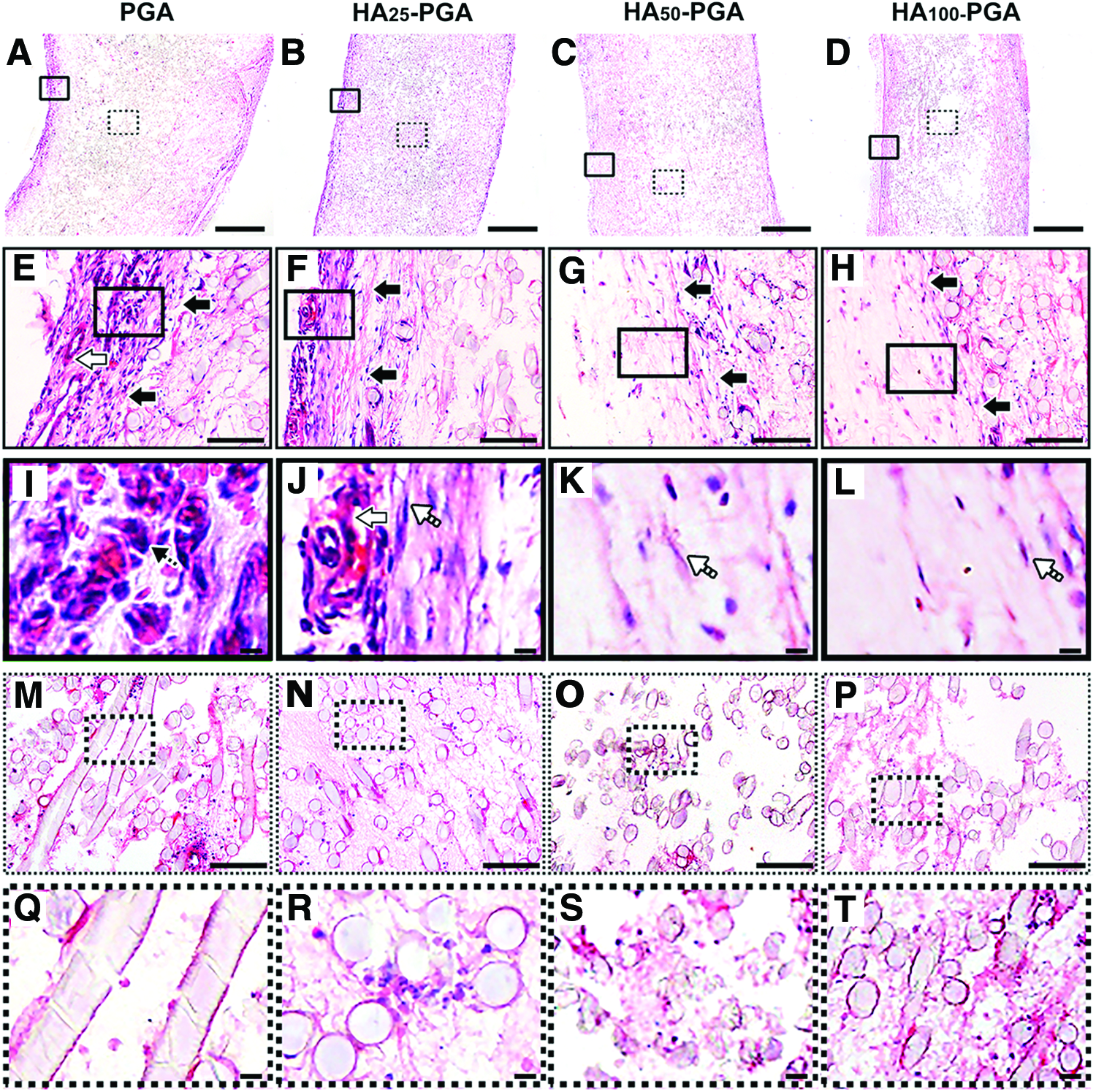
In vivo histological evaluation of host response to implanted noncoated scaffold (PGA) and scaffolds coated with HA at the concentrations of 0.25% (HA25-PGA), 0.5% (HA50-PGA), and 1% (HA100-PGA). H&E staining images of vertical section at 2 weeks postimplantation.
More cells penetrated into the scaffolds at 4 weeks than at 2 weeks. As shown in Figure 5, compared to noncoated scaffold, more cells penetrated into the inner part of HA-coated scaffold. In general, significantly more fibrotic tissues were formed in coated scaffold than in noncoated scaffold. In addition, cell necrosis evidenced by cell fragments was obviously observed in the inner part of noncoated scaffold (Fig. 5M, Q). By contrast, inflammatory cells along with host fibroblasts were found deeply penetrated into the inner part of HA-coated scaffold (Fig. 5P, T), indicating that the microenvironment created by HA coating allowed the survival of penetrated cells, which is in sharp contrast to the microenvironment of control scaffold (Fig. 5M). All these evidences indicated that HA coating on PGA scaffolds could restrain acute inflammatory reaction in vivo at early stage, yet favor tissue formation at later stage, suggesting improved biocompatibility. We thus choose HA100-PGA as the experimental group for the following experiments.
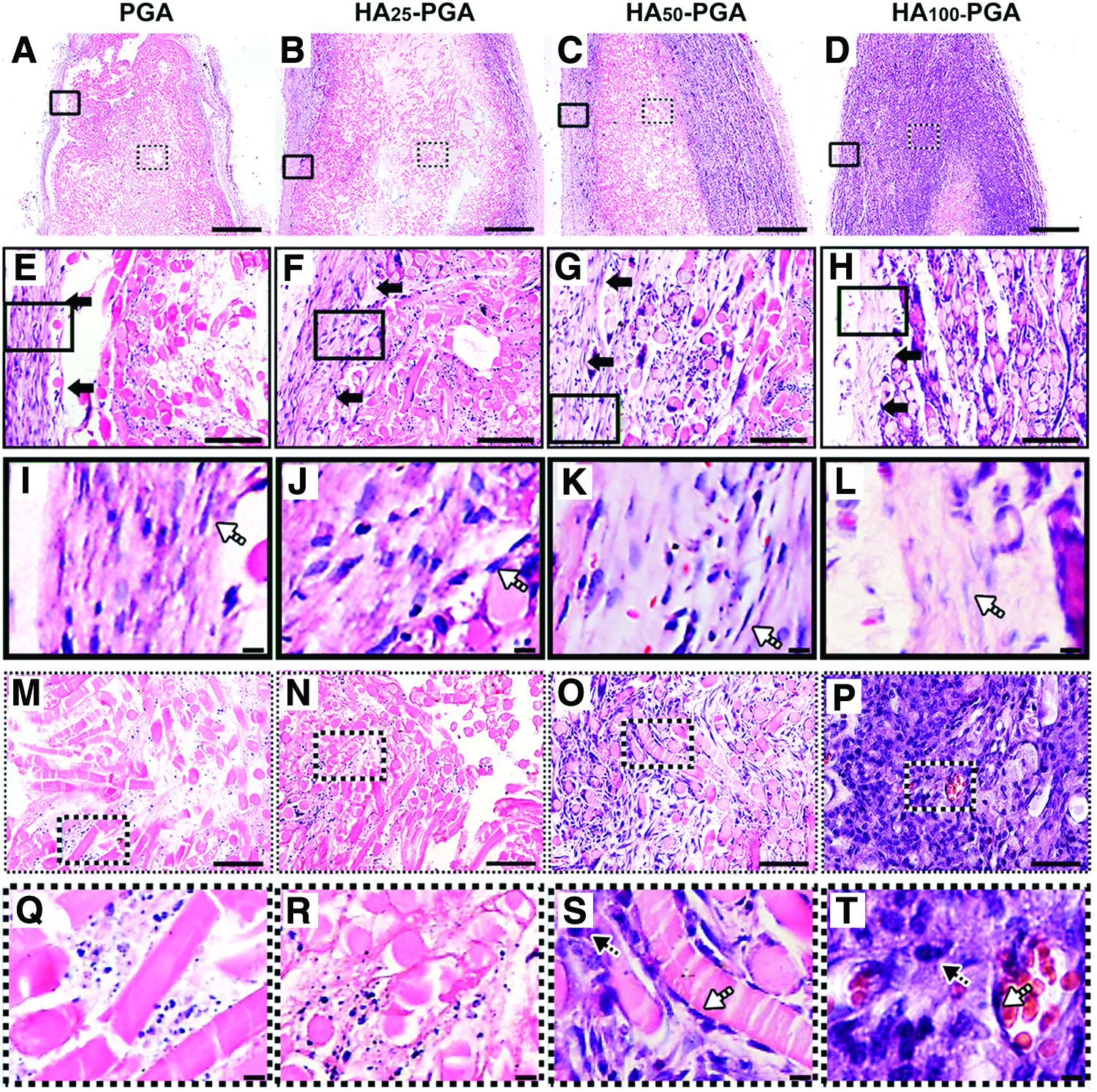
In vivo histological evaluation of host response to implanted noncoated scaffold (PGA) and scaffolds coated with HA at the concentrations of 0.25% (HA25-PGA), 0.5% (HA50-PGA), and 1% (HA100-PGA). H&E staining images of vertical section at 4 weeks postimplantation.
The effect of HA coating on cell adhesion to the scaffold
Cell adhesion efficiency is one of the important aspects of scaffold biocompatibility. This study showed that HA coating of PGA scaffold could significantly enhance the adhesion efficiency from 82.6% (noncoated control) to 96.8% (HA100-PGA) with significant difference between two groups (p < 0.05) after 24 h of culture of scaffold with dermal fibroblasts as shown in Figure 6A.
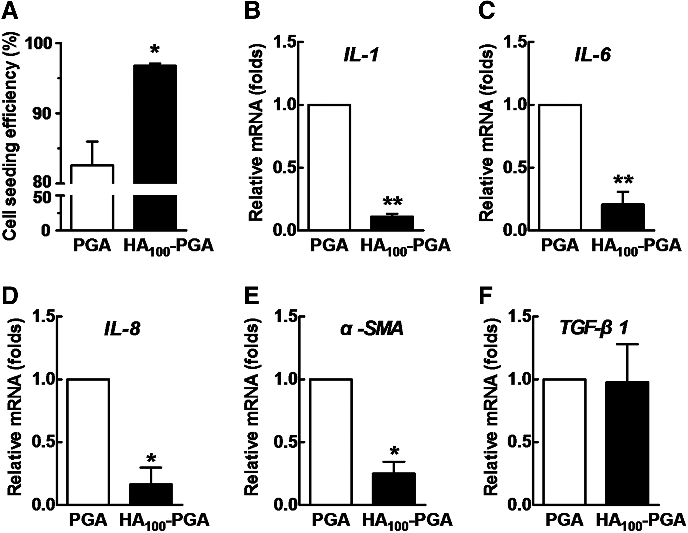
In vitro biocompatibility evaluation assay. Human dermal fibroblasts were seeded on noncoated (PGA) and HA-coated (HA100-PGA) scaffolds for their adhesion efficiency
Effect of HA coating on the gene expression of inflammatory cytokines
To further examine the effect of HA coating on scaffold biocompatibility, qPCR was performed on the RNA extracted from the dermal fibroblast seeded on the scaffold at day 7 post-cell seeding. As shown in Figure 6B–F, when fibroblasts were cultured on HA100-PGA, the gene expression levels were significantly lower than those of control group, including IL-1 (11.0% ± 2.0% of control, p < 0.01), IL-6 (20.7% ± 10.0% of control, p < 0.01), IL-8 (16.4% ± 11.6% of control, p < 0.05), and α-SMA (24% ± 8.1% of control, p < 0.05). However, the mRNA level of TGF-β1 showed no statistically significant difference between two groups (p > 0.05). These molecules represent common markers of inflammatory or fibrotic process, and reduction of the gene expression of these molecules through HA coating indicates the improved biocompatibility.
HA coating enhanced engineered cartilage formation in vitro
After 4 weeks of in vitro culture, neocartilage was formed in both noncoated group and HA-coated group (HA100-PGA) with lacuna structure formation revealed in H&E staining. Toluidine Blue staining also reveals cartilaginous matrix deposition in both groups with relatively stronger staining in HA-coated group, indicating better cartilage formation in HA-coated group (Fig. 7A). Quantitative analysis also showed that total collagen (Fig. 7B) and total GAG (Fig. 7C) contents were significantly higher in HA100-PGA group than in PGA group (p < 0.05). With four more weeks of in vitro culture, relatively more mature cartilage was formed in both groups with stronger Toluidine Blue staining in HA-coated group (Fig. 7A). This was also supported by findings that more total collagen and total GAG were produced at 8 weeks than at 4 weeks for both groups (Fig. 7B, C, p < 0.05). Quantitatively, more GAG and total collagen were produced in coated group than in control group as well at 8 weeks time point (Fig. 7B, C, p < 0.05). qPCR analysis also revealed significantly higher gene expression levels of collagen II (Col II, Fig. 7D, p < 0.01), aggrecan (Aggr, Fig. 7E, p < 0.05), and Sox 9 (Fig. 7F, p < 0.05) in coated group than in control group at both the fourth week and eighth week time points. For collagen X, qPCR analysis showed its significantly upregulated gene expression level in HA100-PGA group than in PGA group at 8 weeks (p < 0.01), but not at 4 weeks (p > 0.05) as shown in Figure 7G.
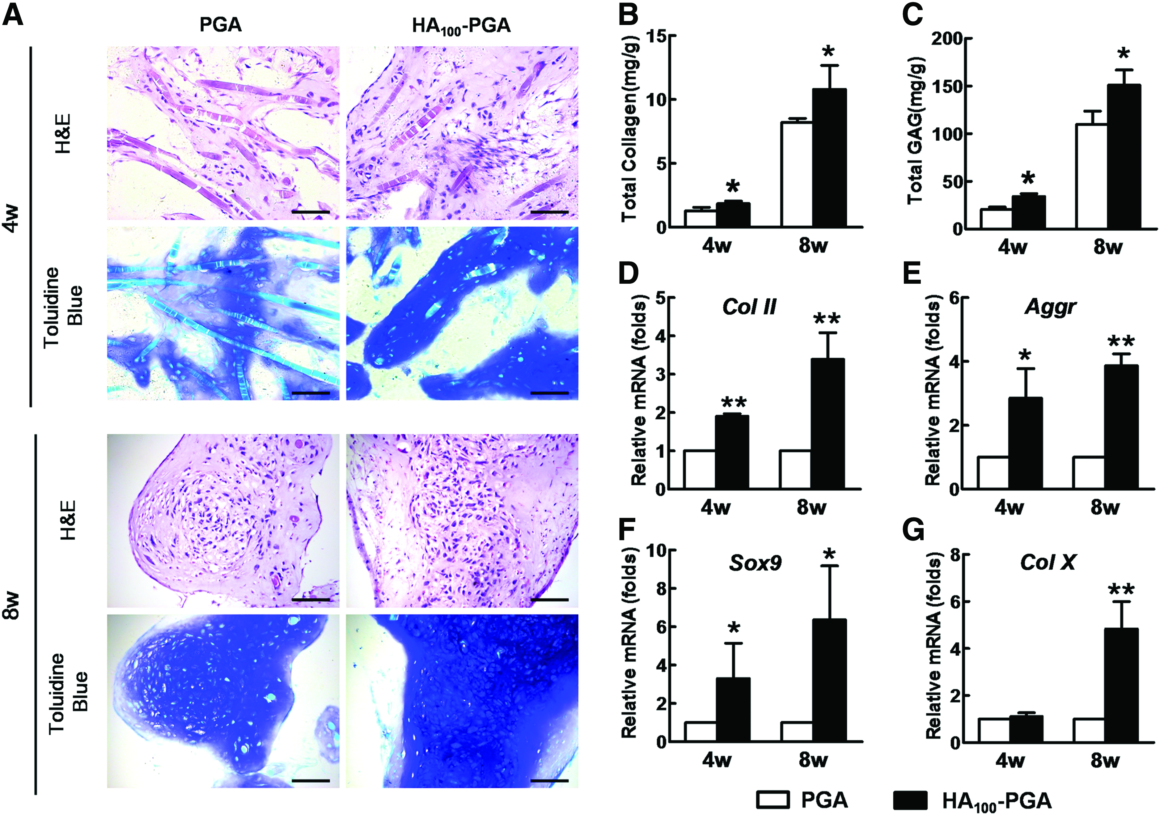
Characterization of cartilage in vitro engineered with noncoated (PGA) and HA-coated scaffold (HA100-PGA).
HA coating favored engineered cartilage formation in vivo
After tissue harvesting, there seemed no significant difference in the gross view (Fig. 8A vs. D) and in the histology (Fig. 8B, C vs. E, F) of engineered cartilages of both groups. Nevertheless, quantitative analysis showed relatively higher total GAG content in coated group than in control group (Fig. 8G, p < 0.05), but not collagen content (Fig. 8H).
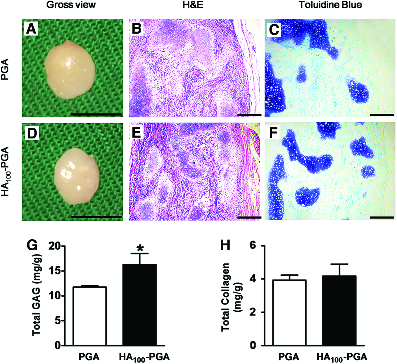
Characterization of the cartilages in vivo engineered with noncoated (PGA) and HA-coated scaffold (HA100-PGA). Both groups of cartilages were evaluated with gross view
Discussion
PGA nonwoven fibers are one of the most commonly used synthetic scaffold materials for tissue engineering. It has been widely applied for engineering cartilage, 7 tendon, 8 blood vessel, 4 peripheral nerve, 9 skin, 10 tendon, 6 etc. and succeed in engineered tissue formation and repair. The obvious disadvantage is the released acidic degradative product, which causes host inflammatory response and impairs the quality of engineered tissue. This phenomenon was observed in a pilot study as early as in 1998, which showed that PGA fibers could cause fibrotic response upon in vivo implantation in a pig model for cartilage engineering, and which showed less biocompatibility than pluronic hydrogel. 22 To overcome this shortcoming, our group has developed in vitro engineering approach for cartilage, 18 blood vessel, 23 and tendon engineering 8 to wash away acidic degradation product and partially degrade the scaffold during in vitro culture so as to possibly reduce inflammatory response after in vivo implantation.
In addition, we explored the possibility of generating pH neutralized poly(α-hydroxy acid) fibrous scaffold through the incorporation of tripolyphosphate nanoparticles into fibrous PLGA scaffolds for tissue engineering, which showed improved biocompatibility to seeded adipose-derived stem cells. 24 Similar approaches were also reported to enhance the biocompatibility of poly(α-hydroxy acid) based scaffolds. 14 However, the biosafety concern of incorporated chemical compounds will remain an issue for its clinical translation.
In this study, we explored the possibility of enhancing PGA biocompatibility by combining the strengths of both synthetic and natural scaffold by coating nonwoven PGA scaffolds with HA, a most commonly used extracellular matrix (ECM) molecule. As shown in Figure 4, in vivo implantation of cell-free PGA scaffold caused significant host inflammatory reaction, which led to early infiltration of inflammatory cells (small sized with round nucleus) at the peripheral area and fast vascularization (Fig. 4E) after 2 weeks of implantation. By contrast, HA coating could significantly attenuate this acute inflammatory reaction, particularly in high concentration HA-coated scaffold (HA100-PGA), where less amount of inflammatory cells was infiltrated and no obvious vascular structure was observed (Fig. 4H). With 2 more weeks of implantation, more inflammatory cells were found surrounding the peripheral area and penetrating inside the inner portion of HA-coated scaffolds (Fig. 5B–D), particularly in high concentration HA-coated scaffold (HA100-PGA, Fig. 5P, T). By contrast, fragmented cell debris was observed inside the inner part of noncoated scaffold, although live cells remained observable at the peripheral area (Fig. 5A, E, M, Q). This phenomenon indicates that HA coating improves the microenvironment that allows for cell survival, although it remained to be able to stimulate inflammatory reaction.
The fragmented cell debris observed in the central part of noncoated PGA is likely caused by the acidity of degraded PGA fibers. Indeed, the results of in vitro degradation assay revealed sharper drop of pH value of control PGA than HA-coated PGA during in vitro hydrolysis (Fig. 3A). Principally, cells have to produce their own matrices after contacting non-natural materials to create their own native cellular microenvironment, 25 and this interaction is also likely to induce undesired cellular response when less biocompatible biomaterials are used. As shown in Figure 6, when using an in vitro model, 26 culture of fibroblasts with uncoated PGA fibers induced significantly upregulated gene expression levels of IL-1, IL-6, and IL-8. In addition, α-SMA, a marker of myofibroblasts which often occur in fibrotic tissue, was also highly expressed. However, HA coating could apparently attenuate the gene expression of these molecules, indicating that HA coating exerted a beneficial effect on the initial stage of cell–material interaction.
This finding was also supported by other studies. For example, in an inflammatory model of interstitial cystitis, HA was found able to significantly suppress IL-6 and IL-8 secretion and increase the secretion of sulfated GAGs both in vivo and in vitro levels. 27 Oliviero, F.'s study also showed that the presence of HA (1500–3200 kDa) was able to reduce cytokine release in the monocytic cell culture stimulated with calcium pyrophosphate crystals or lipopolysaccaride. 28
HA is a natural ECM-derived molecule that exists in a variety of tissue types, particularly in cartilage. A previous study showed that pretreatment of PGA/PVA fibers with 0.1% sodium hyaluronate (w/w) (MW = 1.5 × 10⁶ Da) helped reduce the descending pH value in an in vitro assay. 29 Similar results were also observed in this study (Fig. 3A), which was likely attributed by HA neutralizing effect and, thus, prevented PGA fibers from acid-induced disintegration of the scaffold. Besides, HA coating also facilitated adhesion of seeded cells on scaffold materials as HA can mediate the binding between cells and the materials through HA and CD44, a receptor for HA as supported by Figure 6A. Another report also revealed that HA could improve in vitro substrate adhesion ability and proliferative activity of human chondrocytes. 30
However, the beneficial effect of HA coating might be more prominent in cell-free scaffold based tissue regeneration than cell-seeded approach in vivo. Although significant difference was found in host response to implanted PGA fibers with or without HA coating (Figs. 4 and 5), the difference in host response and tissue formation was not significant for in vivo engineered cartilage, as both HA-coated and noncoated PGA fibers seeded with chondrocytes all formed engineered cartilages with no significant difference in gross view (Fig. 8A, D), histology (Fig. 8B, E), and Toluidine Blue staining (Fig. 8C, F), although total GAG production was relatively higher in coated group than in control group (Fig. 8G). We expected that, when implanted, in vivo environment favors extracellular matrix production of seeded cells, which render the beneficial effect of HA coating less significant shortly after in vivo implantation.
Compared to in vivo tissue engineering, the beneficial effect of HA coating was relatively significant for in vitro tissue engineering using cartilage as an example. As shown in Figure 5, although there was no significant difference in tissue maturity and cartilaginous matrix production (Fig. 7A) at histological level, significantly higher amounts of total collagens (Fig. 7B) and total GAGs (Fig. 7C) were produced in HA-coated group than in noncoated group. Furthermore, much higher gene expression levels of collagen II (Fig. 7D), aggrecan (Fig. 7E), and Sox9 (Fig. 7F) were observed in HA-coated group than in noncoated group. These results further reveal the beneficial effect of HA coating on engineered tissue formation at subcellular levels, which may eventually improve the tissue quality of engineered tissue in the long run. Similar beneficial effect was also found in HA modified PLGA scaffolds. 31
In this study, some shortcomings remain. For example, due to different volumes of scaffolds being tested in different assays, the porosity of different sized scaffolds is likely different, although same porosity scaffold was used in both experimental and control group in each of the tests. Although significant change was observed in cell behaviors between coated and noncoated scaffold, the HA coating might be unstable due to the water soluble nature. A stable HA coating may further enhance the biocompatibility of polymer-based scaffolds.
Furthermore, the mechanism regarding how HA reducing acidity of PGA degradative products remains unclear, the likely hypothesis may include: (1) When sodium salt of HA is dissolved in a buffer solution (pH 7.2), it will usually raise the pH of the solution because it was originally produced by adding NaOH to HA solution; and (2) HA (particularly for aged samples) may contain a few glucosamine residues, which have free amino groups (pK = 7.5), and may function as a buffer compound attenuating pH dropping.
In summary, nonwoven PGA scaffolds have been widely used as a scaffold for engineering various types of tissues, including cartilage, 15 tendon, 32 blood vessel, 4 peripheral nerve, 9 and skin. 10 In addition, they were shown able to in vitro and in vivo engineer tissues and repair related tissue defects. Although these reports indicate the feasibility of nonwoven PGA scaffolds as a tissue engineering scaffold, the disadvantage of poly(α-hydroxy acid) fibrous scaffold was also widely reported for their undesired acidic degradative products, which could affect its biocompatibility to seeded cells, host tissue, and engineered tissues. 33 Although no significant difference in the structure of engineered tissues was observed, the improved gene expression of cartilaginous markers, as well as increased total collagen and GAG production, indicates that the biocompatibility of this type of scaffold can be further enhanced through the coating of natural ECM-derived molecules, which is likely to reduce acid triggered scaffold disintegration, reduce inflammatory host response, and improve cell adhesion and tissue formation.
Conclusion
In this study, we presented that 1% HA solution could effectively coat PGA fibers and proved that coated PGA exhibited less inflammatory reaction, better cell adhesion, and less gene expression of IL-1, IL-6, IL-8, and α-SMA. It also decreased the acidity of degradation products in vitro. Furthermore, coated PGA could engineer better cartilages in vitro with higher content of total collagen and GAG, as well as higher gene expression levels of collagen II, aggrecan, and Sox9. Hence, the data indicated that HA coating could significantly enhance the biocompatibility of this traditional scaffold material, which could work as a compelling ameliorant for PGA fibers.
Footnotes
Acknowledgments
This study was supported by National Natural Science Foundation of China (31170937, 31470943) and National “863” (2012AA020507).
Disclosure Statement
No competing financial interests exist.