
Abstract
Decellularization of tissues and organs has high potential to obtain unique conformation and composition as native tissue structure but may result in weakened tissue mechanical strength. In this study, poly(glycerol-sebacate) (PGS) elastomers were combined with decellularized aorta fragments to investigate the changes in mechanical properties. PGS prepolymer was synthesized via microwave irradiation and then in situ crosslinked within the decellularized aorta extracellular matrix (ECM). Tensile strength (σ) values were found comparable as 0.44 ± 0.10 MPa and 0.57 ± 0.18 MPa for native and hybrid aorta samples, respectively, while elongation at break (ɛ) values were 261% ± 17%, 7.5% ± 0.57%, and 22.18% ± 2.48% for wet control (native), decellularized dried aortae, and hybrid matrices, showing elastic contribution. Young's modulus data indicate that there was a threefold decrease in stiffness compared to decellularized samples once PGS is introduced into the ECM structure. Scanning electron microscopy (SEM) analysis of hybrid grafts revealed that the construct preserves porosity in medial layer of the vessel. Biocompatibility analyses showed no cytotoxic effects on human abdominal aorta smooth muscle cells. Cell studies showed 98% activity in hybrid graft extracts. As a control, collagen coating of the hybrid grafts was performed in the recellularization stage. SEM analysis of recellularized hybrid grafts revealed that cells were attached to the surface of the hybrid graft and proliferated during the 14 days of culture in both groups. This study shows that introducing an elastomer into the native ECM structure following decellularization process can be a useful approach for the preparation of mechanically enhanced composites for soft tissues.
Introduction
T
As a widely studied approach, decellularization is a well-known method that eliminates or reduces immune system-related rejections through post-transplantation. By this approach, cells and cellular materials, which lead to the immune rejection of the natural sourced tissues, are removed.7,8 Decellularization protocols use physical, chemical, and biological agents, or their combinations.9–13 One of the best advantages of decellularization is that it preserves natural tissue's unique structure, however; it should be noted that insufficient chemical exposure may result in ineffective decellularization, while the increased amount can lead to an extracellular matrix (ECM) structure that is weak in mechanical properties and may disrupt specific protein binding sites. For instance, widely used ionic detergents such as sodium dodecyl sulfate (SDS) with a proven efficiency in removing cellular antigens from the tissues and organs can affect the structural conformation of the natural tissue and may lead to deformation and weakening of the mechanical properties.14,15 It can be noted that some natural or synthetic crosslinkers were reported to prevent the structural weakening that occurs as a result of the agents used in obtaining decellularized matrices.16–18
On the contrary, synthetic polymers have also been reported as a good source for scaffolding material in vascular tissue engineering due to their durability and strength. Despite good mechanical properties, cell attachment to polymeric matrices can be poor when compared to naturally derived scaffolds. Until now, many synthetic polymers such as e-PTFE (expanded poly(tetrafluoroethylene)), PET (poly(ethylene terephthalate)), and poly(glycerol-sebacate) (PGS) were utilized as vascular graft materials. PGS is a biodegradable elastomer attracting interest with its promising mechanical properties and has been reported as a preferred polymer in the repair of soft tissues. The mechanical behavior can be tuned by altering molecular weight and crosslink density. However, it is often hard to prepare structures to mimic ECM unique properties such as nanostructure and cell response in an advanced level by using synthetic materials. Therefore, it would be a good approach to combine the advantages of decellularized matrices with synthetic materials.
In this study, we propose a combined soft tissue engineering matrix by in situ crosslinking of a biodegradable prepolymer within a decellularized tissue as an aorta repair material. The hybrid aorta grafts prepared by the method of this study hold great promise in defeating the problem of weakening in the mechanical properties of decellularized matrices following harsh chemical processes. The sheep aorta grafts were decellularized by a common ionic detergent method to prepare cell-free ECM matrices and a prepolymer of PGS was then impregnated to the lyophilized decellularized aorta fragments and crosslinked in vacuum for 10.5 h. Combining decellularized aorta with PGS elastomers not only enhanced the mechanical properties but also biocompatibility due to the incorporated decellularized ECM unique structure, topography, chemistry, and hierarchical reconstruction.
Materials and Methods
Decellularization process
Freshly slaughtered sheep aortae obtained from the slaughterhouse (Kazan Meet Combination, Ankara, Turkey), 1 × 4 cm aorta fragments, were prepared for the decellularization process. Sheep aorta grafts were dissected and excessive blood was removed by washing with phosphate-buffered saline (PBS). One of the well-established SDS protocols with excessive exposure was applied. 19 Samples were incubated in hypotonic 10 mM Tris Buffer (pH 8) (Sigma, Germany) with 0.1% EDTA (Amresco) for 1 h at room temperature and then exposed to 1% SDS for 48 h under an orbital shaker at 37°C temperature (Gerhardt, Germany). Decellularization solution was refreshed at the end of the first 24 h. The aorta samples were then washed with PBS for 2 days in an orbital shaker by refreshing PBS every 12 h to remove residual chemical agents. Subsequently, decellularized samples were kept at −80°C for further analysis.
Biochemical and histological characterization of decellularized aortae
Decellularized and native aorta tissues were fixed in 10% buffered formalin (Sigma, Germany) for at least 24 h at room temperature. After fixation, some tissue pieces were obtained from different parts of the decellularized samples for histological examination. The process continued with dehydration, and xylene (Merck, Germany) was applied for 1 h as a tissue hardening and clearing agent. Samples were paraffin embedded and then the blocks were kept at 4°C. Multiple cross sections with 5 μm thickness were obtained from anterior to the media of the samples using microtome (Leica, Germany) and then stained with hematoxylin and eosin (H&E) and Verhoeff-Van Gieson staining. Dimethyl methylene blue (DMMB) (Sigma, Germany) assay was performed to quantify sulfated glycosaminoglycan content of decellularized tissues. The fragments were then freeze-dried (Labconco, FreeZone 6 plus) and 10 mg decellularized and native tissue samples (n = 5 each) were exposed to enzymatic digestion overnight with 1 mg/mL Proteinase K (Sigma, Germany) in 100 mM ammonium acetate (BDH Analar, United Kingdom) solution at 60°C until all tissue pieces are digested successfully. DMMB reagent was added to samples and absorbance was read at 525 nm immediately afterward. Values measured were normalized by dry weight of each sample. PicoGreen (Quant-iT™ PicoGreen® dsDNA Assay Kit; Invitrogen) assay was applied to the same digested samples according to the manufacturer's instructions, and absorbance values were read between 485 and 528 nm.
Preparation of PGS-acellular aorta hybrid grafts
PGS synthesis is carried out in two stages: prepolymerization and curing. In this study, PGS elastomers were synthesized from equimolar (1 M each) glycerol (Sigma, Germany) and sebacic acid (Sigma, Germany) monomers. Our group has reported a microwave-assisted synthesis route for PGS synthesis for the first time, in which the prepolymerization step is done in a microwave reactor. 20 This approach drastically reduced the synthesis time and eliminates the use of purge gases. Briefly, monomers were loaded onto a glass Petri dish and the reaction was carried out at 650 W for 5 min (1 min × 5) with 10-s breaks after each 1-min microwave exposure. The decellularized tissue fragments were then embedded into the prepolymer. It can be noted that the use of wet decellularized samples should be avoided since this may prevent PGS prepolymer's penetration. Also, hot prepolymer may deform the freeze-dried samples. Therefore, the crosslinking stage was performed with the decellularized tissue fragments by using cooled PGS prepolymer in a vacuum oven operated less than 10 mbar and at 150°C for 10.5 h. Characterization of the PGS-acellular aorta hybrid grafts was performed with histological, mechanical, and scanning electron microscopy (SEM) analyses.
Mechanical evaluation of hybrid grafts
Mechanical properties of native, decellularized, and hybrid aorta fragments (n = 5) were evaluated by tensile tests. Native and freeze-dried decellularized samples were kept at −80°C, while hybrid graft fragments were kept at room temperature until evaluation. The tensile test was carried out in a universal tester (Zwick/Roell Z250, Germany) under 0.2 N load with a loading speed of 5 mm/min to rupture. Each sample was fixed to the clamp of the equipment with filter paper to prevent dislodgement. At the end of the test, the software provided a stress (%)–strain (MPa) diagram, and results of tensile strength (σ, MPa), elongation at break (ɛ, %), and Young's modulus (E, MPa) were obtained.
Cell culture
Human abdominal aorta smooth muscle cells were cultured to investigate cytotoxic potential of prepared PGS-acellular aorta hybrid grafts. For this purpose, expanded human abdominal aorta smooth muscle cells (1 × 10⁶, at passage 2) were thawed within 1 min at 37°C water bath, seeded to T25 culture flasks, and subsequently placed in the 37°C, humidified 5% CO2 incubator. Cultured smooth muscle cells medium [DMEM low glucose (Capricorn, Germany), 10% fetal bovine serum (FBS) (Capricorn, Germany), 1%
Last, the PGS-acellular aorta hybrid grafts were recellularized with human abdominal aorta smooth muscle cells to examine the achievement of cell adhesion to the constructs. For this purpose, both collagen-coated (control, 3.2 mg/mL collagen) and uncoated samples were prepared and cells were seeded to the hybrid grafts (0.3 × 10⁶ cells/cm2) at passage 4 and then placed into the CO2 incubator. It should be noted that this study was performed only for comparison since collagen coating may induce clot formation in vivo. To verify cell attachment, the Live/Dead stained constructs were monitored with confocal microscopy following the days 4, 7 and 14. Constructs were also fixed with 2.5% glutaraldehyde for SEM analysis.
Results
Decellularized matrix characterization
The effectiveness of the decellularization process was evaluated with H&E fundamental histological staining. It should be noted that the elastic fibers, which have an active role in the tight and elastic form of the vascular tissues, and collagen ensure vessel integrity. However, while a tighter and denser structure was observed in the control aorta (Fig. 1A), the structure became slightly looser and there was an increase in the distance between the fibers after decellularization. Following the 48 h of decellularization process with 1% SDS (Serva, Germany), no residual cells were observed in the tissue based on the HE staining (Fig. 1B). Based on Verhoeff-Van Gieson collagen and elastin staining assay, the elastic fibers in tissue contained dense/tight elastin (black) in the control aorta (Fig. 1C) cross section, while there was a substantial decrease (Fig. 1D, arrows) after decellularization. Likewise, there was a decrease in the amount of collagen (yellow) in the tissue following the decellularization process. Changes are observed in the three-dimensional structure and conformation of the matrix after the decellularization process.
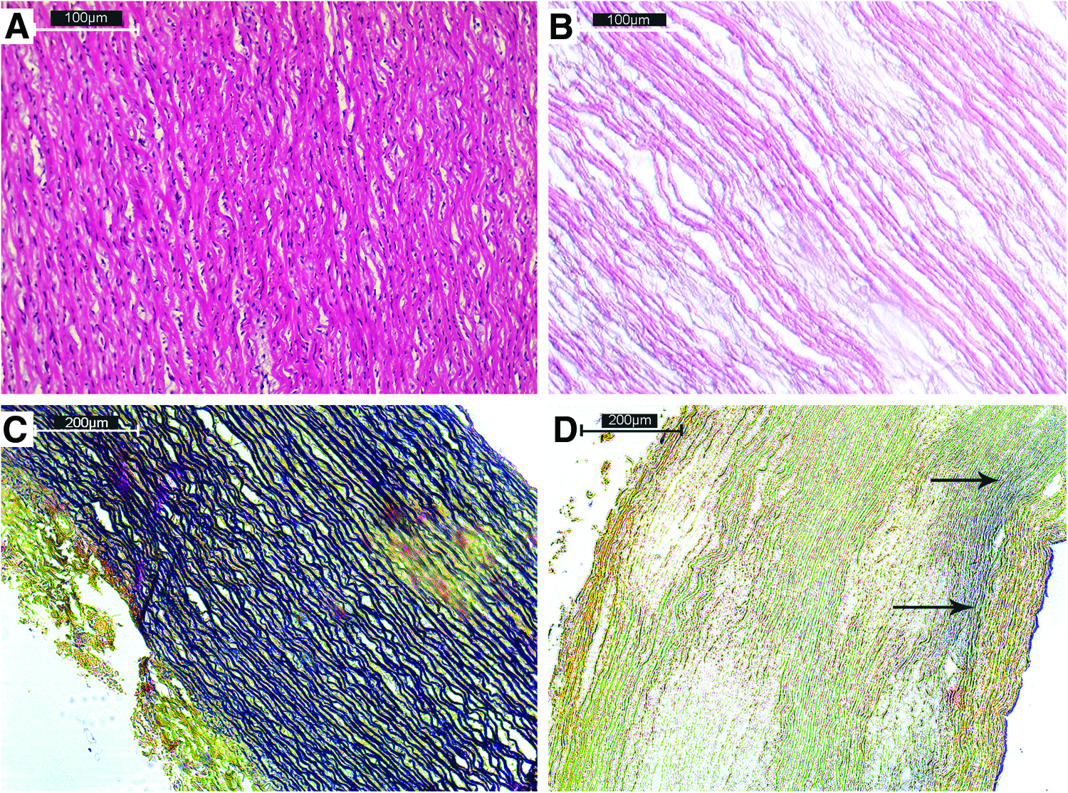
Histological and biochemical evaluation of decellularized aorta samples.
Evaluation of hybrid constructs
Both manual and microtome cross sections of the PGS-acellular aorta composite fragments were monitored with a light microscope. PGS elastomer (yellow) was found penetrated to the thickness in the manual cross section (Fig. 2A, B) and pores were observed in patches. In the manual cross section of the Verhoeff-Van Gieson elastic/collagen staining of the hybrid samples, fibers were slightly thicker as a result of PGS incorporation (Fig. 2C, D) when compared to postdecellularization staining (Fig. 1C, D). In addition, pore structure was observed in the hybrid fragments in the SEM images (Fig. 3C, D). SEM images of the PGS elastomer and the PGS layer, which covers the surrounding area of the vessel, can be seen in Figure 3A and B, respectively.

Manual

SEM images of
Mechanical evaluation
In the evaluation of the mechanical properties, the unprocessed control aorta, decellularized aorta (wet), lyophilized aorta (dry), and PGS-aorta composite construct images were presented (Fig. 4A–D). In Table 1, again, tensile strength (σ), elongation at break (ɛ), and Young's modulus (E) values were given for each group (sample size = 2.2 × 9.1 mm, sample area = 20.02 mm2, n = 5 with standard errors). Tensile strength (σ) values were found as 0.44 ± 0.10 MPa and 0.57 ± 0.18 MPa for native and hybrid aorta samples with no statistically significant difference (p = 0.4206), even though there was an ∼23% increase compared to initial strength. The elongation at break (ɛ) values were found as 7.5% ± 0.57% and 22.18% ± 2.48% for wet control (native) and hybrid matrices (p = 0.079), showing a significant difference (p > 0.05) and a roughly threefold increase and an elastic contribution of the elastomer. The Young's modulus data indicate that introducing PGS into the ECM increased the elasticity of the aorta samples drastically (p = 0.0159) from 34.7 ± 4.7 MPa (for decellularized samples) to 10.3 ± 3.05 MPa (for the hybrid grafts).
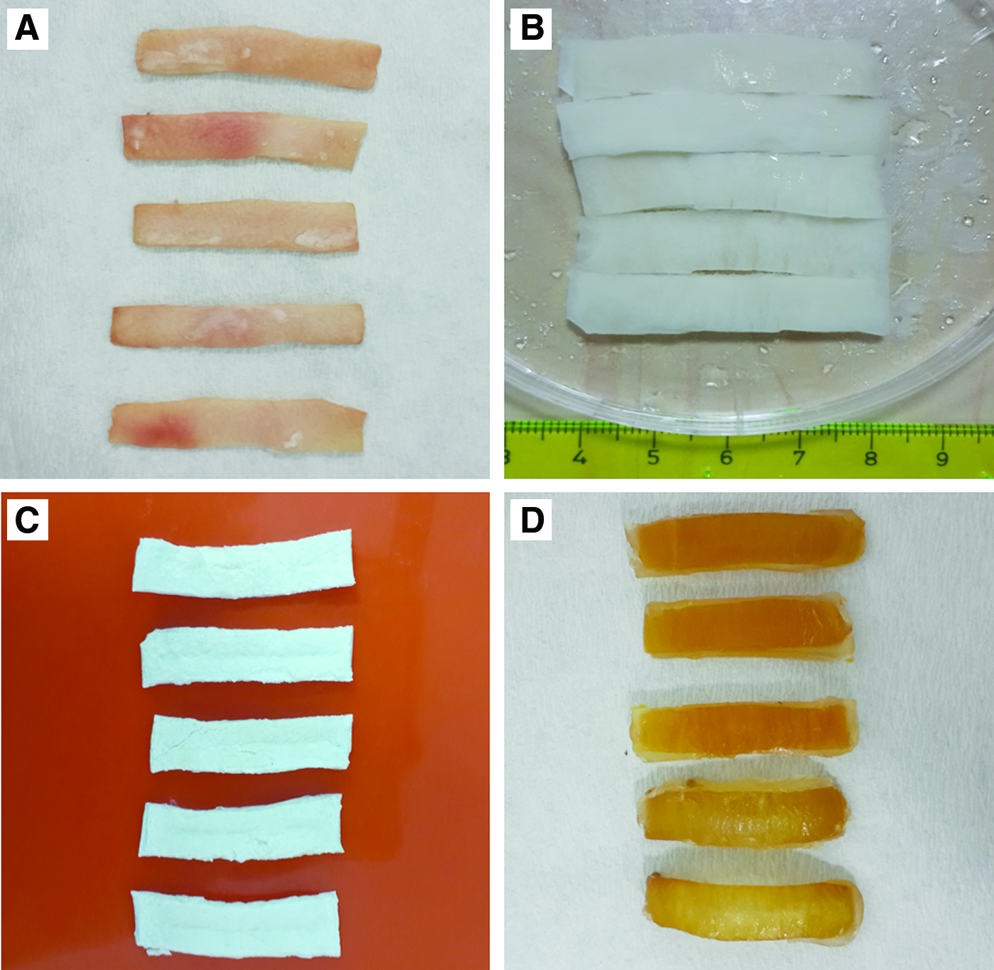
Photographs of
SD, standard deviation.
Biocompatibility analysis
Based on the Live/Dead analysis results by the elusion method (0.1 g/mL), following cell culture with human abdominal aorta smooth muscle cells (P4) in 0.02 × 10⁶ cells/well cell density, most of the cells were alive on the 1st, 4th, 7th, and 14th day. In terms of proliferation success, there was no significant difference between control and test groups (p > 0.05 according to the t-test performed by using cell counts obtained via ImageJ software) on the 1st, 4th, and 7th days; however, smooth muscle cells were unable to count due to overconfluency on the 14th day (Fig. 5). Also, based on alamarBlue cell activity test results (Fig. 6), no significant difference (p > 0.05) was found by using t-test (GraphPad Prism 5 software) in the cell activities between the control and test group on the 1st, 4th, and 14th day (p = 0.1896, p = 0.1009, and p = 0.2478, respectively). Only on Day 7, the difference was meaningful (p = 0.0057*). The results showed that the PGS-acellular aorta hybrid graft fragments did not show cytotoxic effects on the selected cells.
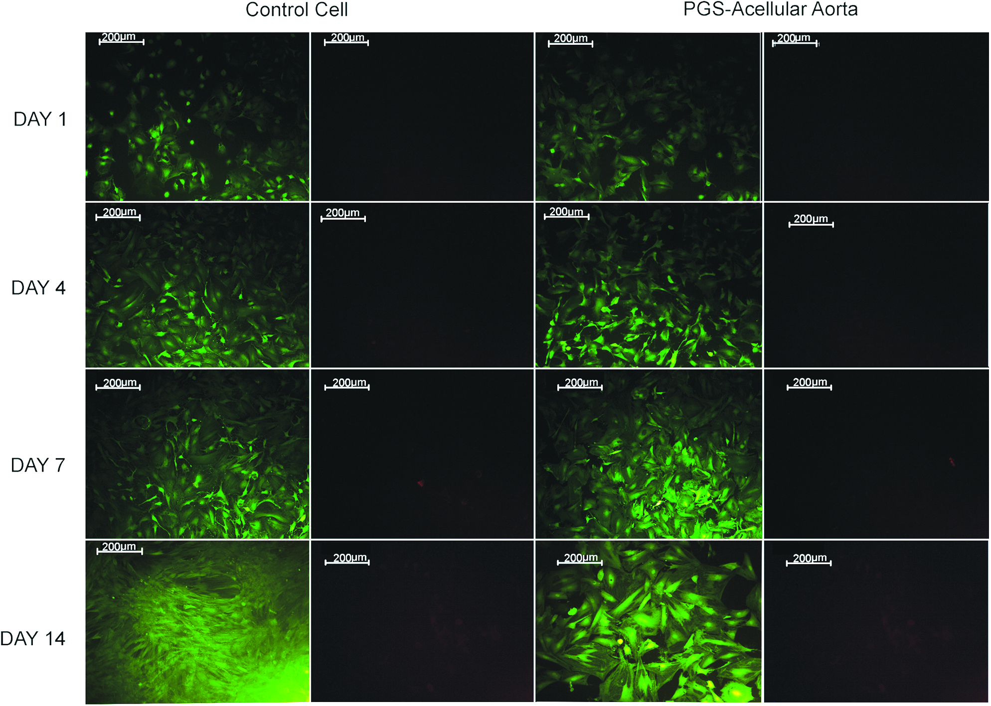
Confocal microscopy images from Live/Dead cell viability assay for hybrid constructs.

AlamarBlue biocompatibility analysis results of hybrid constructs at day 1, 4, 7, and 14.
In both groups (collagen coated and uncoated), the cells were alive and a small number of dead cells were observed (Fig. 7). Cell morphologies were the same and nearly in the round-shape form in both groups. On day 7, SEM analysis was performed (Fig. 8). On the 14th day of the static cell culture, viable cells were seen in low fluorescence emission in the coated and uncoated constructs with a small number of dead cells. Based on SEM results, cell–cell interactions (arrows) were observed in both coated and uncoated groups. However, cell proliferation was higher in the collagen-coated group on the following 7th and 14th day and the coated PGS-acellular aorta hybrid graft surfaces were covered with a cell layer and the cells were observed to secrete their own extracellular matrices. In the recellularization process, which was performed by static cell culture, the cell adhesion success was found sufficient in the collagen-coated and uncoated PGS-acellular aorta hybrid grafts.

Confocal microscopy examination of hybrid constructs with human abdominal aorta smooth muscle cells at days 4 and 14 following recellularization.
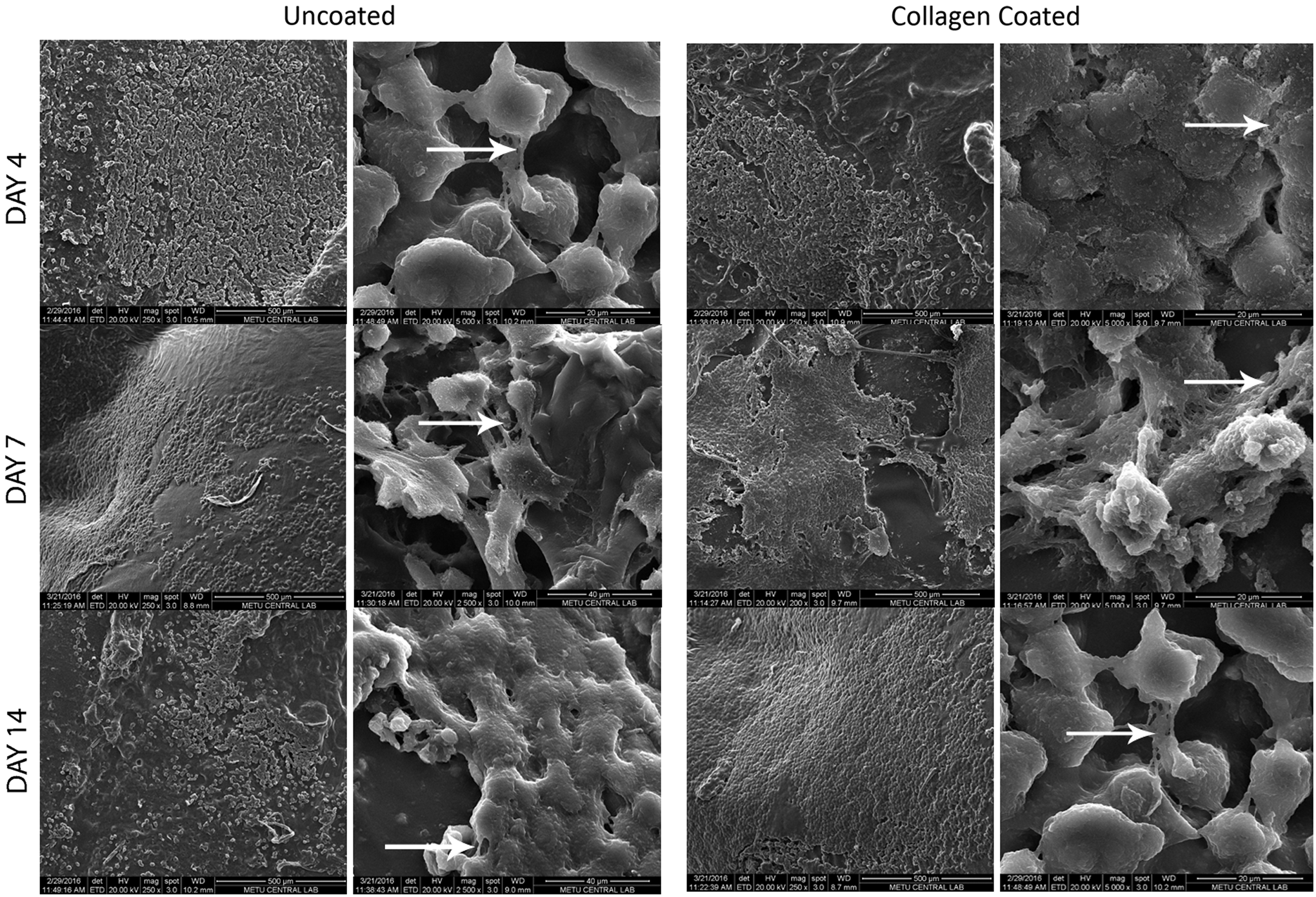
SEM images of cells on uncoated and collagen-coated recellularized hybrid constructs at days 4, 7, and 14.
Discussion
In decellularization, the fundamental goal is to effectively remove the cell and cellular materials, which create an immunogenic effect and then protect the unique three-dimensional structure and the components of the native tissue constructs obtained. Besides that, the mechanical properties of the ECM structure should be protected and promote restructuring of tissue following decellularization. Unfortunately, due to the harsh chemicals used in the decellularization protocols, there is always a loss in the mechanical properties of the final ECM, and constructs cannot maintain their long-term stability and may face blood leakage or rupture in the event of an aorta repair. 21 As an anionic detergent, SDS with its negatively charged groups can disrupt the noncovalent bonds in the protein structure, expand the gaps between the collagen chains, deteriorate the integrity of fibers, and cause a decrease in the mechanical strength of the tissue. 9 The acellular matrices obtained by decellularization of the cardiovascular tissues are also expected to maintain their hemodynamic properties. Therefore, obtaining a smooth surface is another goal. In cardiac tissue engineering, many tissues (cardiac valves, aorta) have been decellularized by several methods so far and the effect of the decellularization process on the mechanical properties of the tissue scaffold was evaluated. Recently, a crosslinking of the ECM matrix has been reported to overcome decreased strength and long-term stability occurred following decellularization. 16 The changes in the structure and quantity of the proteins and glycosaminoglycans that constitute the matrix after the decellularization process significantly alter the matrix function. Large quantities of elastin and collagen, which are responsible for mechanical endurance and flexibility in the vessel tissue, are lost. The decellularization process can also affect the links between the GAG chains bounded to the vessel wall that constitute a network for endurance and adhesion sites for growth factors. 22 The results of our SDS-based decellularization process demonstrate that there was also a decrease in the amount of collagen and elastin in the tissue after decellularization.
In this study, a combinational approach to obtain enhanced vascular grafts is presented. For this purpose, decellularized aorta constructs were combined with the PGS elastomer with high flexibility and endurance to produce hybrid aorta grafts. The crosslinking protocol (dehydrothermal crosslinking) is also a known procedure especially used in collagen-based scaffold fabrication and leads to minimal ECM deformation during the 10.5 h of treatment. 23 The homogeneous distribution of an elastomeric phase within the weakened ECM was shown to increase the mechanical properties of aorta fragments. Attributed to its synthesis route, the biocompatible feature of the elastomer yielded a hybrid tissue construct capable of cell attachment. PGS elastomer can also restore the surface characteristics, that is, could provide a smooth inner surface to the construct. The biodegradation profile of the selected PGS elastomer was also deemed as a spacer for tissue growth throughout the repair process. The following recellularization studies show good cell attachment and proliferation onto the hybrid aorta grafts. Kumar et al. reported an ultimate tensile strength of 1.5 MPa and a Young's modulus of 1.4–11.1 MPa for native blood vessels. 24 Schneider et al. also reported a Young's modulus value of 5.36 MPa with a strain of 54.21% for native tissue, while these values were 3.65 MPa and 42.44% for decellularized counterparts. When synthetic materials are used as the scaffold material, higher tensile strength values (>10 MPa) can be achieved. 25 The mechanical properties obtained in this approach were comparable to a study in which the Young's modulus value was reported as 7.1 MPa for a bilayered scaffold prepared from a synthetic material. 26 When compared to the decellularized grafts, there was approximately a threefold increase obtained both in elasticity and strain values of the hybrid grafts prepared in this study.
The degradation studies performed by using elastomer alone revealed a 2/3 decrease in weight after 90 days in Ringer solution (n = 3, data not shown). It can be speculated that this decrease may be compensated through the neo-ECM formation by cell activity in vivo. The elastomer used may also block the pores within the native ECM following decellularization and provide a decellularized matrix with a smaller surface area for degradation. However, neo-tissue formation and degradation rate can be tuned and synched by changing the elastomer/decellularized tissue ratio, molecular weight of the prepolymers, and crosslinking density.
To the best our knowledge, it is the first time an elastomer is crosslinked within a decellularized aorta matrix to overcome the problem of attenuation in structural and mechanical features brought along by the used chemicals during the cell removal process. The prepolymers can be easily manufactured by our previously reported protocol 20 and manufacturing of hybrid ECM-elastomer constructs can be easily modified for other type of tissues according to physical properties of the tissue structure. Prepolymer properties such as viscosity, molecular weight, and in the following stage the crosslinking density can be altered for different applications too. Therefore, the proposed methodology described herein can provide benefits to tailor the desired properties of acellular matrices by combining them with synthetic elastomers in the search for an enhanced graft material to be used in the repair and engineering of soft tissues.
Conclusion
In this study, PGS-acellular aorta hybrid grafts were designed with the aim of enhancing the mechanical properties of an aorta, especially the elastic behavior that had been lost during the harmful decellularization process. We proved that combining elastomeric PGS polymers with decellularized grafts can provide sufficient strength, recellularization capacity, and handling properties to the final constructs. The basic idea discussed here can be exploited to a full-thickness section by combining different layers of tissue with variable amounts of synthetic materials for diverse requirements of the final constructs. This concept can therefore be modified for other soft tissue applications and may be used in the efforts of providing off-the-shelf combinational products.
Footnotes
Acknowledgments
This study was supported by Hacettepe University (Project No. THD-2015-8859) and BMT Calsis Co.
Disclosure Statement
No competing financial interests exist.