
Abstract
The repair of large long bone defects requires complex surgical procedures as the bone loss cannot simply be replaced by autologous grafts due to an insufficient bone stock of the human body. Tissue engineering strategies and the use of Advanced Therapy Medicinal Products (ATMPs) for these reconstructions remain a considerable challenge, in particular since robust outcomes in well-defined large animal models are lacking. To be suitable as a model for treatment of human sized bone defects, we developed a large animal model in both skeletally immature and mature sheep and made close observations on the spontaneous healing of defects. We warn for the spontaneous repair of large defects in immature animals, which can mask the (in)effectiveness of ATMP therapies, and propose the use of large 4.5 cm defects that are pretreated with a polymethylmethacrylate (PMMA) spacer in skeletally mature animals.
Introduction
T
Moreover, a good quality of the surrounding tissues allows better bone regeneration, but is not always possible, in particular in large defects or infected nonunions with substantial damage of the periosteum. To overcome the challenge of the compromised environment, Masquelet initiated the concept of an induced membrane by a foreign body reaction, for which he used polymethylmethacrylate (PMMA) bone cement.7,8 This allows the creation of a so-called biochamber in which a bone graft or an advanced therapy medicinal product (ATMP), can be implanted. 9 In combination with the diamond concept, this adds a fifth element to the prerequisites for an ideal bone healing, and has been described as the Pentaconcept. 10
The main challenge in evaluating new ATMPs is the use of a suitable model that ideally mimics the real clinical situation and predicts the outcome in the patient. Therefore critical-sized bone defects in large animals are mostly used. A variety of defect models have been described and usually tested as “active implanted” versus “empty control” defects. However there is not always a consensus in literature about the exact definition, nor the ideal way to create large skeletal defects. However, good models for preclinical testing are an absolute prerequisite to bring therapies to the patient.11,12 In the field of bone repair, and this in contrast to other domains of tissue engineering where the biological phenomena are dominant, the geometric and mechanical conditions also play an indisputable role, which needs to be taken into account in the translational research.13,14
To obtain a reliable model of a defect pseudarthrosis, we set up an experiment in both skeletally immature and mature sheep, creating tibial bone defects of both 3.0 and 4.5 cm in immature and 4.5 cm in mature animals, according to the guidelines that critical-size defects should be at least 2 to 2.5 times the diameter of the bone 15 These gaps were left empty during a period of 6 weeks to allow ingrowth of fibrotic scar tissue that was subsequently removed for the implantation of a cement spacer, for another 6 weeks to induce a Masquelet membrane. The way these defects were created, is described in detail, emphasizing that critical size defects have to be interpreted with extreme caution as it is not uncommon that in empty defects, before cement insertion, spontaneous repair occurs.
Materials and Methods
All experiments were performed with approval of the Ethical Committee of the Medanex Clinic (CRO, Diest, Belgium) and in accordance with the guidelines for the care of laboratory animals. Forty Charolais-Swifter sheep were used and divided into 32 skeletally immature (10 months old) and 8 mature (4 years old) sheep (Table 1). After sedation by xylazin (0.1 mg/kg, Xyl-M 2%®; VMD, Arendonk, Belgium) intravenously, anesthesia was installed by ketamine (4 mg/kg, Ketamine 1000®; Ceva Santé Animale, Libourne, France) and midazolam (0.2 mg/kg, Dormicum®; Productos Roche S.A. de C.V., Toluca, Mexico), using a jugularis catheter that could serve for postoperative intravenous administration of medication. For antibiotic prophylaxis, the second-generation cephalosporin ceftiofur (2.2 mg/kg, Excenel®; Phizer Animal Health, Kirkland, Canada) and natriumbenzylpenicilline (40,000 IU/kg, Penicillin®; Kela NV, Hoogstraten, Belgium) were administered. Endotracheal intubation was performed and the anesthesia was maintained with isoflurane, using buprenorphine (0.005 mg/kg, Vetergesic®; Pantheon UK Limited, Wiltshire, United Kingdom) and meloxicam (0.5 mg/kg, Metacam®; Boehringer Ingelheim Vetmedica GmbH, Ingelheim, Germany) for painkilling. The sheep were installed in lateral decubitus on the left side giving a free exposure to the right hind limb that was carefully clipped, scrubbed, and draped. An Ilizarov fixator (Medicalplastic, Milano, Italy) was installed to the right tibia (Fig. 1). Proximally, one and a half ring were fixed to the bone with three 1.8 mm Kirschner wires (Smith & Nephew, Memphis, TN) and three (one lateral and two medial) 4 mm self-tapping Apex screws (Stryker, Selzach, Switzerland), both for 3.0 and 4.5 cm defects. Distally, two 1.8 mm wires and two medial 4 mm Apex screws were anchoring the bone to one ring that was temporally interconnected with the proximal ring with two threaded rods for the 3 cm defects. For defects of 4.5 cm, an additional distal half ring with one 1.8 mm Kirschner wire and one 4 mm Apex screw were used. The wires were tensioned to 130 kg, using the dynamometric Ilizarov spanner. Subsequently, a medial incision was made at mid-diaphyseal level and a tibial segment of 3 cm was outlined in 12 young, skeletally immature sheep and of 4.5 cm in 20 young and the 8 adult sheep. At this stage, the periosteum was excised as far as the exposure allowed. At the proximal and distal level, 3.2 mm drill holes were made. An osteotomy was performed with a chisel at both ends and the fragment removed (Fig. 2A). Finally, the remnants of the periosteum blended to the fascia covering the muscles at the lateral side of the tibia were removed meticulously by excising the entire fascia.
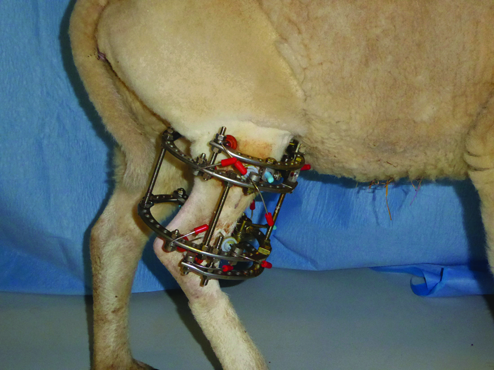
Right hind limb of sheep with circular external fixator to stabilize the tibial bone defect.

Outline of 4.5 cm tibial defect just before removal
In all animals (n = 40), the wounds were simply closed leaving the cavity empty. The rings were firmly interconnected with four threated rods and all parts of the fixator were carefully controlled to avoid any mechanical instability. Finally, the alignment of the bone fragments was checked under image intensifier and the images collected.
After the operation, all animals were allowed to move freely and bear full weight on the operated limb. Antibiotics were continued intravenously for 5 days. Meloxicam, the nonsteroidal anti-inflammatory drug already started during the surgical procedure, was continued for 5 days at a dose of 0.5 mg/kg.
Radiographic assessment in an anteroposterior and lateral direction was performed immediately postoperatively and at 6 weeks, just before reintervention, and assessed according the presence of bone in the defect. The amount of bone was expressed as a percentage of the surface of the defect in both planes. At this stage, in all sheep, in as far as the cavity was not closed spontaneously, on which occasion no further surgery was undertaken, the defect sites were debrided from scar tissue and filled with a cement spacer (Fig. 2B, C). For this Masquelet procedure, a PMMA spacer was prepared (Palacos®; Heraeus Medical, Wehrheim, Germany) and just before hardening, inserted in the bone defect, thereby slightly draping the cement around the bony edges. During hardening of the PMMA in the defect site, the cement spacer was continuously cooled with a solution of NaCl 0.9% (B.Braun Melsungen AG, Melsungen, Germany) to avoid thermal damage to the surrounding tissues. The cement was carefully covered by closing the subcutaneous tissue followed by skin sutures. These animals were followed for another 6 weeks and again radiographically evaluated at removal of the cement, on which occasion, a biopsy of the membrane was taken (Fig. 2D, E). Subsequently, all animals were treated with an ATMP implantation as part of a larger ongoing experiment, which is beyond the scope of this publication (Fig. 3).

Schematic overview of the experimental design of the surgical intervention for a 3 and 4.5 cm defect in a sheep tibial defect model. ATMPs, advanced therapy medicinal products.
Statistical analysis was performed using the two-tailed Fischer's exact test with the significance level set at p < 0.05 (GraphPad Software, Inc., La Jolla, CA).
Results
All sheep recovered well and were able to bear weight on the operated hind limb. Apart from some superficial pin tract infections that could be treated with intravenous antibiotics or local disinfection, there were no complications. Alignment of the limb was maintained on all occasions and there were no problems with the fixator hardware. The radiographic assessment on day one showed a complete empty cavity in the 40 sheep (32 young and 8 adult).
In the 12 young animals with a 3 cm empty defect, a substantial bone repair was noticed in 6 of them with >50% filling after 6 weeks (Fig. 4). These six animals were not further operated and followed for another 2 years with radiographic assessment at 3 monthly intervals, showing a complete filling of the defect. The other six animals stayed free of any significant bone formation and at reintervention, a fibrotic block of scar tissue was excised to allow the insertion of a cement spacer. From the 20 young sheep with a 4.5 cm defect, new bone formation at the posterolateral position of the defect was noticed in 5 animals filling <50% of the defect surface, whereas 15 had no signs or only a very limited amount of bone repair. In all 20 sheep, the scar tissue and the occasional bone ingrowth were removed at 6 weeks before cement implantation (Fig. 5A).

Empty 3 cm defect in skeletally immature animal at operation

Comparison of 4.5 cm tibial defect in skeletally immature
In the eight adult sheep with the 4.5 cm defect, no bone formation at all or a limited bone outgrowth at the bone edges could be noticed, and at reintervention, the cavities were completely filled with a block of fibrotic scar tissue that was excised for the insertion of the spacer (Fig. 5B) (Table 1).
All cement spacers were removed after another 6 weeks and at this stage, the animals were used for further experiments aiming at defect reconstruction with ATMPs, the results of which are subject to further analysis and do not contribute to this report.
The Masquelet membrane formation was adequate in all animals, regardless of defect size, and at histological analysis, a fibrovascular membrane was found on each occasion. The radiographic analysis at removal of the spacer did not show any relevant bone formation around the spacer. The difference in outcome for mature versus immature animals with a 4.5 cm defect was not significant at the p < 0.05 level (p: 0.2808); the comparison between a 3 and 4.5 cm defect in skeletally immature animals was found to be not significant (p: 0.2499). The results in immature 3 cm defects compared to mature 4.5 cm defects showed a significant difference (p: 0.0419).
Discussion
One of the biggest challenges in defect reconstruction research is the translation of experimental data to the clinic. Because surgeons are usually confronted with very large defects, models that are relevant for the patients who are in need of treatment have to be developed. If complex reconstruction procedures become necessary, ATMPs can bring a solution, but to study their value in clinically relevant conditions, representative large animal defects, mimicking the clinical situation as recommended in the European Medical Agency (EMA) guidelines, should be used. 16 FDA directories suggest that the chosen models should relate to the overall product development and the clinical product should be tested in the preclinical animal model in its final formulation rather than an equivalent one, for which in this particular case, “human size-like” defects are desirable. 17
For the use of large so-called critical-size defects in animals of appropriate size such as goats and sheep, uttermost care should be taken.12,18 Apart from the size, suggested to be 2 to 2.5 times the diameter of the bone, other parameters have to be taken into account such as the age of the animal (skeletally immature vs. adult) and the way the defect is created, for example, by careful removal or not of the periosteum. 19 Also, the fact whether a defect reconstruction is studied immediately after its creation (the optimal fresh defect) or after some delay allowing ingrowth of fibrosis and mimicking the real clinical situation of a defect nonunion, might alter the outcome. The anatomic localization that is chosen should also be equal to the clinical situation as in practice most defect nonunions are localized in the long weight-bearing bones, especially the tibia. Also, the time to follow-up should not be underestimated as early bone formation might not be entirely clinically relevant and early promising results might finally turn out not to be successful. On the other hand, slow onset regeneration can progressively evolve and lead to a well-maturated callus. Moreover, there is the phenomenon of spontaneous bone regeneration, which can occur in a model and thus interfere with the implant that is examined, and this “background noise” can lead to overenthusiastic conclusions about the netto effect of ATMPs. It can to some extent be responsible for the alleged new bone formation of experimental treatments and in case such models would be used for direct ATMP insertion, the evaluation of the newly formed bone could be falsely positive and lead to exaggerated optimistic conclusions. 20 Neglecting the rules of good preclinical testing could create unrealistic expectations and is probably the reason why so many bright ideas and novel developments in tissue engineering never come from bench to bedside as the translational research is poorly or incorrectly performed.
Finally, the fixation method can also interfere with the bone restoration, in a negative way due to an insufficient stability or even positively due to collapse and compression forces on one side, promoting healing due to the partially reduced defect size.
Reichert et al. made a substantial contribution to the study of large animal defects and concluded that many reports in the literature have serious shortcomings, making them unreliable as a model for translational studies. The authors pointed out that the ideal defect is a midshaft tibial gap of minimum 3 cm in adult sheep, for which they recommend an age between 7 and 9 years as the morphologic characteristics of the bone mimic the adult human situation best at this age. 21
A critical analysis of our results also shows that defect length is important but should be correlated with the age of the animal. If, according to the standard, as described on the site of American Standards for Testing and Materials ASTM, bone defects with a length of 2 to 2.5 times the diameter of the bone are taken (leading to 3.0 cm in immature and 4.5 cm in mature animals) a different outcome between both groups is noticed if left untreated. 15 While none of the eight adult sheep had any relevant bone repair and thus a real critical-size defect, half of the young animals with a 3 cm defect showed a healing tendency with finally a complete bridging. Even a 4.5 cm defect in young animals gives a partial regeneration in one quarter of the animals as illustrated in Figure 4A. Despite careful removal of the periosteum, the biological potential of young animals is overwhelming, leading to a spontaneous repair even in a compromised environment, that is, biologically deprived from osteoprogenitor cells and with critical dimensions. As the circular fixator maintains the reduction throughout the experiments without any collapse or loss of reduction, this good mechanical stability allows an optimal function of the biological processes with unforeseen regenerative capacities as demonstrated in 11 of the 36 skeletally immature animals, although they strictly fulfilled the requirements of critical-size defects. Even statistical analysis should be interpreted carefully. There is no significant difference between the mature versus immature animals; nevertheless, in some of the immature animals, partial bone regeneration is present. In case more limited groups are used, statistical differences could become significant. Between 3 cm immature versus 4.5 cm mature sheep, there is a significant difference from which we would recommend for the use of adult models. If an immature model would be used for the study of bone reconstruction by immediate insertion of an ATMP, the results would be overshadowed by the spontaneous bone formation and lead to potentially false interpretations. On the other hand, the same conditions in an adult animal did prove to be reliable and can be considered a real critical-size defect.
The insertion of cement after 6 weeks of fibrotic ingrowth, thereby mimicking the real clinical condition of a compromised environment compared to a membrane induction in a freshly created defect as done in most studies, is more reliable, but needs further experiments on a potential difference in the biological activity of the membrane and the subsequent capacity for bone regeneration. 22
Finally, for an optimal study of bone reconstruction, we suggest that the fixation technique should barely interfere with the gap where the biological repair processes should take place, and we believe that external fixation is most reliable to study the behavior of the critical-size defect. Moreover, it allows a full interpretation of the newly formed bone in vivo using conventional radiological and even CAT-scan imaging. 23 To overcome the shortcomings of unilateral fixation, which sometimes does not allow to maintain a good reduction or full weight bearing, circular fixation is a perfect alternative as shown in our experiments. Indeed, mechanical stability and maintenance of alignment and defect size were never a problem and allowed free use of the limb and unrestricted walking of the animal mimicking the clinical situation.
Taken together, the presented data clearly suggest that any interpretation of bone formation in critical-sized defects has to be done with regard to both the biological situation and mechanical environment and failure to do so, could create the impression of promising therapies that finally will not work in surgeons' hands.
Conclusion
The translation of laboratory-based approaches for bone defect repair to a relevant clinical situation will only succeed if correctly performed studies in a reliable large animal model demonstrate that the proposed strategy is successful. This approach will avoid failures and impede that patients become prone to unnecessary and avoidable morbidity.
To create an absolute critical defect, the authors recommend the use of the tibia of adult sheep, the creation of a defect of sufficient size (4.5 cm) with careful resection of the entire periosteum, and a latency period of 6 weeks to allow ingrowth of fibrosis, followed by a cement spacer during 6 weeks. To avoid interference with the fixation, we prefer an external circular fixator that allows a perfect stabilization with preservation of reduction and full weight bearing without restrictions. Only if all these parameters are taken into account and strictly fulfilled, we speak about a real critical-size defect. This ideal defect/nonunion model is not only suitable but also indispensable for clinically relevant translational investigations, leading to reliable conclusions.
Footnotes
Acknowledgments
This work was supported by an IWT-TBM grant from the Belgian government, titled “Regenerative therapy for congenital pseudarthrosis of the tibia in patients with neurofibromatosis type 1.”
Disclosure Statement
No competing financial interests exist.