
Abstract
Three-dimensional (3D) bioprinting is a promising technology for manufacturing cell-laden tissue-engineered constructs. Larger tissue substitutes, however, require a vascularized network to ensure nutrition supply. Therefore, tailored bioinks combining 3D printability and cell-induced vascularization are needed. We hypothesize that tailored hydrogel blends made of agarose-type I collagen and agarose-fibrinogen are 3D printable and will allow the formation of capillary-like structures by human umbilical vein endothelial cells and human dermal fibroblasts. Samples were casted, incubated for 14 days, and analyzed by immunohistology and two-photon laser scanning microscopy. The 3D printability of the hydrogel blends was examined using a drop-on-demand printing system. The rheological behavior was also investigated. Substantial capillary network formation was observed in agarose-type I collagen hydrogel blends with concentrations of 0.2% or 0.5% collagen and 0.5% agarose. Furthermore, storage moduli of agarose-collagen blends were significantly increased compared to those of the corresponding single components (448 Pa for 0.5% agarose, 148 Pa for 0.5% collagen, and 1551 Pa for 0.5% agarose-0.5% collagen). Neither the addition of collagen nor fibrinogen significantly impaired the printing resolution. In conclusion, we present a tailored hydrogel blend that can be printed in 3D and in parallel exhibits cell-induced vascularization capability.
Introduction
D

Opposing bioink requirements in terms of biofunctionality and 3D printability are summarized in this model. The 3D printing process demands hydrogel solutions with a viscosity that guarantees dispensability, a stiffness that ensures construct stability, and degradation properties that enable a long-term cell culture. At the same time, the hydrogel contains the viable cells and must facilitate cell spreading and angiogenesis. 3D, three dimensional.
For the formation of capillary-like networks, materials that support embedded cells with regard to adhesion, proliferation, migration, and alignment are required. Such scaffolds are often fabricated of protein-based hydrogels such as fibrin or collagen, which offer a promising opportunity for long-term application in donors.15–17 Sufficient angiogenesis in fibrin-based constructs with coculture of endothelial cell and fibroblasts has already been demonstrated in several studies revealing prevascularization.18–20 The addition of angiogenesis-supporting cells like dermal fibroblasts increase the long-term stability of capillary-like structures compared to mono-culture with human umbilical vein endothelial cells (HUVEC) only. Moreover, in type I, III, and IV collagen hydrogels, angiogenesis can be achieved for varying applications like bone tissue regeneration, vascularized soft tissue flaps, and skin.21–23 Biofabrication of hydrogels consisting of fibrin and collagen is typically processed by molding. To date, fibrin and collagen are not optimized for 3D drop-on-demand printing due to excessively long gelation times or pronounced viscoelastic behavior. 24
This study investigated the suitability of hydrogel blends of agarose, type I collagen, and fibrinogen for both 3D printing and capillary-like network formation in vitro. We hypothesized that tailored blending of hydrogels results in hybrid materials allowing both applications. The hydrogel combinations were examined for homogenous distribution of hydrogel compartments during the casting process, as well as for 3D printability, hydrogel swelling, degradation, and rheology. In addition, HUVEC and human dermal fibroblasts (HDF) were embedded in the hydrogel blends and capillary-like structure formation analyzed after 14 days of cultivation by histology, immunohistochemistry, and two-photon laser scanning microscopy (TPLSM).
Materials and Methods
Cell isolation and culture
HUVEC were isolated from umbilical cords provided by the Department of Gynecology and Perinatal Medicine (RWTH Aachen University Hospital) as approved by the local ethics committee (EK 218/14). HUVEC were isolated following established protocols. 19 HUVEC were used up to passage 4. HDF were isolated from juvenile foreskins provided by the Department of Urology (RWTH Aachen University Hospital) as approved by the local ethics committee (EK 218/14), following established protocols. 19 For seeding, the cells were cultivated in DMEM and used up to passage 6.
Hydrogel molding
HUVEC and HDF were trypsinized (Trypsin EDTA; Pan Biotech), counted (CASY Cell Counter; Schärfe Systems), and molded directly according to the protocols listed below. With a total volume of 350 μL hydrogel-cell suspension, each hydrogel was molded in 24-well plates (Greiner Bio-One) with a dispenser pipette. Cell concentrations were set to 3 × 10⁶ for HUVEC and HDF/mL hydrogel each. Hydrogels were incubated for 14 days at 37°C and 5% CO2 with EGM-2 medium supplemented with 0.16 mg/mL tranexamic acid (Cyclokapron; Pfizer). All trials were carried out for n = 3. For abbreviations, see Table 1 and Supplementary Table S1 (Supplementary Data are available online at
Pure fibrin and collagen hydrogels were used as controls (concentration in weight per volume).
Agarose-type I collagen blend
Agarose solution (22 mg/mL; Sigma-Aldrich Agarose, low gelling temperature) was prepared to reach an agarose concentration of 2 and 5 mg/mL in final hydrogel blends, respectively. 0.5 mL 10× DMEM, 0.5 mL DMEM, and 100 μL of 1 M sodium hydroxide (NaOH; Sigma-Aldrich) were added to 4 mL collagen stock solution. Two different stock solutions were chosen to achieve final concentrations of 2 mg/mL (4 mg/mL, Collagen G, Biochrom for a final concentration of 2 mg/mL) and 5 mg/mL (9.9 mg/mL, FibriCol; Advanced BioMatrix). For molding, both solutions were mixed. Polymerization of agarose was induced for 5 min at 4°C. Pure collagen was allowed to polymerize for 30 min at 37°C and 5% CO2.
Agarose-fibrinogen blend
Agarose solution was prepared as described above. Fibrinogen stock solution was diluted with tris-buffered saline (TBS; Sigma-Aldrich) to a concentration of 22 mg/mL and mixed with agarose and cell suspension to a final concentration of 12.5 mg/mL fibrinogen and 5 mg/mL agarose. Agarose gelled at 4°C for 5 min.
Fibrin gel
Fibrin hydrogels were molded with a final concentration of 5 mg/mL fibrinogen (from human plasma; Merck Millipore), 3 U/mL thrombin (thrombin from bovine plasma), and 3.75 mM calcium chloride (CaCl2; both Sigma-Aldrich) in the fibrin gel. All chemicals were dissolved and diluted in TBS. For molding, cell suspensions, thrombin, and CaCl2 were pipetted in wells and mixed thoroughly with fibrinogen stock solution. Fibrin was allowed to polymerize for 30 min at 37°C and 5% CO2.
Type I collagen gel
For type I collagen gel preparation, 4 mL of the collagen stock solution (9.9 mg/mL, FibriCol; Advanced BioMatrix for a final concentration of 5 mg/mL) was diluted with 100 μL 1 M NaOH, 0.5 mL 10× DMEM, and 0.5 mL 1× DMEM. The collagen solution was polymerized for 30 min at 37°C and 5% CO2.
Printing system
Hydrogel solutions were dispensed in a microvalve-based drop-on-demand printing process. A custom-built air pressure-driven printer with four print heads as previously described was used. 25 The printer head was heated using a Peltier element. The printing process was controlled using a software platform developed in-house.
Printability of cell-free hydrogels
Cell-free hydrogel blends were printed using the following parameters: 0.5 bar printing pressure, 300 μm valve diameter (SMLD 300 G; Fritz Gyger), and 450 μs valve opening time. Columns of hydrogels and hydrogel blends (AGR0.5, AGR0.5COLL0.5, and AGR0.5FIB1.25, n = 4) were printed at constant printer head position by printing each droplet on top of the previous one on a custom-made moveable platform making use of the supporting effect of perfluorocarbon (PFC, perfluorotributylamine, and Fluorinert Electronic Liquids FC-43; 3M). PFC supports soft hydrogels by floating due to its high level of buoyant density. 8 The PFC was cooled to 10°C to accelerate the gelation of agarose.
Height and width of the printed hydrogel columns were measured using ImageJ. Droplet volumes of AGR0.5, AGR0.5COLL0.5, and AGR0.5FIB12.5 were measured by printing 100 drops of a cell-free hydrogel blend on a Petri dish with defined weight. After weighing, the droplet volume was calculated by assuming an approximate density of 1 g/mL for all hydrogel blends.
Rheological characterization
All hydrogels and hydrogel blends applied in this study were rheologically characterized by measuring the shear moduli and phase angles of gelled hydrogel samples (n = 4). Therefore, 1.2 mL of each gel was suspended on the platform of the rheometer (Kinexus ultra+, Malvern) at room temperature. After the 4° cone was lowered onto the hydrogel solution, the temperature was decreased to 4°C and held for 5 min for all agarose-containing gels. Then, the temperature was directly increased to 37°C to initiate/accelerate the gelation of collagen/fibrin for 30 min. Strain-controlled amplitude sweeps were performed at 1 Hz from 0.01% to 100% strain. The linear viscoelastic region (LVR) was determined to end when the shear modulus reached 90% of its initial value. Since for pure fibrin the shear modulus constantly increased, the end of its LVR was set to a strain of 1%. The phase angle φ was calculated from the ratio of the loss to storage modulus.
The gelation of collagen within an already gelled agarose network was investigated by preparing cell-free samples of AGR0.5 and AGR0.5COLL0.2. After agarose had formed a stable gel, a constant shear stress was applied on the samples, while the temperature was increased to 37°C. Meanwhile, the shear modulus was measured over time.
Hydrogel swelling and degradation
Swelling and degradation behavior of cell-free hydrogel blends (AGR0.5COLL0.5 and AGR0.5FIB1.25) were assessed over 14 days under sterile conditions according to the incubation time of the cell-laden samples. For each measurement point of degradation, four samples (each 500 μL) were molded into 24-well plates (VWR). On day 0, all hydrogel samples were weighed before submersion in the medium (EGM-2) and incubated at 37°C and 5% CO2. On each measuring day, the samples were weighed again and subsequently lyophilized (Christ Alpha 1–2 LD plus; Martin Christ Gefriertrocknungsanlagen). The pH value of the surrounding medium was measured (InLab Micro; Mettler). Dried samples were weighed and the mass loss was calculated as follows:
To determine hydrogel swelling, four samples were prepared on day 0, submerged in the medium, incubated, and weighed on each measurement date. The swelling rate was calculated as follows:
Transmission electron microscopy imaging
Cell-free samples of all hydrogels and hydrogel blends were fixed in 3% glutaraldehyde solution overnight, subsequently dehydrated with ethanol, and embedded in epon. A 90 nm thick slice was cut with a microtome (EM UC 6; Leica Microsystems). Imaging was conducted on a transmission electron microscope (TEM) (EM 906; Carl Zeiss) at 60 kV.
Histology
Hydrogel blends were fixed in Carnoy's solution, dehydrated, and embedded in paraffin. Samples were cut into 3 μm sections, which were stained for trichrome following a previously published protocol.20,26 Images were taken with a light microscope (Axio Imager D1; Carl Zeiss Microscopy).
3D angiogenesis and evaluation
Samples were fixed in ice-cold methanol for 30 min and kept in phosphate-buffered saline for 3D imaging with a TPLSM (FV1000MPE; Olympus). HUVEC were stained with mouse anti-CD31 (PECAM-1, 1:100; Sigma-Aldrich) and Alexa Fluor® 594 goat anti-mouse IgG (1:400; Acris Antibodies). Each sample was imaged at randomly chosen spots with a stack depth of 140 μm. The 3D stacks were imaged with a 25× magnification water immersion lens (Olympus XLPLN 25× WMP-SP, NA 1.05). The dimensions were set to 500 × 500 × 140 μm with a resolution of 0.497 μm for x and y position and 1 μm for z position. Furthermore, HUVEC interaction with collagen streaks was investigated by TPLSM imaging, visualizing collagen by second harmonic generation at a wavelength of 840 nm (blue) and CD31-stained HUVEC (red). As control, pure cell-free agarose hydrogels were imaged using second harmonic generation phenomenon at a wavelength of 840 nm.
The analysis and evaluation of 3D stacks were first performed with AutoQuant X3 deconvolution and afterward ImagePro-Analyzer 7.0 software (both Media Cybernetics) was used for further image evaluation. Resulting values for volume, cell surface area, length, and branching points were statistically evaluated. All four parameters are characteristic for the examination of capillary-like vessel network formation in hydrogels.
Statistics
Statistical analysis was performed using a one-way analysis of variance with Tukey's post hoc tests using SPSS software. A value of p < 0.05 was considered statistically significant.
Results
Printability of hydrogels
In principle, all tailored hydrogel blends containing 0.5% agarose showed a basic 3D printability (Fig. 2a). The droplet volumes of the agarose-based hydrogels were 92.4 ± 3.2 nL for AGR0.5, 64.3 ± 7.8 nL for AGR0.5COLL0.5 (−30.4%), and 72 ± 7.5 nL for AGR0.5FIB1.25 (−18.6%). The differences in droplet volumes between AGR0.5 and the hydrogel blends were significant (Fig. 2b). Furthermore, the measured droplet volumes of each hydrogel blend differed significantly. Printed hydrogel columns were between 15 and 18 mm in height and between 3 and 6 mm in width. No significant deviation was detected between height and width of pure agarose columns and those of the hydrogel blends.

The measurement of column height and width for printed AGR0.5, AGR0.5COLL0.5, and AGR0.5FIB1.25
Rheological characterization
The rheological characterization of the agarose-collagen blends AGR0.5COLL0.2 and AGR0.5COLL0.5 revealed a significant increase of both the storage and loss moduli compared to those of the single components agarose and type I collagen. Representative curves of the shear moduli for AGR0.5COLL0.2, AGR0.5COLL0.5, AGR0.5, COLL0.2, and COLL0.5 are shown in Figure 3. Regarding the pure hydrogels, the storage modulus G′ was 447.5 ± 99.0 Pa for AGR0.5, 326.6 ± 64.7 for FIB0.5, 40.0 ± 2.2 Pa for COLL0.2, and 148.3 ± 24.2 Pa for COLL0.5. However, for the hydrogel blends AGR0.5COLL0.2 and AGR0.5COLL0.5, storage moduli 531.8 ± 178.1 Pa and 1551.2 ± 394.8 Pa were measured. Compared to all other hydrogels and hydrogel blends, the elastic shear modulus of the combination AGR0.5COLL0.5 was significantly increased compared to all other measured materials. Furthermore, G′ of AGR0.5COLL0.2 was significantly higher than the elastic shear modulus of COLL0.2. In addition, the viscous component G″ of the shear modulus of AGR0.5COLL0.5 was 136.1 ± 39.2 Pa and significantly increased compared to all other hydrogels.
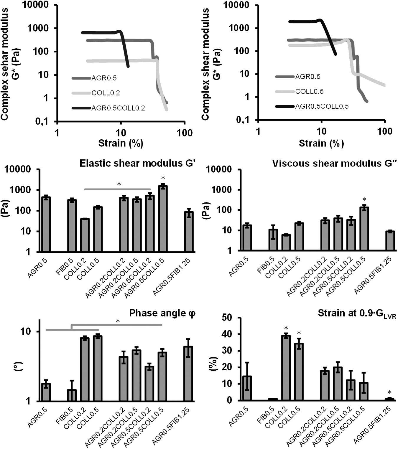
Rheological investigation of all hydrogels and hydrogel blends revealed that blending agarose and collagen led to a significantly altered shear modulus and phase angle compared to pure agarose and collagen. Blending had no significant influence on the strain at which the blended samples first showed signs of hydrogel network damage, the end of the linear elastic region. The graphs illustrate mean ± standard deviation (n = 4).
The addition of collagen to agarose led to a significantly increased phase angle between G′ and G″ when comparing AGR0.5COLL0.5 (5.0° ± 0.6°) to pure agarose (1.8 ± 0.2) and fibrin (1.5 ± 0.53). However, the phase angle of AGR0.5COLL0.5 was still significantly lower than those of COLL0.2 (8.1° ± 0.5°) and COLL0.5 (8.6° ± 0.6°). The strains at which the shear modulus reached 90% of its initial value (indicating the end of the LVR of both collagen hydrogels) were significantly higher compared to all other hydrogels and hydrogel blends. For both collagen hydrogels, these critical strains were 39.1% ± 1.4% for COLL0.2 and 34.3% ± 3.1% for COLL0.5. For pure agarose at a concentration of 0.5%, a critical strain of 14.6% ± 8.3% was measured. For the agarose collagen blends, this value even decreased to 12.3% ± 5.7% for AGR0.5COLL0.2 and 10.7% ± 6.1% for AGR0.5COLL0.5. Furthermore, the strain at the end of the LVR of AGR0.5FIB1.25 was significantly lower compared to all hydrogels and hydrogel blends (0.87% ± 0.8%). Since the shear modulus of FIB0.5 increased continuously with increasing strain, the shear moduli G′ and G″ and the phase angles were calculated between 0% and 1% strain.
The shear modulus of a pure agarose control sample at 0.5% was not remarkably changed by temperature increase throughout the entire experiment (ca. 200 Pa). In contrast, the shear modulus of an agarose-collagen blend sample was initially higher (800 Pa) at 25°C and then increased with the temperature curve reaching a plateau at ∼2000 Pa (Fig. 4). A hydrogel column of AGR0.5COLL0.5, which was incubated in parallel, showed a shift from transparency to turbidity.
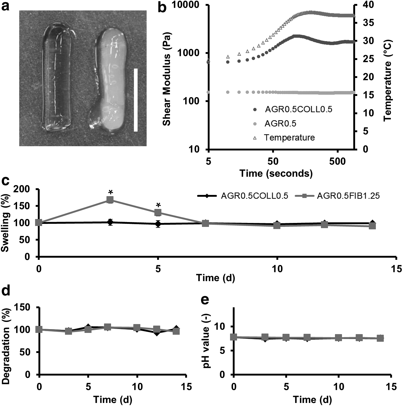
Gelation of collagen within the hydrogel blends AGR0.5COLL0.2 and AGR0.5COLL0.5, respectively, was proved by visual examination and rheology. A printed column of AGR0.5COLL0.5 showed characteristic turbidity after incubation
Hydrogel swelling and degradation
Substantial water uptake was measured for AGR0.5FIB1.25 (Fig. 4). The wet weight of the hydrogel blend AGR0.5FIB1.25 showed a significant increase of 67.6% ± 8.9% on day 3 (compared to AGR0.5COLL0.5) and 30.2% ± 10.0% on the fifth day. At this point, the hydrogel swelling further decreased until day 7, when a significant difference in swelling could no longer be measured. The collagen-containing hydrogel blend AGR0.5COLL0.5 did not show a remarkable water uptake over a period of 14 days. No degradation could be measured for the sample weight of AGR0.5COLL0.5 or AGR0.5FIB1.25. Furthermore, the pH value in the surrounding medium remained almost constant for all four hydrogel blends at a physiological level ranging between 7.4 and 7.8 during the complete testing period.
Transmission electron microscopy imaging
Control images of AGR0.5, COLL0.5 and FIB0.5 revealed the morphology of the individual materials (Fig. 5 and Supplementary Fig. S1). The agarose fibers, which were organized in a dense network, were found to be thinner and more equally dispersed than the collagen fibers. Fibrin formed a dense network consisting of thin fibers.
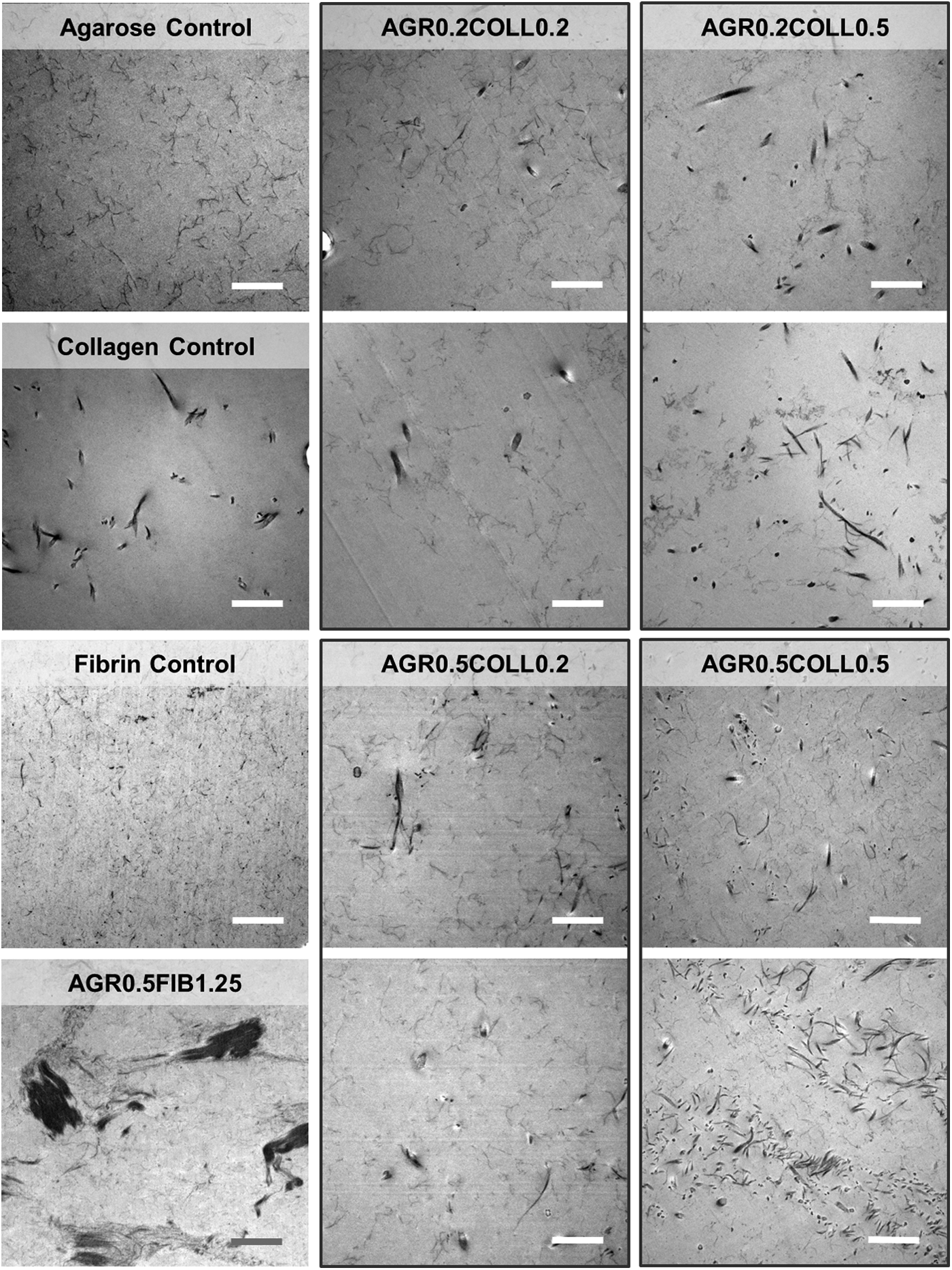
TEM imaging revealed the microstructure of hydrogel blends and their components. Blending of agarose and collagen resulted in coexistence of fibers from both gel types. However, for the hydrogel blends, especially AGR0.5COLL0.5, occasionally distinct collagen streaks were found and both materials appeared to be separated into two different phases. TEM images of AGR0.5FIB1.25 revealed agglomeration of fibrinogen (black areas). Scale bar represents 1 μm. TEM, transmission electron microscopy.
Blending of agarose and collagen resulted in a coexistence of different fibers from both gel types. However, for the hydrogel blends, especially AGR0.5COLL0.5, distinct collagen streaks were occasionally found. TEM images of AGR0.5FIB1.25 revealed two phases consisting of either agarose fibrils or agglomerated protein.
Capillary-like network formation in hydrogel blends
Capillary-like structure formation was only observed in hydrogel blends containing collagen. AGR0.5COLL0.5 showed the highest tubule formation, followed by ARG0.5COLL0.2 (Fig. 6). In these samples, HUVEC aligned and sprouted into three-dimensionally branched tubules, whereas only few cells remained clustered. However, compared to fibrin as positive control, both hydrogel blends showed less capillary-like structure formation. Interestingly, the hydrogel blend containing fibrinogen (AGR0.5FIB1.25) showed no capillary-like network formation at all. In the other hydrogel blends, AGR0.5COLL0.2, AGR0.2COLL0.2, and AGR0.2COLL0.5, no aligned cells were found either. The type I collagen control could not be evaluated since the hydrogel fully contracted during cultivation. Regarding structure volume and surface area, a significantly higher value was observed for the fibrin control in comparison to the group of AGR0.2COLL0.2 and AGR0.2COLL0.5, as well as AGR0.5FIB1.25 (all summarized as “round cells” showing no capillary-like structure formation) (Fig. 6). Higher values for structure volume and area correlate with a higher proangiogenic potential of the fibrin scaffold compared to the blends. In addition, a significant difference was observed for AGR0.5COLL0.5 in comparison to samples with only round cells for structure volume and total cell surface area of all detected cells. Direct comparison of the FIB0.5 and AGR0.5COLL0.5 revealed the following results: with a focus on cell volume, we achieved a mean value of 3013.8 ± 1320 μm3 (FIB0.5) and 2022.8 ± 601.3 μm3 (AGR0.5COLL0.5). Regarding cell surface area, fibrin had a value of 1438.9 ± 660.4 μm2 and AGR0.5COLL0.5 had a surface area of 868.4 ± 278.9 μm2. Focusing on structure length and branching points, the fibrin gel control had in both cases a significantly higher value compared to the analyzed hydrogel blends AGR0.5COLL0.5 and AGR0.5COLL0.2. For the evaluation of structure length and branching points, round and clustered cells were not taken into account. Regarding branching points, it was further observed that AGR0.5COLL0.5 as well as AGR0.5COLL0.2 had significantly different values in direct comparison to each other.

Three-dimensional cocultivation of HUVEC and HDF in printable hydrogel blends consisting of agarose and collagen versus nonprintable fibrin gel control. Images show CD31/Alexa Fluor 594-labeled capillary-like structure formation using two-photon laser scanning microscopy. Scale bar: 300 μm. Structure volume, surface area, structure length, and branching points were chosen as parameters to evaluate the quality of capillary-like structure formation. All four parameters provide information about the formation rate of capillary-like structures, the structure thickness, as well as the rate of network branches. Values were determined by image processing of two-photon laser scanning images through ImagePro® analysis. HDF, human dermal fibroblasts; HUVEC, human umbilical vein endothelial cells. Color images available online at
AGR0.5COLL0.5 hydrogel blends showed lumen formation in TPLSM cross sections with a diameter of 5 to 25 μm (Fig. 7). Furthermore, increased diameter and lumen formation in fibrin gels compared to AGR0.5COLL0.5 blends were observed (Fig. 7).
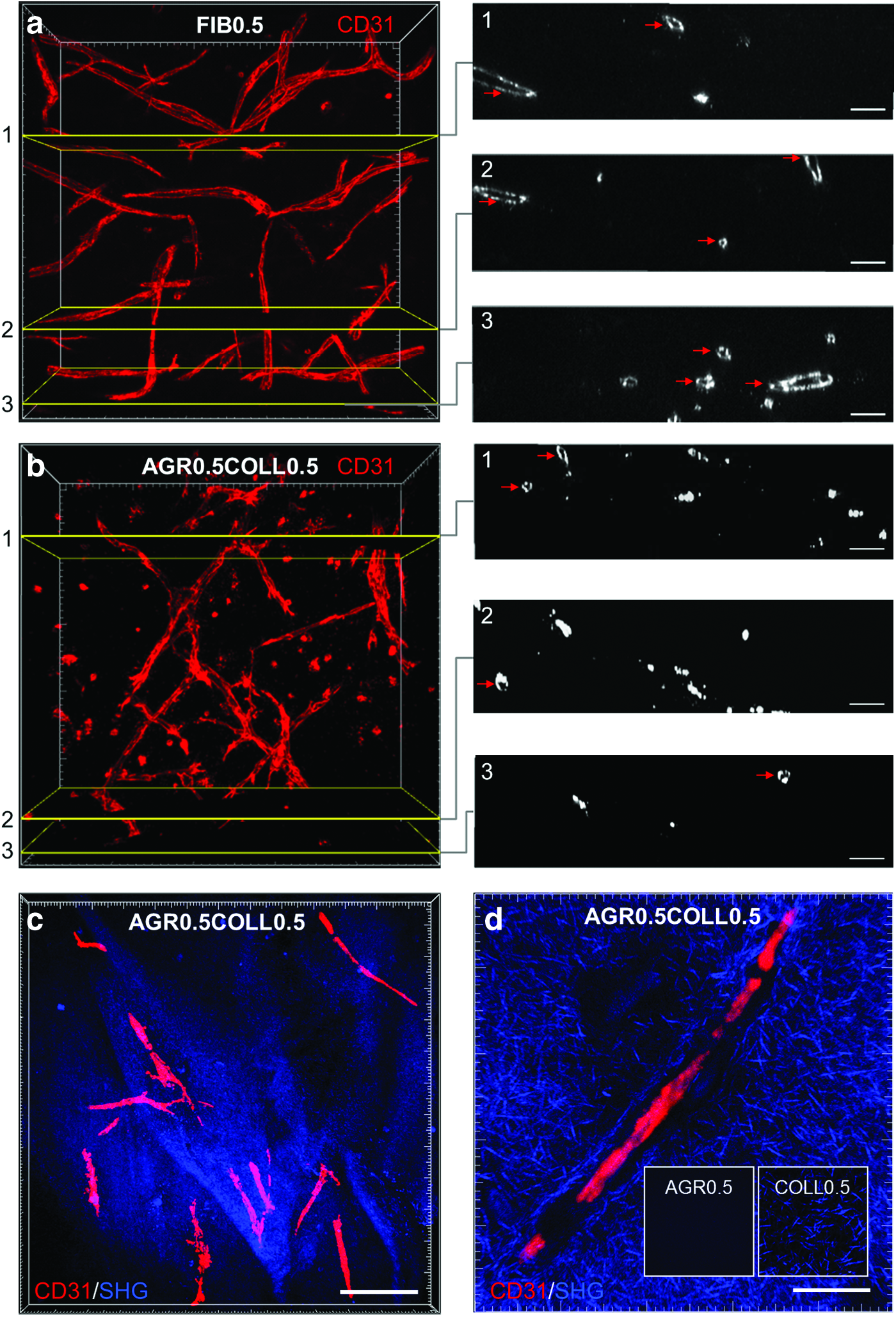
Cross sections of TPLSM image stacks visualize the formation of hollow capillary-like structures in FIB0.5 and AGR0.5COLL0.5 indicated by red arrows
Additional focus was set on the distribution of collagen within the hydrogel blends visualized by trichrome collagen staining since only homogenous dispersion would guarantee cell attachment to RGD motifs throughout the entire composite matrix. Light imaging confirmed a homogenous collagen distribution in collagen-containing hydrogel blends regardless of agarose and collagen concentrations (Fig. 8). With increasing collagen content, the staining was stronger, while increasing agarose concentration led to decreased collagen signal. Since the agarose control had no trichrome-positive binding sites, the sample was almost transparent. With regard to the images from TPLSM, collagen could easily be detected due to its autofluorescence using second harmonic generation phenomenon. Pure type I collagen gels showed intertwined collagen fibril networks compared to pure agarose hydrogels. In pure agarose, no signal was detected at a wavelength of 840 nm. Some capillary-like cell formations (red) were found to align along collagen streaks (blue), which were occasionally observed. Images of angiogenic structures revealed cellular tubules surrounded by a dense network of collagen fibrils (Fig. 7).
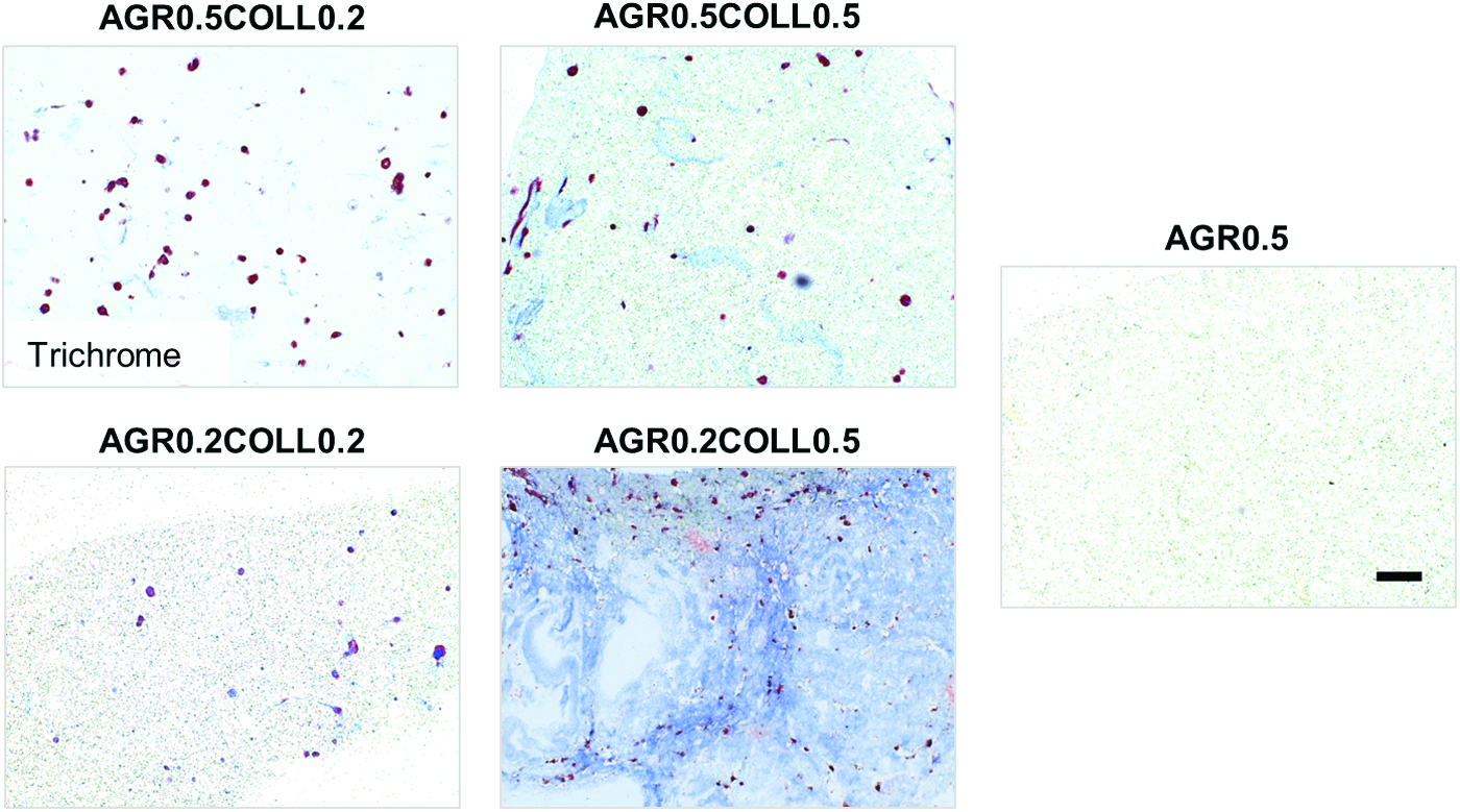
Trichrome staining was performed on paraffin sections of hydrogel blends and fibrin control to examine homogenous collagen (blue) and agarose distribution in hydrogel blends after cultivation over 14 days. Red Chromotrop 2R composition of trichrome solution visualizes cytoplasm of HUVEC and HDF embedded in hydrogel matrix. Scale bar represents 50 μm. Color images available online at
Discussion
The main challenge in the development of suitable bioinks is the combination of partly contradictory requirements in terms of sufficient biofunctionality and 3D printability. To date, only a few studies have been published that present a suitable 3D-printable material with respect to resolution and construct stability, and promote cell adhesion, spreading, and migration.27,28 Therefore, this study elucidates to which extent the blending of agarose with fibrinogen or type I collagen can offer a promising solution to this problem.
The composition of the hydrogel blends was carefully considered based on the observation that cell sprouting is strongly dependent on the hydrogel stiffness and the presence of cell adhesion motifs. 29 In terms of material characteristics, capillary-like structure formation relies on several factors such as hydrogel stiffness, composition, and microstructure. It was demonstrated that endothelial cell migration and capillary network formation are inhibited with rising fibrin hydrogel scaffold concentration.30,31 Therefore, bioink development must balance the intricate interplay of these parameters to enable 3D printing as well as to ensure angiogenesis. Based on recently published results, the concentration of agarose was set to maximum of 0.5% allowing basic 3D printability as well as cell sprouting when blended with type I collagen. 10 For collagen, a maximum concentration of 0.5% and for fibrinogen, a higher final concentration of 1.25% could be achieved. Since a significant increase in the stiffness of the resulting hydrogel blend was expected, it was decided not to polymerize the fibrinogen to fibrin. However, AGR0.5FIB1.25 did show severe water uptake and loss of shape during the assessment of hydrogel swelling (Fig. 4). Therefore, it is assumed that the noncross-linked protein is not sufficiently immobilized within the hydrogel blend. No blend showed signs of significant degradation over a period of 14 days submerged in endothelial cell culture medium. Furthermore, the agarose-collagen samples kept their initial shape very well, which indeed qualifies this combination for long-term incubation subsequent to the cell-laden printing procedure. 32
This study has proven that the printability of agarose was not impaired by the addition of either collagen or fibrinogen. Furthermore, the reduced droplet volume of the dispensed hydrogel blends indicates that blending results in an increase in viscosity, which might even improve printing resolution.
Dynamic mechanical analysis revealed that blending of agarose with either 0.2% or 0.5% collagen resulted in a significantly increased shear modulus. Similar results were also reported in earlier studies.10,12 As TEM images revealed a network of finely interpenetrating agarose and collagen fibrils, this finding suggests that both hydrogel networks mechanically reinforce each other. The strain marking the end of the LVR at 90% of the initial shear modulus could not be lengthened by hydrogel blending.
For pure agarose or type I collagen hydrogels, different rheological network properties and gelation processes have been reported. Collagen monomers form a 3D hydrogel at physiological temperature and pH by formation of collagen fibers from thin filaments that are noncovalently entangled, 33 whereas agarose undergoes temperature-induced coil-helix transition characterized by formation of single or double helices. 34 This step is followed by aggregation of double helices, which then are interconnected by flexible agarose chains. A major difference in the network properties of these two materials is that single fibers of pure collagen hydrogels can slide and bend independently. 35 When blending agarose and collagen, the individual fiber motion is restricted and the movement of neighboring fibers appears coupled. 36 Thus, the addition of agarose leads to a restriction of cell-induced deformation and remodeling of the matrix. Even though the stiffness of a hydrogel was found to hinder cell spreading and migration and AGR0.5COLL0.5 was the stiffest hydrogel blend investigated in this study, significant capillary-like network formation was identified within this material.30,37 However, cell migration as a prerequisite for angiogenesis might have been enabled by an alteration of the viscoelastic properties induced by blending of agarose with collagen. For viscoelastic materials, the phase angle φ represents the lag between an applied shear stress and the resulting shear strain and indicates energy dissipation. 38 As the phase angle of AGR0.5COLL0.5 was found to be significantly increased compared to that of pure agarose and even AGR0.5COLL0.2, it is assumed that the addition of collagen makes the hydrogel blend more viscous and thus more susceptible to cell-induced deformation. Therefore, a direct correlation between the manifold formation of angiogenic structures and the significant increase of the hydrogel blend's phase angles is shown. It is furthermore presumable that the viscoelastic properties of pure fibrin are locally altered due to the already reported protease-mediated degradation processes. 39 This might explain the initial low phase angle of FIB0.5 and the superior capillary-like network formation during culture. However, this should be further investigated in a subsequent study analyzing the rheological properties of cultured samples at different time points.
As the observed shift from transparency to turbidity and the increase of the storage modulus of the sample are characteristic of the gelation of collagen, it was confirmed that collagen polymerizes even when blended with agarose, both in printed and nonprinted samples. This finding highlights the overall suitability of agarose-collagen blends for tissue engineering applications since the presence of a fully developed collagen network is highly beneficial for cell signaling and guidance. 40
An increase of the collagen content correlating with an increase of RGD sequences resulted in enhanced capillary formation. This was proven by the analysis of parameters evaluating the quality of capillary-like network formation as total cell volume, cell surface area, structure length, and branching points. 41
Agarose-based hydrogel blends that combine 3D printing and capillary-like network formation resulting from a coculture of endothelial cells and fibroblasts have not been investigated up to now. In contrast to the blending approach considered in this study, several studies have proposed the direct printing of perfusable channels to obtain a vascularized construct. The applied fugitive material was subsequently washed out and in a subsequent step, the resulting channels were populated with endothelial cells.42–45 However, cocultivation of HUVEC and HDF enables long-term stability of formed capillary-like structures due to mechanical, fibroblast-induced support. Promising results concerning capillary-like network formation were achieved for AGR0.5COLL0.2 and AGR0.5COLL0.5 hydrogel blends, which also seemed promising from the printing perspective.
As a result, this study demonstrates that 3D printability and angiogenesis are not mutually exclusive. Further steps include the printing of high aspect ratio constructs with directly embedded angiogenesis-performing cells.
Footnotes
Acknowledgments
This study was supported by Deutsche Forschungsgemeinschaft, Bonn (Grant Nos. FI 975/23-1, JO 764/4-1). We thank Prof. Dorothea Rohrmann, Department of Urology, RWTH Aachen University Hospital, for provision of juvenile foreskins. In addition, we thank the Department of Gynecology and Perinatal Medicine, RWTH University Hospital, for kind provision of umbilical cords. This work was supported by the Core Facility “Two-Photon Imaging,” [Interdisciplinary Centre for Clinical Research (IZKF Aachen)] within the Faculty of Medicine at RWTH Aachen University. We thank Dr. Michael Vogt, operation manager of the Core Facility, for his help with the Two-Photon experiments. Furthermore, we thank Mrs. Natalie Hepp for her technical support.
Disclosure Statement
No competing financial interests exist.
References
Supplementary Material
Please find the following supplemental material available below.
For Open Access articles published under a Creative Commons License, all supplemental material carries the same license as the article it is associated with.
For non-Open Access articles published, all supplemental material carries a non-exclusive license, and permission requests for re-use of supplemental material or any part of supplemental material shall be sent directly to the copyright owner as specified in the copyright notice associated with the article.