
Abstract
Nonviral transfection has important implications on gene therapy because of its safety. In particular, polyfection and nucleofection are two widely used systems for nonviral gene delivery. Their potential depends on the transfection efficiency achieved, which is influenced in turn by the type of cells transfected and by the plasmid that carries the gene of interest. The efficiency of transfection by polyfection or nucleofection in human fibroblasts and keratinocytes was evaluated in this study. Transfections were performed with plasmids containing a gene of interest (human cathelicidin antimicrobial peptide) and two reporter genes (red or green fluorescent protein) that included or not an internal ribosome entry site (IRES). The efficiency was measured by flow cytometry in terms of percentage of cells expressing the reporter gene; viability of transfected cells was also evaluated. It was found that nucleofection was more efficient than polyplexes for transfecting fibroblasts, while no significant differences were found between both systems of transfection when applied to keratinocytes. Regarding the viability of fibroblasts after transfection, values were high in both systems. In contrast, keratinocytes were more sensitive to nucleofection. It was also noted that both types of cells decreased reporter gene expression when IRES sequence was located upstream of the reporter gene, suggesting a negative effect on the expression of this gene. These results confirm that the transfection efficiency depends on the type of cells and the system used.
Introduction
G
Although cell transfection has been widely studied in the literature, it is necessary to empirically determine the technical requirements for a given cell culture type as pointed out by Hengge and Volc-Platzer. 5 In this article, the focus is in the transfection of fibroblast and keratinocyte cell types.
Many researchers are currently developing nonviral transfection systems, which have been regarded as safer than systems based on viruses. Two of the more widely adopted are polyfection and nucleofection. 6 On the one hand, polyfection refers to the transfer of genes into cells through cationic polymers, mainly linear or branched polyethylenimine (PEI). The PEI has a high number of surface primary amino groups that interact with DNA, forming polyplex particles protecting DNA from degradation before reaching the nucleus. These kinds of systems are the most simple and economic ones and their use has become popular due to their low cytotoxicity and relative effectiveness in vitro. 7 On the other hand, nucleofection is based on the transient hydrophilic pore formation in the cell membrane. Nucleofection has been described as more efficient with respect to chemical systems, and any mammalian cell can be transfected by nucleofection.
An important step in cell modification is achieving high efficiencies in transfection. That is why efficiency is a key factor in the comparison of different transfection methods. Expression of reporter genes introduced in plasmid vectors is used for measuring the transfection efficiency and could allow tracking of transfected cells. However, some products of gene expression are metabolized extracellularly (e.g., CAMP gene expression). In these cases, it is necessary to separate the reporter gene expression from the interest product. Introducing internal ribosome entry site (IRES) sequences in the vectors enables the expression of two genes controlled by one single promoter in target cells. 8 In bicistronic vectors containing the IRES sequence, the first gene (upstream IRES) is translated in a cap-dependent manner and the second one (downstream IRES) in an IRES-dependent manner. These systems have been widely used for in vitro and in vivo applications since their discovery, although it has been reported that the expression levels of genes located downstream of IRES sequence are lower than those levels expressed by genes located upstream. 9 This means that low levels of a reporter gene located downstream of the IRES sequence can result in an underestimation in the expression of the gene of interest; the latter is generally located upstream of the IRES sequence and with a translation cap dependent, 10 which in the translation, serves as a “molecular tag” that marks the spot where the 40S ribosomal subunit is to be recruited.
In this study, primary human skin cells, fibroblasts, and keratinocytes had been genetically modified by polyplexes and nucleofection with a plasmid vector containing a gene of interest (CAMP) and a reporter gene (red fluorescent protein [RFP] or green fluorescent protein [GFP]). IRES sequences were included in the CAMP plasmid backbones in an upstream position with respect to the reporter gene. The efficiency of transfection and the viability of transfected cells were compared:
• Between the two different types of cells (fibroblasts and keratinocytes) • Between the two transfection systems, polyfection and nucleofection. In all cases, three nucleofection commercial kits were compared: Mirus from Ingenio and L and V from Lonza. • Between the transfection systems with and without the IRES sequence.
The expression of CAMP gene in fibroblast was further evaluated using PCR analysis. The reporter genes were expressed by both types of skin cells even when IRES sequence was included in the plasmid vectors. An interesting application of these findings could be the production of autologous skin three-dimensional organotypic cultures based on cells with the aforementioned modifications. These enhanced organotypic cultures could be more efficient in the treatment of skin wounds.
Materials and Methods
Plasmidic DNA preparation
Lentivector with bicistronic IRES-tRFP (destination vector PS100080 from OriGene) allows both the reporter gene and the gene of interest expression. PrecisionShuttle system was used to introduce the CAMP sequence coding to the hCap18/LL-37 expression; this sequence was obtained from a pCMV6 vector (entry vector RC208872 from OriGene). CAMP sequence was verified into PS100080 (Macrogen Inc.). From now on, the modified plasmid will be called PS100080-CAMP vector. All plasmids were amplified into competent Escherichia coli and purifications were performed by using a plasmid maxi kit (Qiagen EndoFree®). PmaxGFP® (Lonza) was used as the non-IRES-containing plasmid control.
Primary human fibroblast and keratinocyte cell culture
With prior informed consent, approved by the Bioethics Committee of the Faculty of Medicine from University of Antioquia, primary human keratinocytes and fibroblasts were isolated from remaining skin obtained from surgical procedures. Collection, expansion, and cryopreservation of keratinocytes and fibroblasts were performed by explants 11 and/or enzymatic digestion of the skin samples, according to the protocols established by the Group of Tissue Engineering and Cellular Therapy. Skin samples were mechanically fragmented and enzymatically digested by using trypsin/ethylenediaminetetraaceticacid (Sigma-Aldrich). Cell pull obtained was cultured in two different media to obtain fibroblasts or keratinocytes. 11 Cells subcultured between one and five times were used for all experiments.
Polyplex system transfection: linear polyplexes (linear PEI polyplexes)
The transfections were performed by using linear PEI (LPEI 25 kDa; Polysciences). The procedure was based on a protocol described by Hsu and Uludag. 7 Some changes were introduced; the medium of transfection was the same used to culture the keratinocytes without antibiotic. In brief, polyplexes at nitrogen/phosphate ratio = 19 (mol from units of LPEI/mol from nucleotides of PS100080-CAMP or pmaxGFP) were formed in a buffered saline solution before putting them in contact with the cells. Fibroblasts or keratinocytes were subcultured 24 h before transfection and seeded at a density of ≈3 × 10⁴ cells/cm2. The replacement of the cell medium was performed with the polyplexes (containing 1 μg of the plasmid) previously diluted in the medium of transfection. Polyplexes were allowed to be in contact with the cells during 6 h, after cell medium was replaced by complete fresh medium and incubated overnight until cytometry analysis.
Nucleofection system transfection
Transfection by nucleofection of primary human fibroblasts and keratinocytes was performed by using the Nucleofector™ II/2b (Amaxa; Lonza). Different kits of electroporation were evaluated: Cell Line Nucleofector® Kits L and V (Lonza) and Ingenio® Electroporation Kit (Mirus). For fibroblast and keratinocyte programs, U-30 and T-018 were used, respectively. Protocols established by each kit were followed: electroporation volumes of 100 μL were used to transfect 1.5 × 10⁶ cells with 2.5 μg of the plasmid (PS100080-CAMP or pmaxGFP), recommendations of the normal protocols for nucleofection.
Flow cytometry
Measures were performed in a BD LSRFortessa™ cytometer. Agglomerated cells were excluded by adjusting cytometer voltage. Ten thousand events were counted by each measurement. Analysis of results obtained by flow cytometry was done so that the population of autofluorescent cells was excluded from the positive population by the expression of the reporter gene (RFP or GFP). For this, a diagram was plotted with fibroblasts or keratinocytes that were not transfected (negative control). The type of diagram used was a “pseudocolor”: Comp-FITC versus Comp-PE-A. Cells that were above the fluorescence intensity of 103 and simultaneously near to the line at 45° were considered autofluorescent.
Fibroblast RNA isolation, cDNA synthesis, and quantitative real-time PCR (polyplexes)
Total RNA from in vitro-cultured human fibroblasts transfected with polyplexes was isolated with TRIzol reagent (Gibco). RNA integrity was assessed using agarose gel electrophoresis stained with ethidium bromide. cDNA was synthesized from 2 μg of total RNA by using the RetroScript kit (Qiagen, Basel, Switzerland) according to the manufacturer's manual.
Quantitative real-time PCR (RT-PCR) was performed by using 2 μL of cDNA and 18 μL of SYBR Green PCR master mix (Thermo Fisher Scientific). CAMP-specific PCR primers were used to generate 100-bp fragments. All primers were synthesized by Macrogen (forward primer: 5′ CGCCAAAGCCTGTGAGCTT-3′; reverse primer: 5′-TTCACCAGCCCGTCCTTCTTG-3′). A number of CAMP copies were obtained through the generation of a standard curve achieved with serial dilutions of PS100080-CAMP plasmid.
Statistical analysis
Fibroblasts and keratinocytes were isolated from three different donors and triplicates of the transfections were made. Nonparametric tests were used. Kolmogorov–Smirnov test was used to compare the results coming from two independent samples: cells transfected by linear polyethylenimine polyplexes and nucleofection. Kruskal–Wallis analysis of variance was used to compare the results coming from three independent samples of cells transfected by nucleofection using kits L, V, or Ingenio.
Results
Nucleofection kit comparison: kits L, V, and Ingenio
Ingenio Electroporation Kit was evaluated in terms of transfection efficiency and compared with the L and V kits from Lonza, which are the recommended kits to transfect human skin cells by nucleofection. The comparison was performed using the pmaxGFP vector (Fig. 1). The idea behind testing the Ingenio Electroporation Kit was to evaluate how low the efficiency was with respect to its economic price.
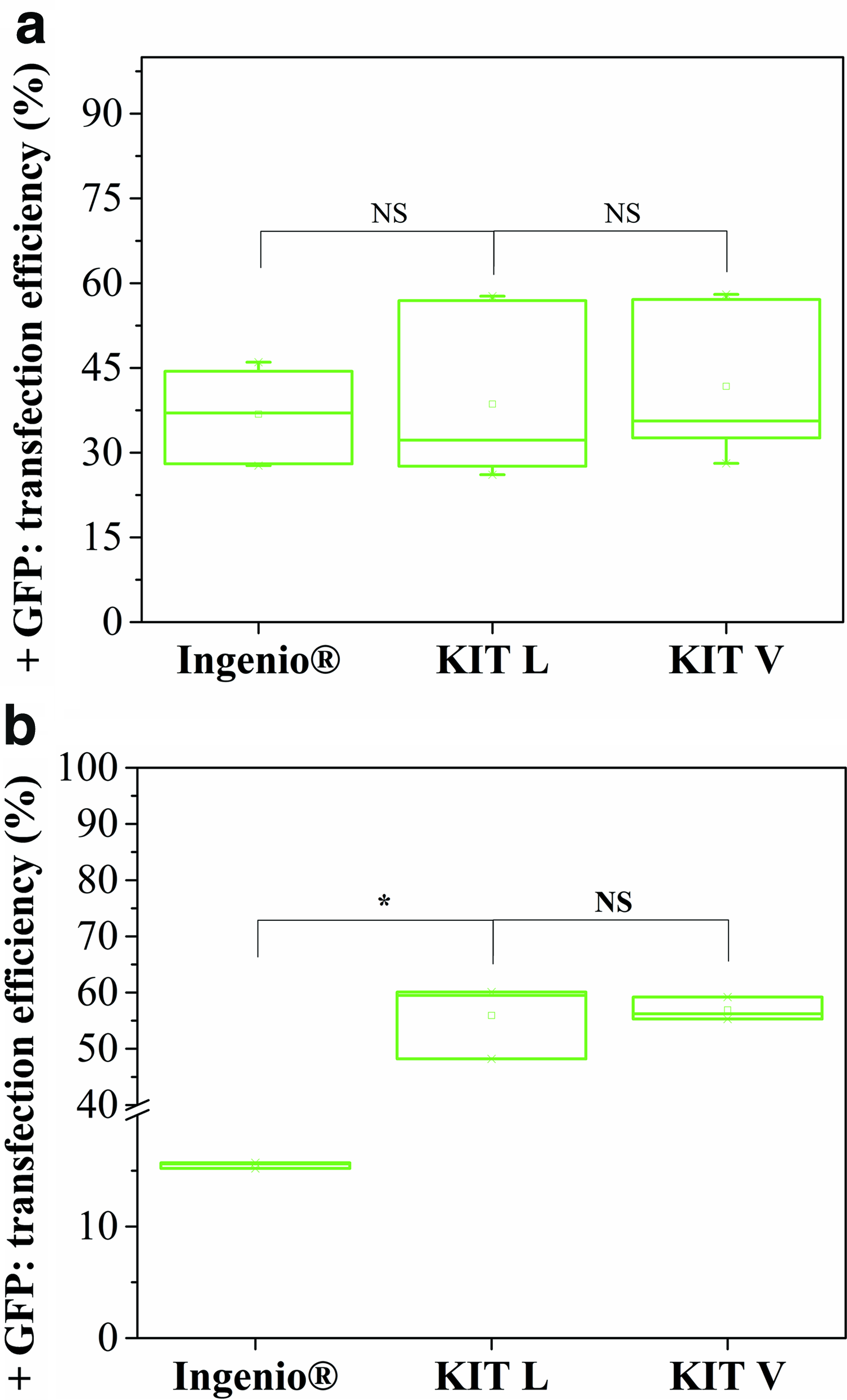
Nucleofection kit comparison. Transfection efficiency in nucleofected cells analyzed by flow cytometry at 24 h postnucleofection.
Efficiency in all cases is defined as the ratio between cells expressing the reporter gene and the total number of cells. Both of these quantities were measured by flow cytometry.
On the one hand, when nucleofection was used in fibroblasts, results from the three kits show no significant differences between efficiency medians. All efficiencies were higher than 26% despite the fact that there were variations inside the samples, even for Ingenio Electroporation Kit. On the other hand, when nucleofection was used on keratinocytes, Ingenio Electroporation Kit had significantly lower efficiency than kits L and V. For keratinocytes, efficiencies of nucleofection performed with Ingenio Electroporation Kit were as low as 15.6%, while for kits L and V, efficiencies were as high as 44.4% and 55.3%, respectively. Therefore, data of three kits of nucleofection were included for fibroblasts, whereas for keratinocytes were not considered those obtained from Ingenio Electroporation Kit tests.
Nucleofection versus linear polyplexes: fibroblasts
Figure 2 shows representative images of fluorescence micrographs and flow cytometry analyses of fibroblasts transfected by nucleofection and polyplexes. In both systems there were transfected fibroblasts expressing the GFP but there were a notoriously higher number of green fibroblasts in nucleofection when compared with the polyplex system.
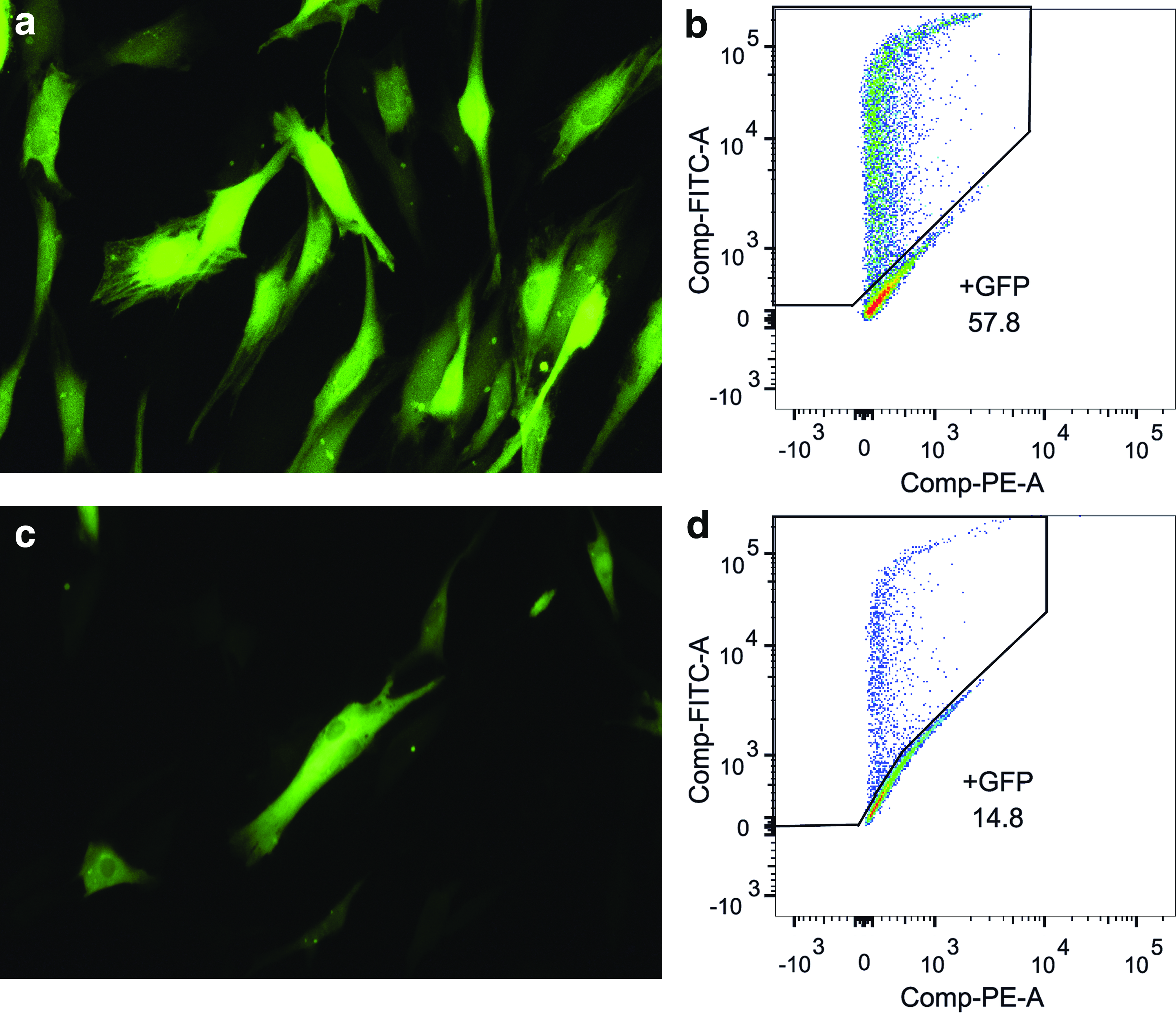
Nucleofection versus transfection with LPP in fibroblasts.
Flow cytometry measures confirmed the optical observations and a quantitative analysis was performed (Figs. 2 and 3).

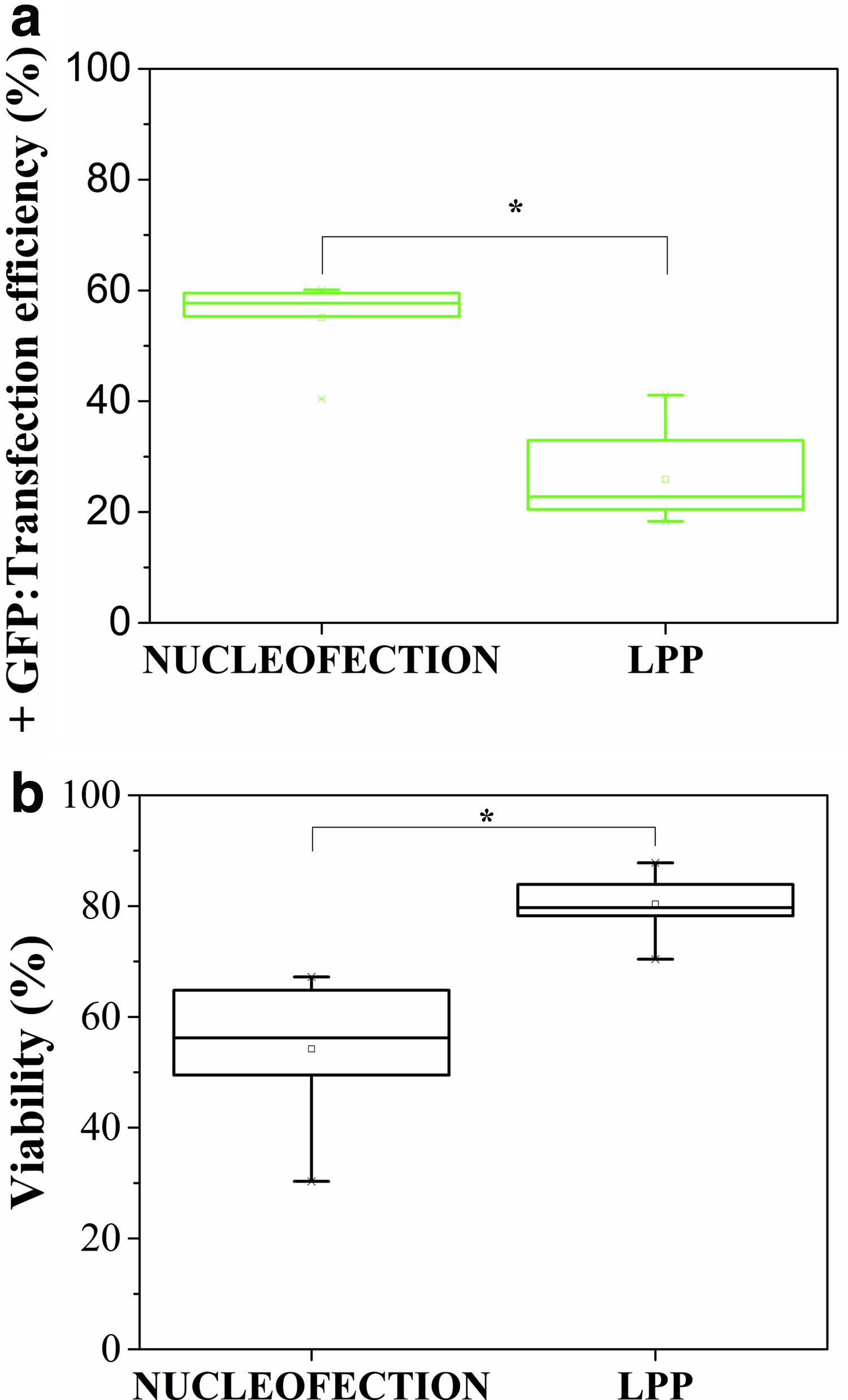
Fibroblasts. Nucleofection versus LPP.
Results of the quantitative analysis are shown as the median values of transfection efficiencies and viabilities of fibroblasts transfected by nucleofection and polyplex systems (Fig. 3). Significant differences were found when both systems were compared. As expected, transfection efficiencies for nucleofection (median = 34.6%) were higher than the ones for the polyplexes (median = 14.8%). Regarding cytotoxicity, viabilities were under 50% for some samples when polyplexes were used, while viabilities were always higher than 68.7% when nucleofection was used.
Nucleofection versus linear polyplexes: keratinocytes
Figure 4 shows representative images of flow cytometry analyses and fluorescence micrographs of keratinocytes transfected by nucleofection and polyplexes. Similar to fibroblasts, in both systems, there were cells expressing the GFP and there were a higher number of keratinocytes expressing GFP with nucleofection. However, in contrast to fibroblasts, a high number of dead cells were observed with nucleofection.

Nucleofection versus transfection with LPP in keratinocytes.
Quantitative results from flow cytometry are summarized in Figure 5.
Keratinocytes. Nucleofection versus LPP.
Efficiencies of keratinocyte transfections were between 40% and 60% when nucleofection was performed, whereas values between 18% and 30% were achieved when polyplexes were used.
Polyplex system: plasmid with IRES sequence versus no IRES sequence
We also evaluated the effect of an IRES sequence in the plasmid vector used to separately introduce both the gene of interest and the reporter gene in both types of cells. As mentioned in the “Introduction” section, it has been described in the literature that the expression levels of genes located downstream of an IRES sequence are lower than those of genes located upstream of them. 9 Since the reporter gene in our plasmid is downstream of the IRES sequence, we evaluated its effect on the expression of the reporter gene by transfecting fibroblasts and keratinocytes with polyplexes. This system showed lower transfection efficiencies in both kinds of cells measured through the expression of the reporter gene.
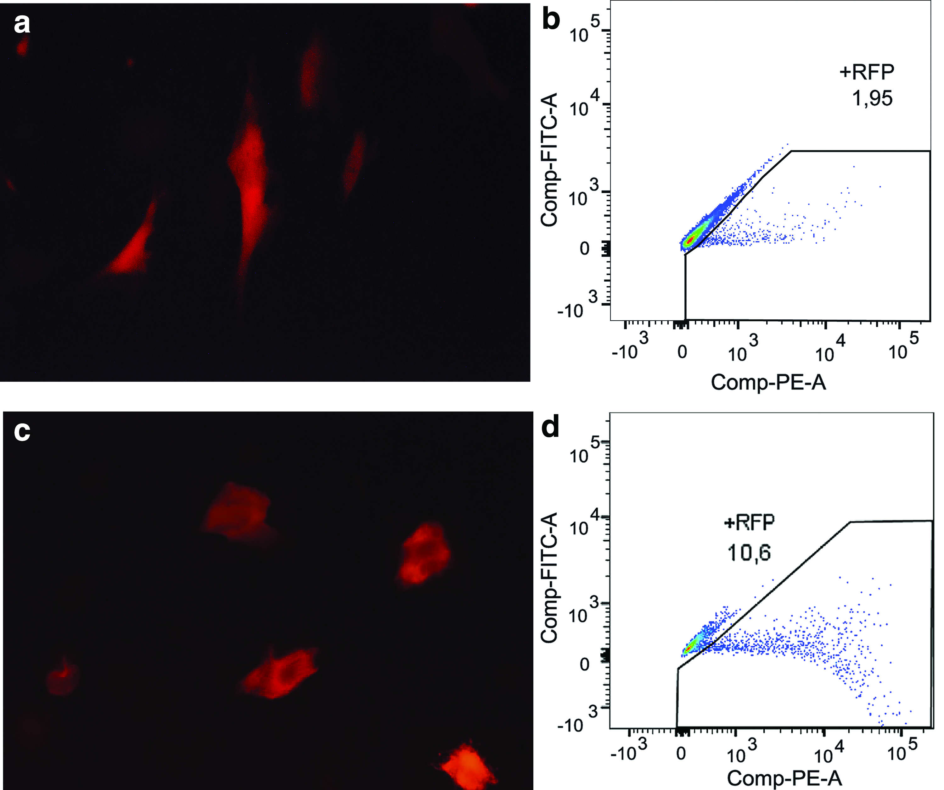
Figure 6 shows representative images of flow cytometry analysis and fluorescence micrographs of fibroblasts and keratinocytes transfected by polyplexes. The plasmid used contains the CAMP gene (interest) upstream the IRES sequence, and downstream of it is located the RFP gene (reporter). A small population of cells (both fibroblasts and keratinocytes) expressing the RFP were qualitatively observed (Fig. 6a, c). Flow cytometry results confirmed the optical observations (Fig. 6b, d).
LPP transfection with a plasmid containing an IRES sequence.
Figure 7 shows the quantitative comparison between the transfection efficiencies and viabilities of fibroblasts and keratinocytes transfected by polyplexes with and without the IRES sequence upstream of the reporter gene (RFP or GFP, respectively).
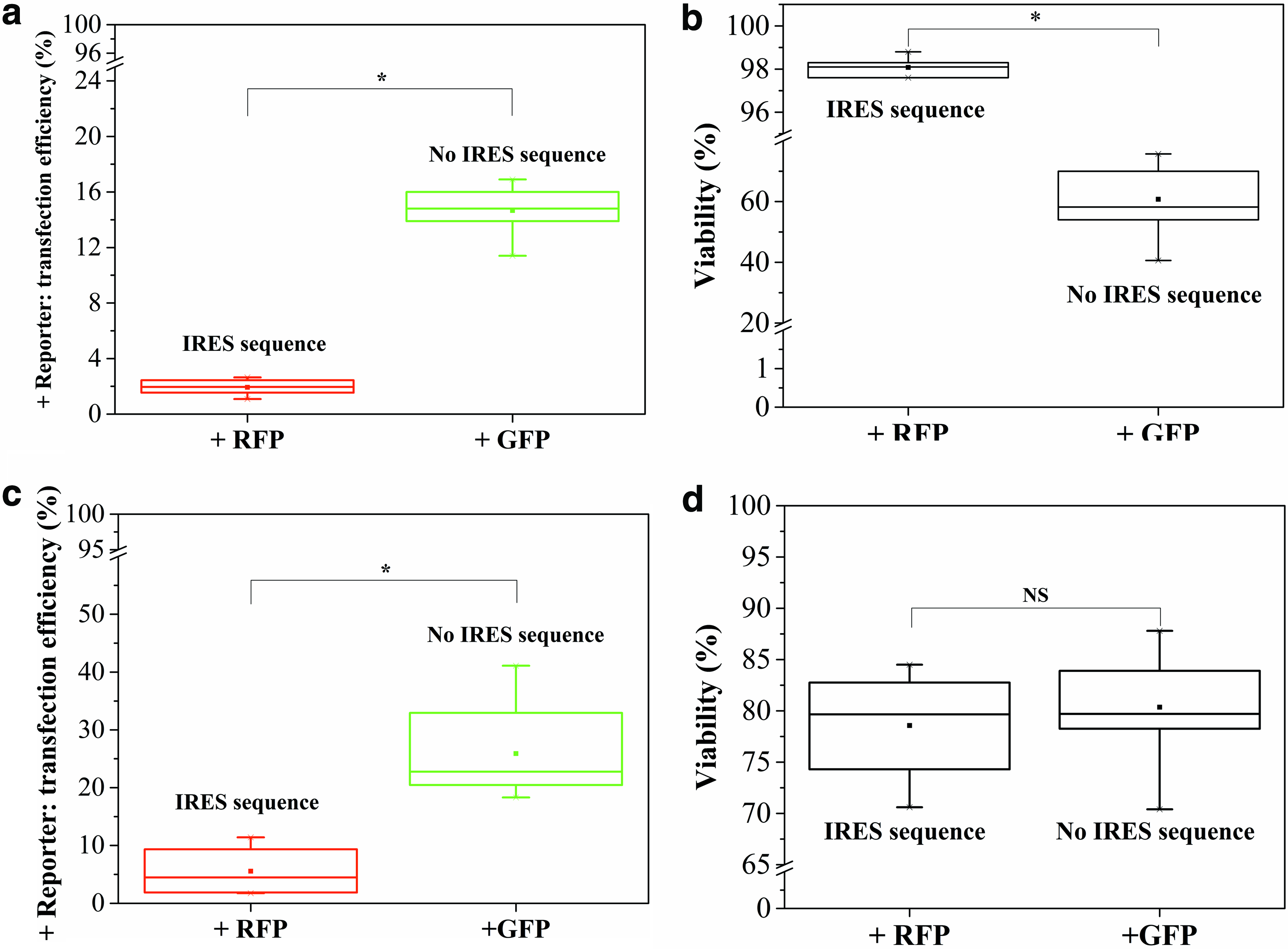
LPP transfection with a plasmid with or without IRES sequence: transfection efficiency and cell viability in
Results showed significant differences between efficiencies of transfection in both types of cells, fibroblasts and keratinocytes, when a vector containing an IRES sequence was used in the transfection. Regarding the expression of the reporter gene, (1) for fibroblasts, an efficiency median of 1.95% was obtained when the plasmid containing IRES was used (+RFP cells) and an efficiency median of 14.8% when the plasmid without IRES (+GFP) was used and (2) for keratinocytes, a similar trend was observed. An efficiency median of 4.5% was obtained when the plasmid containing IRES was used (+RFP cells) and an efficiency median of 22.8% when the plasmid without IRES (+GFP) was used.
Regarding cytotoxicity, significant differences were found for fibroblasts as follows: presence of IRES sequence increased the fibroblast viability from a median of 58.2% to98.1%. Meanwhile for keratinocytes, no significant differences were observed with and without IRES sequence. Viabilities had a median of 79%.
Fibroblast CAMP expression (polyplexes)
Although the polyplex system exhibited low transfection efficiencies through reporter gene expression due to the use of IRES sequence (Figs. 3 and 5), further PCR analysis allowed us to find that the expression of the gene of interest was, in fact, higher than in the controls (nontransfected cells). To confirm this hypothesis, we evaluated the expression of the CAMP gene in fibroblasts transfected with polyplexes, since it is known that these cells do not express the CAMP gene constitutively. Figure 8 shows the results of the CAMP gene-copy number quantification in transfected versus control cells (nontransfected fibroblasts). Overexpression of CAMP gene was observed at 24 and 48 h post-transfection.
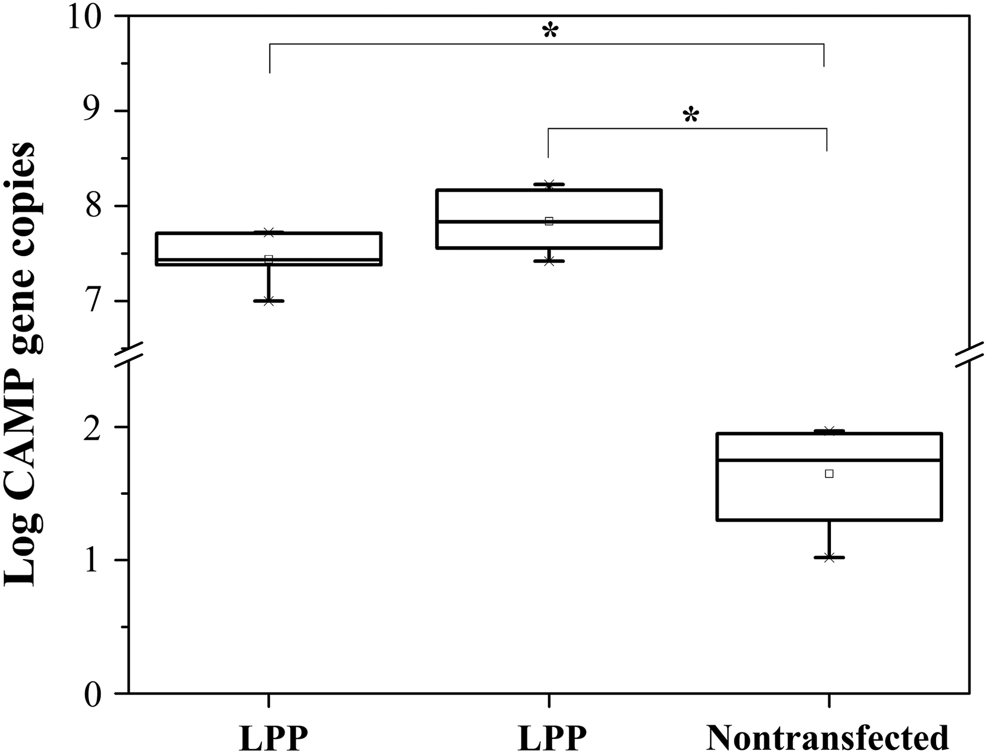
Quantification of CAMP gene expression in human fibroblasts. The cells were transfected with LPP and homogenized with TRIzol for the extraction of total RNA. The number of copies of the CAMP gene was quantified by real-time PCR system (LightCycler® 96; Roche) at 24 and 48 h post-transfection versus controls (nontransfected fibroblasts), K-S test, n = 3, *p < 0.05.
Discussion
Nucleofection has been reported as an efficient transfection system for human cells from primary cultures.12,13 In particular, L and V commercial kits from Lonza have been described as very good options in nucleofection of skin human cells (fibroblasts and keratinocytes). However, there exist other less expensive kits that could be used in the transfection of primary cell cultures. Ingenio Electroporation Kit is one of them, and in this study it was compared with L and V kits from Lonza with respect to nucleofection of primary skin fibroblasts and keratinocytes. Fibroblast results have shown that all three kits were efficient in terms of transfection efficiencies. Obtained efficiencies (Fig. 1a) were higher than other reported transfection systems applied to primary human fibroblasts. 7 In the comparison of nucleofection applied to primary keratinocytes with three different kits (Fig. 1b), results showed a significant decrease in the nucleofection efficiency when Ingenio Electroporation Kit was used. These results could be explained due to the fact that keratinocytes are electrolyte-dependent cells for its differentiation and survival processes, 14 and therefore, these kinds of cells can be more sensitive to the buffers who mediate the electroporation (nucleofection). Kits L and V could contain a supplement that protects cells from electrolyte misbalance, and consequently, that could lead to better transfection efficiency.
In addition to nucleofection, polyplexes are one of the most currently studied systems to genetically modify primary cells in an efficient and economical way. Furthermore, it is well known that results in transfection also depend on the type of cells, so a polyplex system made with LPEI to transfect primary skin fibroblasts and keratinocytes was also evaluated. As expected, efficiencies of nucleofections were higher than those of polyplexes for both types of cells (Table 1).
Higher transfection efficiencies.
Healthier cultures.
LPP, linear polyplexes.
Regarding the cytotoxicity of transfection, it was found that viabilities of fibroblasts had a median of 58% and 80% with polyplexes and nucleofection, respectively (Table 1). Results strongly suggest that nucleofection has higher transfection efficiencies (a) and healthier cultures (b) than polyplexes when applied to primary human skin fibroblasts, as indicated in Table 1.
Comparison between nucleofection and polyplex systems for keratinocytes transfection is not as simple as for fibroblasts. Again, when transfection efficiencies were compared, nucleofection system seems to be better than polyplexes (Table 1). However, even though there were more cells expressing the reporter gene with nucleofection, viabilities decreased significantly (Table 1). Keratinocytes could be more sensitive to nucleofection due to the fact that this type of cells has an active transport of different kind of molecules. These molecules, such as lipids and calcium ions, are required for the keratinocyte normal growth and differentiation processes. 15 In the case that modified skin cells are to be used in tissue engineering, obtaining suitable autologous grafts from healthy cultures is the main goal for tissue regeneration. In particular, skin equivalents built with keratinocytes expressing antimicrobial peptides could have a greater added value. Nevertheless, it is still necessary to have healthy keratinocytes that can differentiate and form stratified epithelia to obtain similar equivalents to healthy skin. Therefore, polyplexes could be an appealing system to preserve the viability of the keratinocytes (Table 1,a) despite their apparent lower efficiency.
In addition to high transfection efficiency and low cytotoxicity in some cases, one needs to obtain the molecule that is expressed from the gene of interest separated from the molecule that is expressed from reporter gene. For example, when the interest product of expression is a peptide, the reporter molecule can interfere with the peptide activity because peptides are short-sized molecules and their functions depend on their tridimensional structure. Another issue that is related to transfection tracking during cell culture is that if the interest product of expression is extracellular and the reporter protein is attached to the product, the reporter cannot be registered during cell culture. As mentioned before, IRES sequence enables separately and simultaneously expression of two genes controlled by one single promoter in target cells.
In this article, we have compared two plasmids, both have a CMV promoter upstream of the reporter genes RFP and GFP. However, only the RFP plasmid contains the IRES sequence upstream of the RFP gene. Even though CMV is a strong promoter in many types of cells, 16 results showed that when IRES sequence is driving the expression of the reporter, it significantly decreases the expression in both types of cells (fibroblasts and keratinocytes) (Fig. 7a, c). As showed by Mizuguchi et al., 10 expression of an IRES-dependent second gene decreases (in most cases between 20% and 50%) with respect to that of the first gene. In fact, statistical analysis shows a significant difference between the efficiencies in terms of the reporter gene expression. This result could represent a problem when one needs to perform an estimation of the expression of the gene of interest through the quantification of cells expressing a reporter gene, 8 since the results of quantification of the copy number of the gene of interest (in this case the CAMP gene) using RT-qPCR, showed an overexpression in the transfected cells.
Overexpression of CAMP gene in fibroblasts transfected with polyplexes was shown to be higher when compared with controls (nontransfected fibroblasts). This result suggests that final delivery of the gene of interest is efficient enough to achieve a high number of gene copies although transfection efficiency measured by expression of the reporter gene is lower. In addition, CAMP gene expression continues to increase 48 h after transfection with polyplexes (Fig. 8), so this system could be attractive to ensure a sustained delivery of gene of interest in human fibroblasts.
Finally, the results showed that viability values were always higher than 70% when IRES sequence was included in the plasmid. For the fibroblasts, it was observed a statistical significant difference in viabilities with and without IRES sequence (Fig. 7b). This can be due to the fact that a lower expression of the reporter gene reduces the cytotoxicity of the reporter gene (RFP).Viabilities of the keratinocytes showed no significant differences (Fig. 7d) and in both cases (with and without IRES sequence) viabilities were high. As discussed before, polyplexes can be more useful in keratinocytes to preserve the viability of the cells when they are transfected.
Conclusion
Results showed that the three kits (L and V from Lonza and Ingenio from Mirus) were efficient in the nucleofection of fibroblasts, while only the L and V kits from Lonza were efficient in the nucleofection of keratinocytes. Nucleofection proved to be more transfection efficient in fibroblasts than polyplexes, but cell viability is best maintained in keratinocytes when a polyplex system is used.
Transfection with the presence of an IRES sequence seems to affect the expression of genes that are downstream of the sequence. Nevertheless, this effect is not necessarily negative, since it allows both (1) the confirmation of the transfection by means of a reporter and (2) the reduction of its expression, consequently decreasing the cytotoxicity of the transfection.
Footnotes
Acknowledgments
The authors acknowledge IPS Universitaria (University of Antioquia) for allowing them to use their facilities and for the obtaining of skin biopsies. This work was financed by Colciencias code number 111556933571 CTO 581-2013. M.I.P.V. is a fellow from Colciencias National Doctoral program, code 727-2015.
Disclosure Statement
No competing financial interests exist.