
Abstract
The purpose of this study is to corroborate the feasibility of an injectable dextran hydrogel, in situ formed through a catalyst-free bioorthogonal reaction, for cartilage regeneration with adult chondrocytes or adipose-derived stem cells (ADSCs). To this end, an injectable dextran (dextran-AN) hydrogel encapsulating the rabbit adult chondrocytes or rabbit ADSCs (rADSCs) or human ADSCs (hADSCs) was successfully prepared by simply incubating the cells with dibenzocyclooctyne-dextran conjugate and azide-dextran conjugate under physiological conditions through a copper-free bioorthogonal reaction without using additional reagents. The possibility of dextran-AN hydrogel/adult rabbit chondrocyte constructs for in vivo chondrogenesis was first assessed in a rabbit osteochondral defect model. Data indicated that the hydrogel was biocompatible against surrounding tissue and could promote adult chondrocyte survival and in vivo chondrogenesis within 8 weeks. Next, in vitro and in vivo chondrogenetic differentiation of rADSCs encapsulating in dextran-AN hydrogel was studied systemically. Compared to the agarose hydrogel as a positive control, the dextran-AN hydrogel elicited comparable ability to support in vitro chondrogenesis of rADSCs within 3 weeks but caused better cartilage regeneration within 6 weeks in a rabbit osteochondral defect model. Furthermore, miR29A downregulating of hADSCs was applied for in vivo chondrogenesis by subcutaneous in situ formation of dextran-AN hydrogel containing the hADSCs in a balb/c nude mouse model. Within 4-week regeneration, the hydrogel group illustrated upregulated mRNA expression level of chondrogenesis-related biomarkers. In addition, improved production of neocartilage extracellular matrices (type-II collagen and glycosaminoglycan) was found for miR29A-downregulatd hADSC group in comparison with miR29A-upregulated hADSC and untreated hADSC control groups. The results of this study show that bioorthogonal dextran-based hydrogel is capable of supporting in vitro and in vivo chondrogenesis of chondrocytes and human stem cells, thus highlighting high feasibility of bioorthogonal hydrogels for clinical translation in stem cell-based cartilage regeneration.
Impact Statement
This work presents a simple and useful protocol to generate biocompatible hydrogel/stem cell constructs for successful cartilage regeneration. Thanks to copper-free bioorthogonal reaction with bioinert features against tissues and proteins; bioorthogonal hydrogels based on biocompatible dextran can be designed to promote vigorous chondrogenetic differentiation of rabbit or human adipose-derived stem cells in vitro and in vivo. This work highlights a high potential of bioinert bioorthogonal hydrogels for clinical translation of stem cell-based cartilage regeneration.
Introduction
A
Hydrogels represent a class of three-dimensional matrices containing crosslinked hydrophilic polymer networks. For their acceptable compatibility and high capability of loading a large amount of water, cells, and growth factors,8,9 hydrogels have been widely studied in cartilage tissue engineering over the past two decades.10,11 An ideal hydrogel system for cartilage repair should hold favorable properties, including biocompatibility, harmless gelation conditions, sufficient stability and tenacity, appropriate porosity, and biodegradability. 12 Injectable hydrogels, also referred to as in situ forming hydrogels, are formed within a period of time after the injection of liquid gel precursors. Because of their facile implementation in cartilage surgery and the capability to adequately fill any irregular-shaped cartilage defect, 13 injectable hydrogels for use in cartilage tissue engineering have received attention over the past few years.
From the chemistry point of view, the methods for injectable hydrogels are classified into physical or chemical crosslinking. Physically crosslinked injectable hydrogels regularly have rather limited applications in cartilage repair, as a result of their relatively low mechanical strength and undesirable instability. 14 In contrast, these issues are not associated for chemically crosslinked injectable hydrogels. 14 As such, a few traditional chemical reactions such as photopolymerization reaction, 8 Schiff-base formation, 15 and Michael-type addition reaction 16 were normally utilized to fabricate chemically crosslinked injectable hydrogels. Although some of the hydrogels were indeed applicable for cartilage repair in an animal model, 17 further clinical translation of these current chemically crosslinked hydrogels remains rather limited.
To our best knowledge, a polyethylene glycol-based hydrogel by photopolymerization reaction was proven to be applicable for human cartilage repair. 18 One dominant reason for the current status is that those traditional chemical reactions for chemically crosslinked injectable hydrogels would badly compromise the proteins and tissues in cartilage. For example, the aldehyde in Schiff-base formation would diminish the bioactivity of proteins. 19 Moreover, the thiol group used in Michael-type addition reaction could degrade the disulfide linkage of growth factors. To translate chemically crosslinked injectable hydrogels for clinical trials of cartilage repair, there is an essential requirement in exploring an alternative chemical reaction which is nonspecific to normal tissue and proteins.
Bioorthogonal chemistry has been studied for biomedical applications over the past few years, as a result of its mild reaction condition without using adverse toxic reagents. 20 Particularly, copper-free bioorthogonal chemistries such as Diels–Alder reaction and strain-promoted azide–alkyne cycloaddition endow an opportunity in the construction of injectable chemically crosslinked hydrogels. 21 Our recent work indicated that injectable dextran-based hydrogels were easily prepared using strain-promoted cycloaddition between dibenzocyclooctyne-modified dextran and azide-modified dextran under physiological conditions. 22 The dextran-based hydrogel (denoted as dextran-AN hydrogel) revealed robust mechanical strength, high porosity, and biocompatibility for rabbit chondrocyte proliferation and chondrogenesis in vitro. Further study on the dextran-AN hydrogel for stem cell-based cartilage repair is however needed to verify potential clinical translation. Furthermore, chondrogenic differentiation of stem cells needs essential growth factors, as well as gene expression regulating. miRNA is a class of noncoding single-stranded RNA molecules encoded by endogenous genes with a length of about 22 nucleotides. A few miRNAs were found to be involved in chondrogenic differentiation of mesenchymal stem cells. For example, miRNA-29 family (miRNA-29a, -29b, -29c) plays a positive role in regulating osteogenesis-related growth factors, which promote bone formation process by Wnt signaling pathway. 23 We also found that miR29a was a valuable assistant to the chondrogenic regulation of human adipose-derived stem cells (ADSCs).
In this study, we developed the protocols to implement dextran-AN hydrogel for in vitro and in vivo chondrogenesis using chondrocytes and ADSCs. Initially, to rationally ascertain the potential of the hydrogel for cartilage tissue engineering, in vivo chondrogenesis of rabbit adult chondrocytes encapsulated in the hydrogel was investigated by filling the chondrocyte/dextran-AN hydrogel into full thickness cartilage defect (reached osteochondral layer) of a rabbit model with a standard surgery procedure. Next, in vitro and in vivo chondrogenesis of rabbit ADSCs (rADSCs) in the hydrogel was examined, and chondrogenesis level related with mRNA markers was characterized and compared to the case using an agarose hydrogel as a positive control. Finally, in vitro and in vivo chondrogenesis of hADSCs in dextran-AN hydrogel was assessed by miR29A downregulated hADSCs. The results of this study indicate high feasibility of bioorthogonal dextran hydrogel for cartilage tissue engineering.
Materials and Methods
Preparation of dextran conjugates
Two dextran conjugates, dibenzocyclooctyne-tagged dextran (Dex-ADIBO) and azide-tagged dextran (Dex-N3) were synthesized as previously reported. 22 Chemical composition of the two conjugates was characterized by 1 H NMR, and the amount of dibenzocyclooctyne and azide groups was calculated to be around 8 and 4 per dextran chain, respectively, according to 1 H NMR analysis.
ADSC isolation and cell culture
All animal experiments were performed after official approval by Animal Care and Experiment Committee of Tongji University School of Medicine (Shanghai, P.R. China). Adult chondrocytes from 8-week New Zealand white rabbits were harvested by standard protocol in our previous report. 22 Primary rADSCs and hADSCs were purchased from Cyagen Co. (Suzhou, P.R. China) and cultured in the stem cell growth medium supplemented by 5% fetal bovine serum, 1% glutamine, 100 U/mL penicillin, and 100 μg/mL streptomycin. In this work, rabbit chondrocytes or rADSCs encapsulated in dextran-AN hydrogel were used to test tissue compatibility and neocartilage matrix production. HADSCs/dextran-AN hydrogel subcutaneously embedded in nude mice were used to examine gene expression related with chondrogenesis.
Preparation of cell encapsulated hydrogels
Two conjugates, Dex-ADIBO and Dex-N3, were dissolved in phosphate-buffered saline (PBS, 0.1 M) at the concentration of 14% and 5% (w/v), respectively, and then the solutions were sterilized by filtering through the filter (0.22 μm; Millipore). Adult rabbit chondrocytes, rADSCs, or hADSCs were harvested and collected in each EP tube for the preparation of cell/hydrogel construct. As a typical protocol, PBS solution (25 μL) containing the cells (2.5 × 10 5 ) and Dex-N3 was gently mixed with the solution (25 μL) of Dex-ADIBO. Next, the mixed solution was dropped in each well of a 24-well plate before gelation. The cells encapsulated in a cool-down heated agarose hydrogel were used as a positive control. After 10–20 min, each cell/hydrogel sample was cultured in a standard chondrocyte or ADSC culture medium (1 mL). For in vitro chondrogenesis, the culture medium was replaced by chondrogenic induction medium having transforming growth factor-β3 (TGF-β3) at the final concentration of 10 ng/mL. 24 The cell/hydrogel constructs were in vitro cultured within 4 weeks in fresh culture medium replaced every other day. In addition, monolayer-cultured ADSCs (untreated group) were used as a control.
Cell proliferation, chondrogenic differentiation, and collagen secretion
The cell survival in injectable hydrogels was analyzed by a Live/Dead Assay Kit (Thermo Fisher) at varied time intervals according to manufacturer's protocol. Under fluorescence microscope, live cells were in green, and dead cells were in red. Cell numbers cultured in hydrogels 1, 2, and 4 weeks postseeding were quantified by DNA quantification (Invitrogen, CA) according to the instruction described.
Chondrogenesis in hydrogel was evaluated by analyzing extracellular cartilage matrix secretion. In brief, cell/hydrogel samples in EP tube were treated by 500 mL of lysis buffer with proteinase K. The samples were incubated at 56°C overnight before glycosaminoglycans (GAGs) and total collagen quantification. For GAG quantification, 100 μL supernatant was mixed with 30 μL of 2.3 M NaCl and 150 μL DMMB solutions. Optical density value of mixed solution was measured spectrophotometrically at 520 nm. For collagen assay, 200 μL of the supernatant and 1 mL of HCl were mixed, incubated at 100°C for 16 h, and dried overnight in a vacuum desiccator. Each dried sample was treated by 500 μL of 0.05 M chloramine-T solution and 500 μL of 15% aldehyde-perchloric acid solution and incubated for 15 min at 60°C. Optical density value of the solution was measured spectrophotometrically at 550 nm, and collagen amount was quantified by a standard curve from a group of standard hydroxyproline solutions.
miRNA-29a transfection
Three RNAs, miRNA-29a sequence (hsa-miR-29a-3p, MIMAT0000086, termed as mimic, UAGCACCAUCUGAAAUCGGUUA), reverse sequence of miRNA-29a (termed as inhibitor, AUCGUGGUAGACUUUAGCCAAU), and shamed miRNA (control) were purchased from RiboBio Co. (Guangzhou, P.R. China). These miRNAs were used for in vitro transfection of hADSCs at a concentration of 33 nM by Lipofectamine 2000 reagent (Invitrogen) according to the manufacturer's protocol. The transfection efficiency of the miRNA-29a was 94.5 ± 0.7%, as determined by flow cytometry (BD) using Cy3-labeled miRNA. Transfected hADSCs were harvested 3 days after transfection.
RNA isolation and real-time quantitative polymerase chain reaction
Differentiated rADSCs in 1% agarose or dextran-AN hydrogel, differentiated hADSCs in agarose, and hADSCs transfected by miRNA (i.e., miRNA-29A, inhibitor or control) were harvested at a time point. Total RNA was extracted using the total RNA Kit according to the manufacturer's instruction (TIANGEN, P.R. China), and thereafter, RNA was reversed transcribed by a PrimeScript 1st Strand cDNA Synthesis Kit (TaKaRa, Japan). Briefly, cDNA using specific primers and the SYBR Green PCR Kit (TaKaRa) were amplified by quantitative real time amplification system ABI 7500 (Thermo Fisher Scientific, MA). Gene expressions of chondrogenesis were evaluated by real time polymerase chain reaction. The primers are showed in Supplementary Table S1 (Supplementary Data are available online at
Surgical procedures and implantation of cell/hydrogel constructs
The anesthesia of rabbit was done by 1 mL/kg 3% pentobarbital. Next, a full thickness cartilage defect (4.2 mm in diameter and 2 mm in depth reached osteochondral layer) was created on the femur trochlea surface with a 4.2 mm diameter drill. The surgery was carefully conducted to construct osteochondral defect. The same procedure was done in two legs of one rabbit. In experimental group, the full thickness cartilage defect was filled by adult chondrocytes or rADSCs/dextran-AN hydrogel construct (25 × 10 4 cell per 50 μL) using a binocular syringe. As a positive control, the same defect was treated by the agarose gel containing chondrocytes or rADSCs. In the blank group, the same defect was washed by PBS. In this way, five New Zealand white rabbits (2.5–3 kg) aged 8 weeks were assigned in each group to provide 10 samples at each time point (n = 10). The animals were sacrificed at 2, 4, and 6 weeks, respectively, postsurgery for cartilage sample harvest. Each sample was sectioned to observe longitudinal cross-section, and the repair level of each sample was graded by the criteria reported previously (Supplementary Tables S2 and S3). 26
In vivo chondrogenesis of hADSCs/dextran-AN hydrogel construct was done by subcutaneous implantation of the construct over 4 weeks. Briefly, miRNA-transfected hADSCs were encapsulated in 1% agarose gel or dextran-AN hydrogel as previously described. After 3-day culturing in chondrogenesis medium in vitro, the cell/hydrogel constructs were implanted subcutaneously in a balb/c nude mouse (male 6–8 weeks) with a sterilized puncture needle.
Histological examination and immunohistochemical staining
Engineered rabbit cartilage bone and subcutaneous cartilage tissue in balb/c nude mouse were harvested. The tissue samples were fixed in 4% triformol, decalcified in EDTA/2Na (subcutaneous samples skipped this step), embedded in paraffin, sectioned (5–10 μm thick), and then deparaffinized with xylene, followed by hydration in ethanol solutions of reducing concentrations (100–70%). The cartilage sections were observed after staining by hematoxylin & eosin (H&E), Masson, and Safranin O & Fast Green staining. The subcutaneous samples were stained by H&E, Masson, and Alcian Blue. Histological grading was performed as previously described. 27
Expression of type-II collagen in regenerated cartilage tissue was examined by immunohistochemical staining. The sections were incubated in goat serum albumin at room temperature for 20 min with nonspecific binding. The sections were then incubated at 4°C for 12 h with mouse anti-rabbit collagen II monoclonal antibody (Abcam, MA) diluted in PBS (1:250) followed by the incubation in FITC conjugated goat anti-mouse antibody (Abcam) that diluted in PBS (1/1000) and DAPI for 30 and 10 min, respectively. After sealing with neutral gum, the sections were visualized under fluorescence microscope (Nikon, Japan).
Statistical analysis
All data are given as mean ± standard deviation. Statistical value was analyzed with SPSS (ver. 22.0.0.0). One-way analysis of variance was performed to find statistical differences, and the difference was considered significant when p < 0.05.
Results
Bioorthogonal dextran hydrogels support in vivo chondrogenesis of rabbit adult chondrocytes
Our previous study pointed out that an injectable dextran hydrogel (dextran-AN) encapsulating rabbit chondrocytes could be formed in situ within around 5 min by simply mixing dibenzocyclooctyne-dextran conjugate (Dex-ADIBO) solution and the chondrocytes suspended in a dibenzocyclooctyne-azide conjugate (Dex-N3) solution under physiological condition (Fig. 1). 22 The structure of the hydrogel scaffold was generated by copper-free, bioorthogonal crosslinking of ADIBO and N3 residues. The hydrogel could effectively retain survival and proliferation of the chondrocytes over 21 days and further supported the formation of new cartilage matrices such as type-II collagen and glycosaminoglycan. To further confirm the possibility of the hydrogel for in vivo chondrogenesis, the dextran-AN hydrogel encapsulating rabbit chondrocytes was in situ formed to fill a full-thickness cartilage defect (reached osteochondral layer) in a rabbit by a standard surgery procedure (Supplementary Fig. S1). After 8 weeks, a new extracellular matrix in the defect was clearly observed by H&E staining assay (Supplementary Fig. S2a), and the matrix possessed the collagen, as characterized by Masson staining (Supplementary Fig. S2b). In addition, immunohistostaining analyses confirmed that the collagen matrix possessed higher type-II expression rather than type-I (Fig. 2). Notably, the newly formed matrix was sufficient enough to fill the foci of the cartilage (arrow indicator), showing efficient cartilage regeneration. In contrast, during the same time, an unrecovered defect tissue could be clearly seen in PBS-treated cartilage defect (blank) group, indicating that cartilage defect without the hydrogel/cell construct failed to regenerate completely by itself. Together, the above data illustrate the effectiveness of dextran-AN/chondrocyte construct for successful cartilage repair in vivo using autologous chondrocytes.
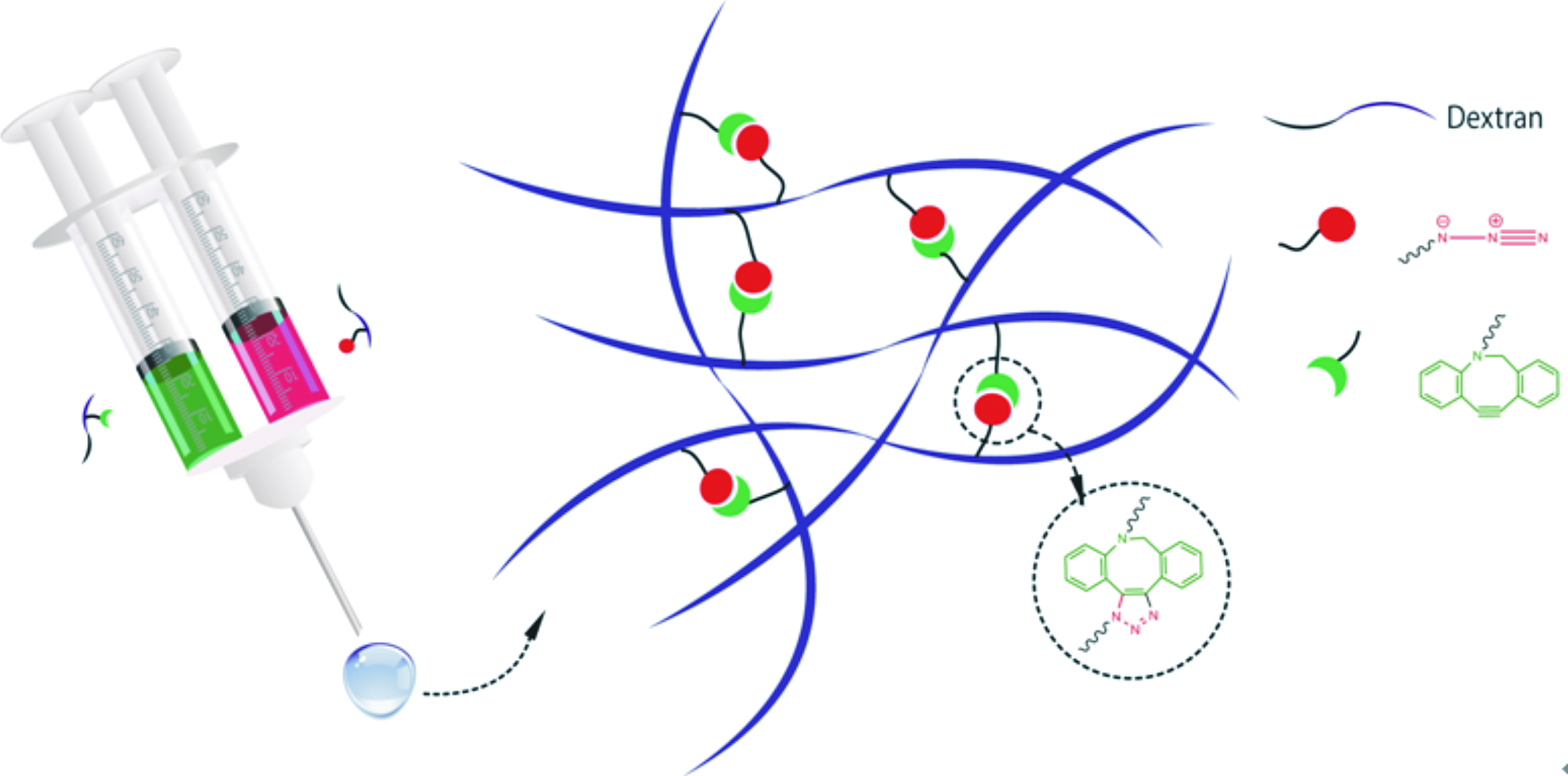
Schematic presentation of injectable dextran-based hydrogel which is in situ formed by strain-promoted azide–alkyne cycloaddition. Color images available online at

Immunohistostaining assay showing COLI
Bioorthogonal dextran hydrogels robustly support proliferation of rADSCs and chondrogenic differentiation in vitro and in vivo
Live-Dead staining assay showed that rADSCs encapsulated in dextran-AN hydrogel within 7 days displayed green fluorescence in most of the cells (Fig. 3a), exhibiting their survival status. Similar phenomenon was also observed for those stem cells in a nontoxic agarose gel (1%) as a positive control. The comparison result confirmed cytocompatibility of dextran-AN hydrogel against rADSCs. Further assays of DNA content of the cells revealed that dextran-AN hydrogel and agarose gel supported the culturing of rADSCs within 2 weeks without compromising their viability (Fig. 3b). Next, the contents of neocartilage extracellular matrices such as GAG and collagen were quantified as a function of culturing time (day 1, week 1, 2, and 3, respectively). It was found that, during 3 weeks, the amount of GAG and total collagen in the dextran-AN hydrogel group markedly increased (p < 0.05) from day 1 to week 1 and thereafter gradually augmented during week 2–3, although not statistically significant (Fig. 4a, b). Such similar tendency was also observed for those cells in the agarose gel as a control. Besides, compared to untreated rADSCs, differentiated rADSCs indicated markedly higher mRNA level of collagen type-II (COLII) and GAG, as typical indicator of elastic cartilage (Fig. 4c). Importantly, in these gels, the mRNA level of collagen type-I (COLI), an unwanted fibrocartilage indicator, was significantly downregulated. More impressively, higher COLII expression was detected in the dextran-AN hydrogel group compared to the agarose gel group. Hence, these outcomes confirm the effectiveness of dextran-AN hydrogel for in vitro culturing of rADSCs.
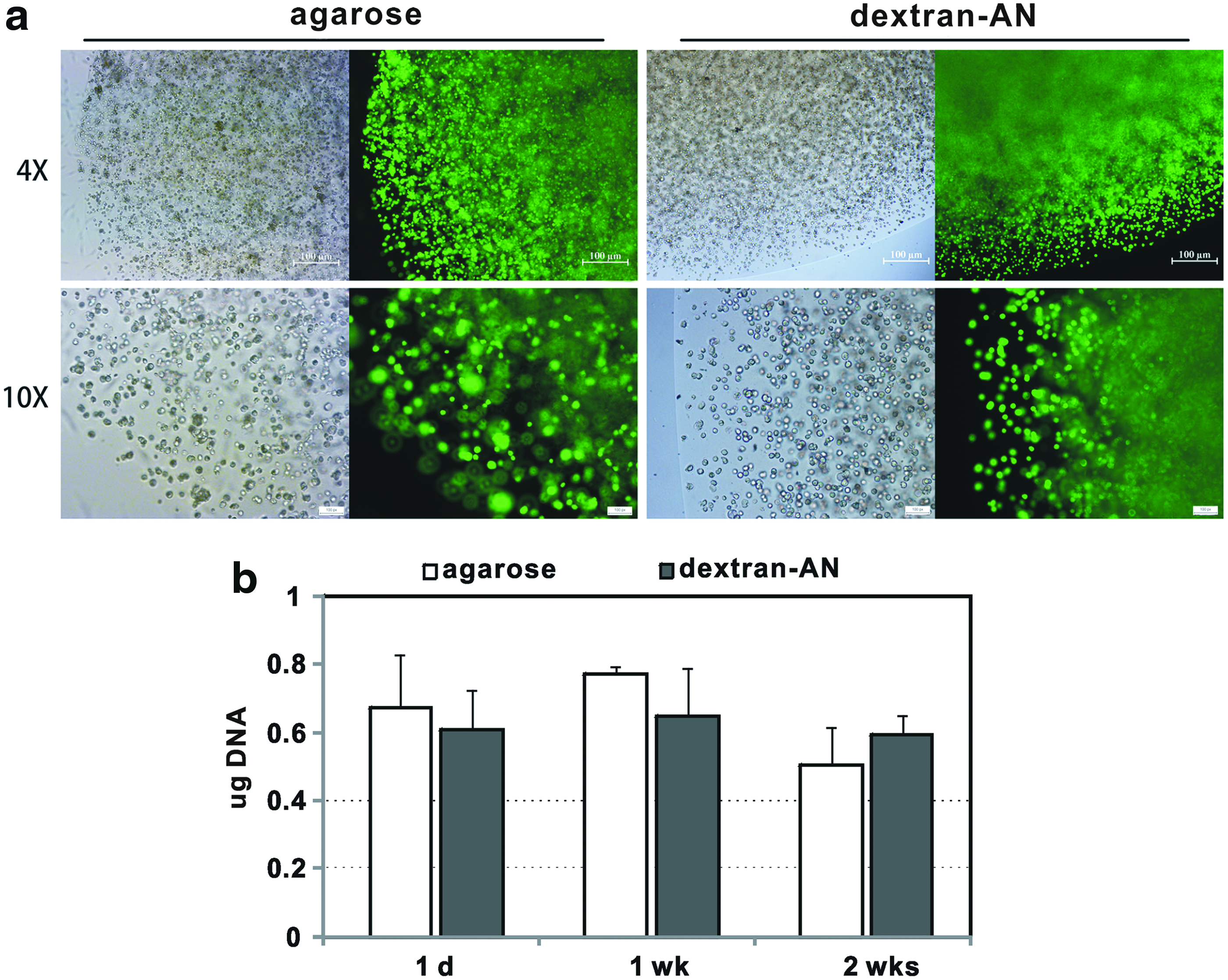
The dextran-AN hydrogel supports in vitro proliferation of rADSCs.
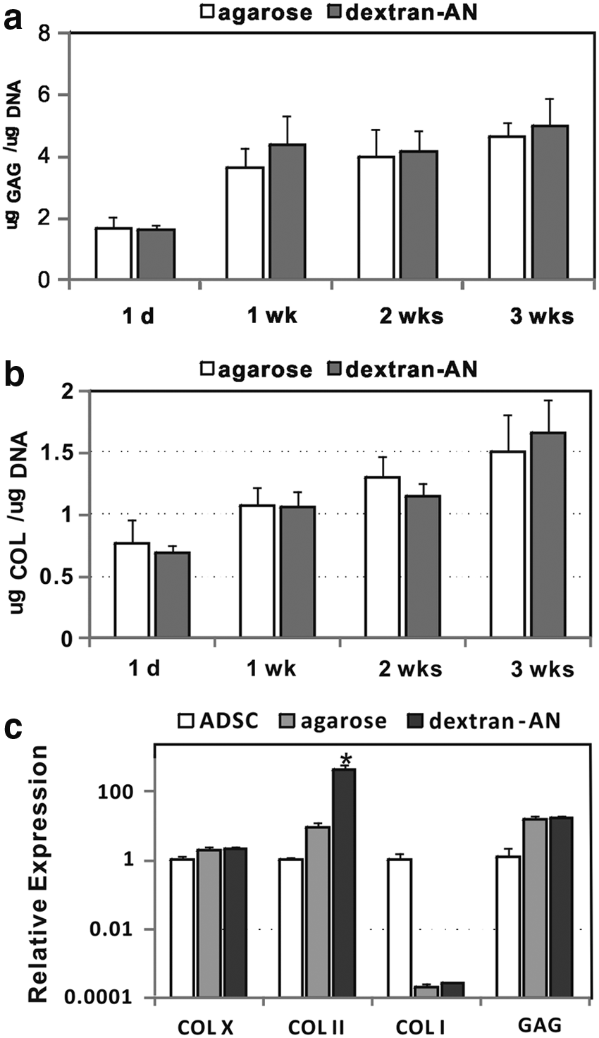
Extracellular matrix quantitative assays of in vitro chondrogenic rADSCs encapsulated in hydrogels.
Next, implantation surgery was conducted with dextran-AN hydrogel encapsulating differentiated rADSCs to fill a full-thickness cartilage defect (reached osteochondral layer) in a rabbit (Supplementary Fig. S1). Macrographic observation showed initial repair of the defect at week 2 for the dextran-AN hydrogel and agarose gel groups, as determined by H&E staining analysis (Supplementary Fig. S3). Furthermore, at week 6, dextran-AN hydrogel group exhibited adequate filling of newly formed cartilage tissue in the foci, and the repair status was better compared to agarose gel group and untreated rADSC (blank) group (Fig. 5a). At week 6, an integration of neotissue with native cartilage was nearly complete in the dextran-AN group. In addition, Masson and Safranin O/Fast Green staining were conducted, where the collagen was stained in blue by Masson assay and GAG in red by Safranin O but the collagen in green by Fast Green. 28 These assays revealed that, at week 6, more area was stained in blue and red in the dextran-AN group compared with the agarose positive control group (Fig. 5b), manifesting better regeneration recovery of the foci region by collagen and GAG in the hydrogel group. This point was further supported by the gross score (Supplementary Table S4) and histochemical staining score of these repaired cartilage tissues (Supplementary Table S5), in which the dextran-AN hydrogel group had markedly better grading compared to the agarose gel control and PBS blank groups. Furthermore, the surface of repaired cartilage foci appeared to be smoother in the dextran-AN group than the agarose control group, implying the elasticity of the regenerated cartilage in the foci. The agarose hydrogel, a physically crosslinked hydrogel, could be decomposed faster in vivo than the dextran-AN hydrogel. As such, the former might undergo adverse loss of ADSCs and differentiated chondrocytes, displaying relatively gross cartilage repaired surface. To further verify the production of newly formed cartilage, COLII immunochemical staining of neocartilage was conducted. Again, the dextran-AN hydrogel group afforded higher level of COLII deposition and more homogenous COLII distribution compared to the agarose control group (Supplementary Fig. S4).
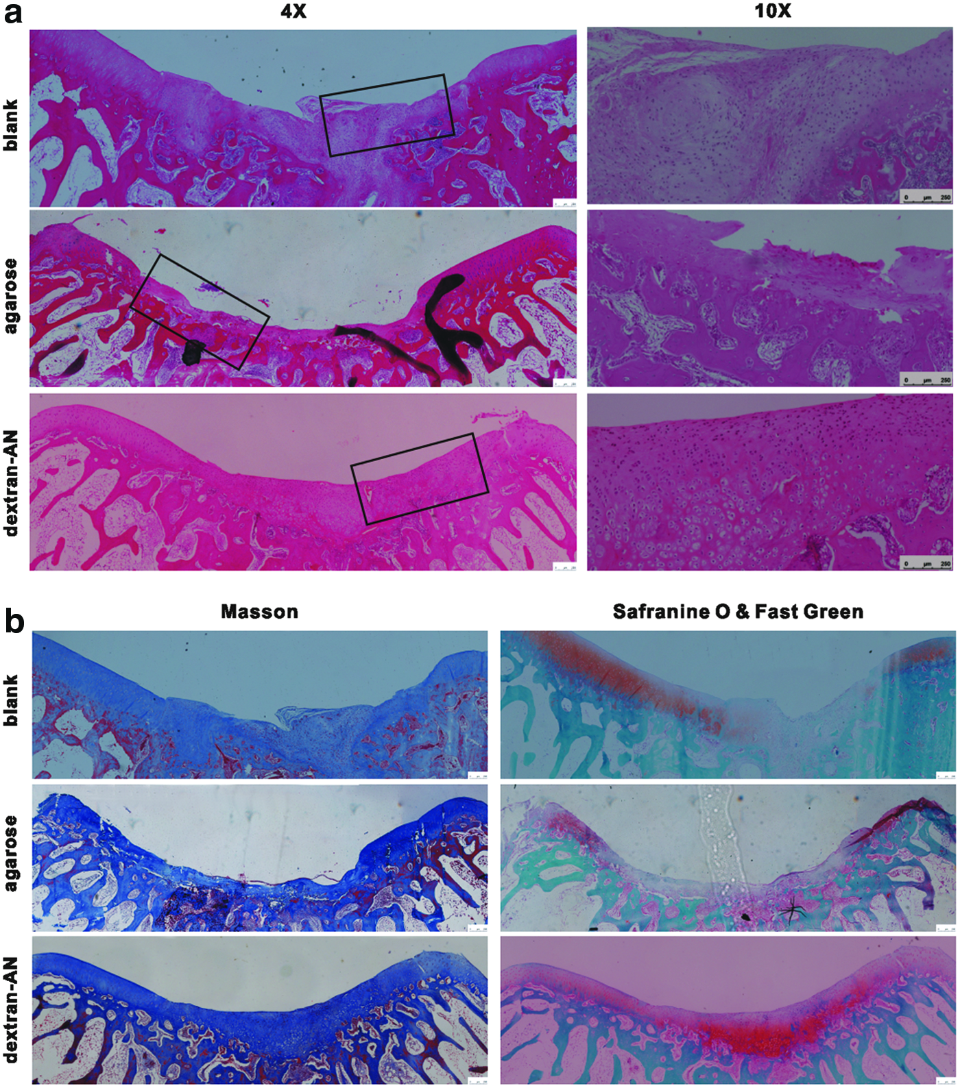
Histochemical staining showing repaired cartilage section at 6-week postimplantation.
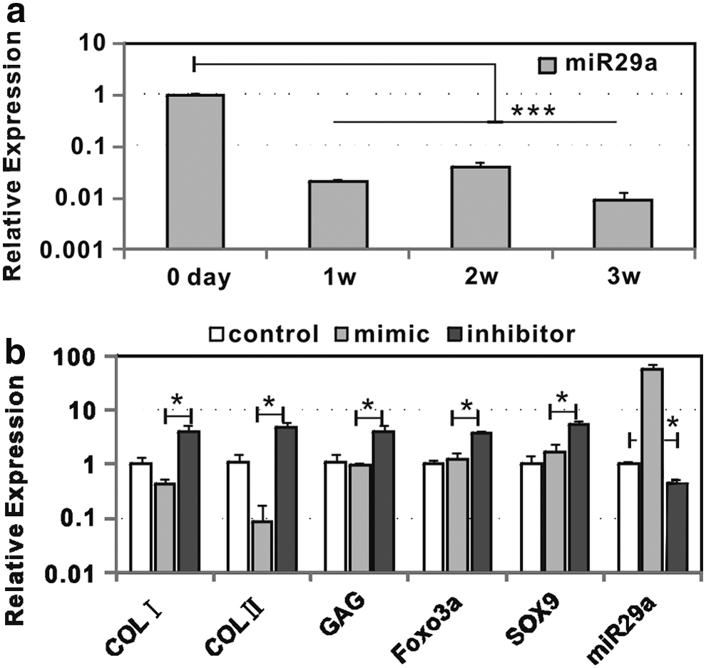
Bioorthogonal dextran hydrogels support in vitro and in vivo chondrogenesis of miR29a-downregulated hADSCs
Chondrogenic differentiation of hADSCs, in vitro seeded in dextran-AN hydrogel, caused significant downregulating of miR29A in the cells during 1–3 weeks of culturing (Fig. 6a). The transfection of hADSCs with miR29a mimic sequence and its reverse (inhibitor) sequence showed that miR29a-downregulating by the inhibitor sequence led to markedly increased mRNA expression level of those chondrogenesis-related biomarkers (COLI, COLII, ACAN, SOX9, and FOXO3a), which suggested that the miR29a downregulating was important to augment the chondrogenesis of hADSCs in dextran-AN hydrogel (Fig. 6b). Furthermore, subcutaneous implantation of miR29a-downregulated hADSCs/dextran-AN hydrogel construct in a balb/c nude mouse model was done. The miR29a-downregulated hADSC (inhibitor) group exerted markedly higher mRNA expression level of typical chondrogenesis-related biomarkers compared to miR29a-upregulated hADSC (mimic) control and untreated hADSC (blank) groups (Fig. 7), illustrating the effectiveness of miR29a-downregulated hADSCs in dextran-AN hydrogel for in vivo chondrogenesis. This point was also supported by histochemical analysis of extracellular matrices (Supplementary Fig. S5), where the inhibitor group exhibited higher content of GAG and collagen deposition in comparison with the mimic control and blank groups. In addition, immunohistostaining assay of COLII indicated higher COLII deposition level for the inhibitor group (Fig. 8). More impressively, the assay revealed that, compared to agarose gel as a positive control, the dextran-AN hydrogel led to higher COLII expression level (Fig. 8), which again supports its effectiveness of the hydrogel for in vivo chondrogenesis of hADSCs.
Chondrogenesis of hADSCs in vitro is downregulated by miR29a.
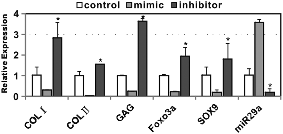
Downregulating miR29a enhances chondrogenic specific mRNA expression in vivo. The expression of chondrocyte-specific mRNA markers in cartilage tissue was detected 4 weeks after subcutaneous implantation of dextran-AN hydrogel encapsulating miR29a-upregulated hADSCs (mimic) or miR29a-downregulated hADSCs in a balb/c nude mouse. The expression of miR29A and other mRNA markers in untreated hADSCs as a control was set as 1.0. *p < 0.05: mimic versus inhibitor.
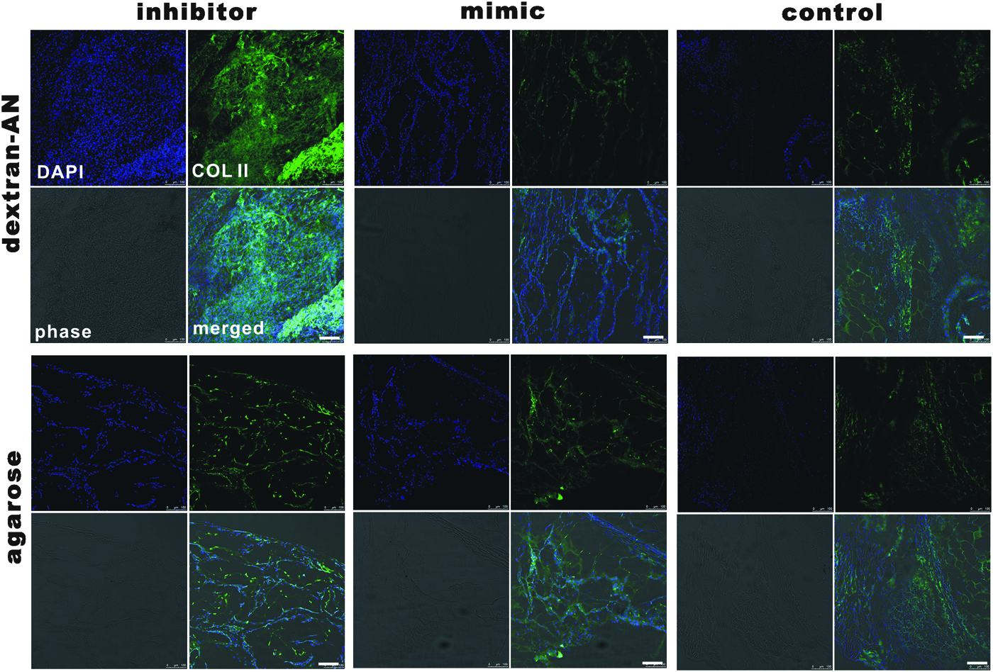
Immunohistostaining assay showing COLII protein expression in the cartilage 6 weeks after subcutaneous implantation of dextran-AN hydrogel containing miR29a-upregulated hADSCs (mimic) or miR29a-downregulated hADSCs (inhibitor). Untreated hADSCs were also used as a control group. (Scale bar 100 μm). The nuclei of chondrocytes were stained by DAPI. Color images available online at
Discussion
Biocompatible injectable hydrogels are effective to maintain phenotype of adult chondrocytes to support their chondrogenesis and are thus indispensable scaffolds for cartilage regeneration. However, the availability of idea injectable hydrogels enabling further clinical translations remains a big challenge. One major reason is that current chemically crosslinked hydrogel systems are not absolutely bioinert to surrounding tissue, due to the fact that those chemical reactions, occurred during in situ hydrogel formation, adversely compromise metabolic activity of cells or bioactivity of growth factors. This issue thus seriously impedes clinical translations of chemical injectable hydrogels, although these hydrogels are indeed suited scaffolds for chondrogenesis in vivo. To our knowledge, only photocrosslinked injectable hydrogels were evaluated in a pilot clinical study. 18 Notably, UV-initiating photocrosslinking reaction may cause undesirable side effect as a result of unconsumed initiators and vinyl groups, as well as the lack of sufficient research data on the biocompatibility of these reactive chemicals against normal tissue. From the chemical point of view, bioorthogonal reaction between bioinert azide and alkyne groups has no side effect on the cells and biomolecules, which is totally differed from traditional reactions such as free radical polymerization, Schiff-base formation, and Michael-type addition reaction. Besides, strain-promoted click chemistry (azide–alkyne cycloaddition) is catalyst free, making such reaction practical to fabricate chemically crosslinked injectable hydrogels.
Although a large number of injectable hydrogels reported was stated with a great promise for supporting the chondrogenesis of adult chondrocytes, only limited reports on in vivo chondrogenesis by chondrocytes had been appeared. 29 This situation is attributed to the reason that most of those chemically crosslinked injectable hydrogels have low manufacturing capacity, as a result of high produce price, and lack favorable features enabling in vivo tests and clinical translations. For example, natural collagen is an off-the-shelf biodegradable matrix widely studied for chondrocyte culturing. However, this biomaterial is very expensive and only available from natural tissue at low produce capacity. Although biodegradable chitosan is available at large scale and low price, it has to be chemically modified due to low solubility under physiological conditions. In this work, we utilize the dextran to serve as a polymeric network matrix of hydrogel, because dextran possesses a few merits, including low price, high produce capacity, high solubility, easy chemical modification of hydroxyl group, and superb compatibility against tissues under physiological condition. In addition, a few studies elicited the potential of dextran-based hydrogels for in vitro chondrogenesis.19,22,30 Thus, injectable dextran-based hydrogels, in situ crosslinked by strain-promoted azide–alkyne cycloaddition, hold a massive feasibility for chondrogenesis and injured cartilage repair. As we expected, the dextran (dextran-AN) hydrogel in combination with bioinert feature of the strain-promoted click chemistry against cartilage tissue displays superb performance to support in vivo chondrogenesis of rabbit chondrocytes within 8 weeks (Fig. 2 and Supplementary Fig S2).
Mesenchymal stem cells derived from adipose represent a promising resource for cartilage tissue engineering for minimal donor site morbidity. It was reported that one gram of adipose tissue contributed ∼70,000 hADSCs after expansion within 2 days. 31 Besides, hADSCs were found to elicit relatively low immunogenicity, as a result of their capability to escape from immune system recognition and inhibit host defence mechanisms. 32 Importantly, under specific culture conditions, hADSCs are capable of differentiation into chondrocyte-like cells in vitro and maintaining chondrogenic phenotype in vivo. 33 These researches manifest that hADSCs are easily accessible and productive seed cells in comparison with autologous adult chondrocytes. Hence, this work herein largely focuses on primary ADSCs for in vitro and in vivo differentiation and chondrogenesis in the dextran-AN hydrogel. In the test of the production rate of GAG and collagen, as two typical matrices of neocartilage, the dextran-AN hydrogel afforded ∼5.0 μg GAG and 1.5 ng collagen per μg DNA, respectively, 3 weeks after in vitro chondrogenesis (Fig. 4). In contrast, lower production rate of GAG was found for those previous systems such as poly(glycolic acid)/polycaprolactone system (∼0.15 μg/μg DNA), 34 poly(lactic-co-glycolic acid)/chitosan (∼0.14 μg/μg DNA), 35 and injectable dextran hydrogel crosslinked using an enzyme (1.8–2.8 μg/μg DNA). 30 Besides, parallel production rate of collagen was observed for the dextran-AN hydrogel and those hydrogel systems reported. These comparison data elicit that the dextran-AN hydrogel is more suitable for ADSC differentiation and chondrogenesis. The lower GAG production for those previous systems is largely attributed to their large pore size and high swelling ratio, which leads to lower level of deposition of extracellular matrix. In addition, these systems probably suffer from unfavorably compromised cell viability due to cytotoxic-free radical produced by UV-initiated photocrosslinking or enzyme-catalyzed crosslinking method. These reasons may rationally interpret that the dextran-AN hydrogel can produce perfect repairing (extracellular matrix deposition without visible foci) in a full-thickness cartilage defect model (Fig. 5). Impressively, in vivo chondrogenesis test reveals better grading in histological staining and gross score in the dextran-AN group compared to the agarose control group (Supplementary Tables S3 and S4). In addition, the grading performance was better in our experiment group compared to that in previous studies.32,34–36 By comparing matrix production and histological grading, the current work strongly suggests the merits and high potential of dextran-AN hydrogel for cartilage regeneration.
Although a few methods are effective for chondrogenic differentiation of primary ADSCs, these approaches normally depend on essential growth factors such as TGF-β3 used in our work and others like bone morphogenetic proteins. 37 Alternatively, functional miRNAs are capable of modulating the chondrogenesis of ADSCs. For example, typical up-expressed miRNAs include miR-23b and miR-153, and down-expressed miRNAs comprise miR-122a and miR-490.38,39 More recently, miR29a was identified to participate downregulation in the chondrogenesis of mesenchymal stem cells. Downregulating of miR29a in the stem cells could upregulate FOXO3a, causing enhanced chondrogenesis. 40 Hence, we utilize miR29A as an assistant to enhance chondrogenesis of hADSCs and neocartilage matrix production in vitro and in vivo (Figs. 7 and 8). Our work offers a practical protocol and useful data to support clinical translation of dextran-AN hydrogel into human cartilage repair.
Conclusion
We have demonstrated that an injectable bioorthogonal dextran-based hydrogel can be successfully implemented to repair a cartilage defect with tissue engineering approach. The biocompatible dextran hydrogel is robust to support in vitro chondrogenesis of adult chondrocytes/hADSCs and efficient in vivo regeneration of full-thickness cartilage defect. This study highlights massive potential of strain-promoted click chemistry for novel injectable hydrogels toward clinical translations of cartilage tissue engineering. Further work may focus on comparative study on cartilage repair performance of this hydrogel system and other scaffold systems used in clinical trials.
Footnotes
Acknowledgments
This work was supported by the National Basic Research Program of China (2014CB964600), Shanghai Municipal Natural Science Foundation (18ZR 1442100), and the National Natural Science Foundation of China (81572632).
Disclosure Statement
No competing financial interests exist.
References
Supplementary Material
Please find the following supplemental material available below.
For Open Access articles published under a Creative Commons License, all supplemental material carries the same license as the article it is associated with.
For non-Open Access articles published, all supplemental material carries a non-exclusive license, and permission requests for re-use of supplemental material or any part of supplemental material shall be sent directly to the copyright owner as specified in the copyright notice associated with the article.