
Abstract
The careful study of cell-based lung repair and regeneration ex vivo may one day provide us with the necessary tools to create a patient-derived alternative to cadaveric donor lungs for transplantation. Many parameters must be monitored and optimized to advance this aim. The use of rat lungs as a small-scale model for lung regeneration is an efficient way to develop the key improvements required for optimal tissue repair and regeneration. In this study, we report the use of a novel high-throughput, automated, multichannel lung bioreactor system, which allows for culture and analysis of rodent scale isolated lungs. In this model, five decellularized rat lungs can be repopulated with human primary endothelial and epithelial cells, cultured under varying perfusion and ventilation parameters in parallel, and analyzed for standardized endpoints. As a proof of principle, we report a multiphase organ culture protocol, which achieves consistent tissue regeneration across lungs at multiple points during culture, and further promotes overall tissue maturation through the application of lung ventilation.
Impact Statement
This work presents methods for ex vivo lung recellularization and biomimetic culture in a high-throughput and consistent manner. These methods allow for the testing of multiple variables, all of which are simultaneously controlled and monitored on a single fully automated pump system, and subsequent assessment of both epithelial and endothelial repair and tissue regeneration. This system provides a controlled environment for tissue repair, wherein key variables can be modified, monitored, reproduced, and optimized to advance the goal of ex vivo tissue regeneration based on native organ scaffolds.
Introduction
L
Bioengineered lung grafts made by combining native extracellular lung matrices with patient-derived regenerative cell populations present an exciting alternative to cadaveric donor lungs for transplantation. 2 To this end, perfusion-decellularized lung scaffolds can be readily prepared and combined with primary or pluripotent-derived cell types. 3 These recellularized constructs can then be matured outside the body in specialized bioreactors, with the aim of recreating higher-level organ function.4,5 Up to 75% reendothelialization has been achieved in decellularized rat lungs, 6 but full vascular barrier function and a completely regenerated epithelium capable of native-like gas exchange remain current challenges within the field.
While much progress has been made, many questions remain in defining the optimal recellularization methods, and ultimately translating this process to a clinically relevant scale. We have yet to define the ideal combination of regenerative cell types, and we continue to work toward defining specific ex vivo biomimetic culture conditions that adequately mature and prepare the lung constructs for transplantation.2,7
In the present report, we describe a novel, high-throughput, and automated multichannel bioreactor system, which supports the simultaneous recellularization and culture of up to five rat lungs in parallel under controlled conditions. Our system allows for the regulation of variable biomimetic conditions, including perfusion flow rates and pressure targets, as well as multiple modes of fluid and air ventilation during lung culture. Regenerating lung tissue can be monitored throughout culture, and assessed for numerous endpoints, including total cellularity, cell fate and phenotype, metabolic activity and changes in gene expression, protein production, glucose consumption, and lactate production. The true novelty of this system lies in its ability to consistently regulate and monitor flow, pressure, and ventilation parameters simultaneously for multiple lungs while reducing the potential error from combining separate systems for each lung and each parameter. We conclude that this system provides a valuable platform to study ex vivo lung repair and tissue regeneration in a more precise manner.
Materials and Methods
Study approval
All experiments were approved by the Massachusetts General Hospital Animal Utilization Protocol (#2014N000261).
Lung decellularization
Rat lungs were decellularized as previously described.3,8 Briefly, cadaveric rat lungs were explanted from male Sprague-Dawley rats (250–300 g, >8 weeks of age, Charles River Laboratories), fitted with pulmonary artery (PA) and pulmonary vein (PV) cannulas, and decellularized by perfusion of 0.1% sodium dodecyl sulfate solution through the PA at 30 cmH2O, followed by washing. Acellular lungs were then fitted with a straight tracheal cannula and mounted in a custom bioreactor with connections to the PA, PV, and trachea (Fig. 1A). Adequate decellularization of lung tissue using this method has been previously confirmed,3,8 and an additional rat lung was decellularized following the same protocol then fixed and stained as a negative control (Fig. 3B, C).
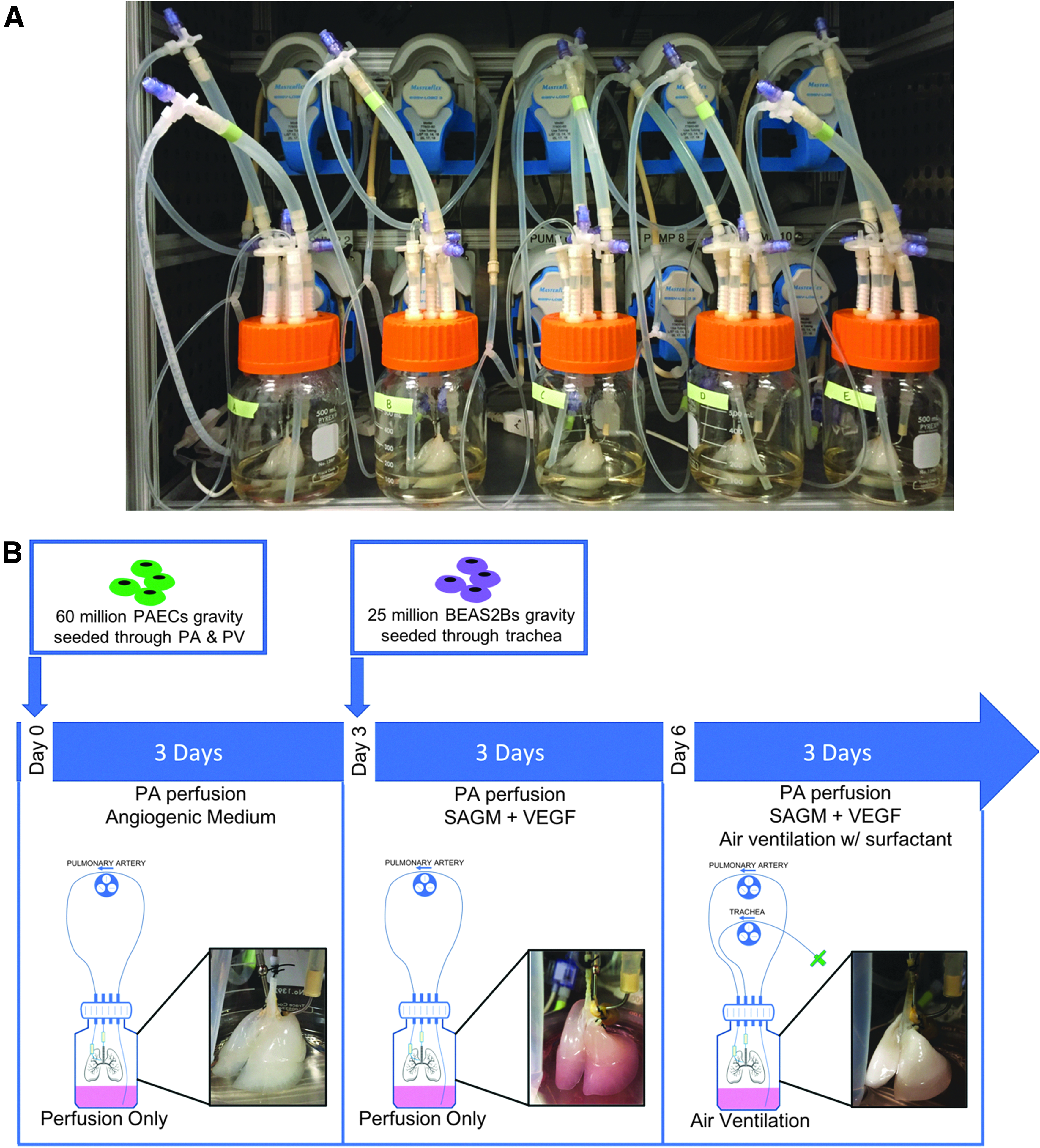
Overview of culture setup and parameters.
Endothelial cell priming, seeding, and culture
Bioreactors were prefilled with angiogenic media, to support endothelial proliferation, as prepared according to the previously published protocol, 6 and lung scaffolds were primed for 2 h by both PA and PV perfusion at 1 mL/min, in a 37°C incubator, where the remainder of the culture experiments were carried out. The multichannel pump system (IVIVA Medical) allowed for continuous monitoring of perfusion pressures and data logging.
Primary human PA endothelial cells (PAECs) (ATCC PCS-100-022) were expanded to ∼95% confluence in PromoCell endothelial cell growth medium 2 (C-22011), trypsinized, and counted. For each lung, 60 million PAECs were resuspended in 100 mL of angiogenic media. Lung perfusion was stopped and 30 million cells in 50 mL cell suspension were delivered to both the PA and PV lines by gravity at a height of 30 cm. Following seeding, each lung was given a nonperfusion static culture period of 2 h. Immediate initiation of vascular perfusion following seeding could interfere with cell attachment. During in vitro culture of PAECs, cells typically adhere to gelatin-coated plastic within 2 h of cell seeding, and previous literature has shown that 2 h of static culture is sufficient for cells to attach before resuming perfusion. 6
Following static culture, the reendothelialized lungs were perfused from both the PA and PV overnight at 1 mL/min. On the following day, PV perfusion was ceased and PV drainage allowed through the open cannula to the inside of the bioreactor. Perfusion was then increased to 4 mL/min to the PA only. On each day of culture, a 2 mL sample of medium was taken for glucose, lactate, pO2, and pH level testing.
Epithelial cell seeding and culture
On day 3 of culture, all angiogenic medium was removed from the bioreactor and tubing and replaced with 70 mL PromoCell small airway epithelial cell growth medium (SAECGM) (C-21070) supplemented with 40 ng/mL vascular endothelial growth factor (VEGF). Bioreactors were then returned to the incubator and PA perfusion continued for 2 h at 4 mL/min.
BEAS2B cells (ATCC CRL-9609), were expanded to ∼95% confluence, trypsinized, and counted. For each lung, 25 million epithelial cells were resuspended in 10 mL SAECGM + VEGF and delivered to airways by gravity seeding at a height of 10 cm. After delivery of the full cell suspension, 3 mL of medium was delivered to clear the tubing of remaining cells and additional medium was added to the bioreactor to bring the overall culture volume to 150 mL. After seeding, the bioreactors were returned to the incubator and given a 1.5-h static culture period for cell attachment, slightly shortened from the length of static culture after endothelial cell seeding because the perfusion does not directly pass through the airways and alveoli and thus is less likely to inhibit cell attachment. Then PA perfusion was reinitiated at 4 mL/min.
Air ventilation
On day 6, after medium was changed, 300 μL of porcine surfactant (Curosuf) was diluted in 10 mL medium and gently injected into the airway. An air-filled syringe was then used to gradually fill the airways with air and recruit the alveoli. Once the lungs were visibly fully recruited, ventilation was commenced using a roller pump generating a 3-s inhale followed by an 18-s passive exhale period, which was gradually reduced to 9 s (5 breaths/min). The tidal volume throughout ventilation was 0.5 mL. These ventilation parameters were selected through a series of recellularized lung culture experiments and designed to deliver a smaller tidal volume and longer inspiratory and expiratory duration relative to spontaneous breathing seen in similarly sized rats so as to be more protective to the regenerating tissue. 9
Data collection
On days 3, 6, and 9 of organ culture, multiple endpoints were analyzed. Tissue was collected at each time point to be fixed by 4% paraformaldehyde submersion for histology as well as stored in RNAlater solution for subsequent quantitative polymerase chain reaction (qPCR) gene expression analysis. To collect the tissue, individual lobes were sterilely tied off and excised. On day 3, the right cranial and right middle lobes were excised. On day 6, the accessory lobe of the right lung was excised. On day 9, the right caudal lobe was excised, after which the remaining left lobe was fixed by gravity perfusion/inflation of 4% paraformaldehyde in phosphate buffered saline (50 cmH2O) through the trachea cannula to give a more native histological picture of the lung in its inflated state.
Throughout culture, the bioreactor software was connected to sterile pressure sensors in line with PA perfusion and airway ventilation. PA and airway pressure data were logged at a rate of 1 data point per second for the entirety of culture, allowing for all pressure data to be analyzed upon the completion of culture.
Resazurin metabolic assay
On days 3, 6, and 9, a PrestoBlue resazurin metabolic assay was run to quantify the overall cellularity of the lungs. 10 A 0.05 mM Resazurin solution (80 mL) was circulated through the PA of each lung for 60 min at a constant flow of 4 mL/min. Samples of medium were taken after 1 h and fluorescence was quantified in triplicate at 560 and 590 nM.
Quantitative polymerase chain reaction
mRNA was isolated (Qiagen RNeasy Plus Kit) and transcribed to cDNA (Invitrogen SuperScript III). Gene expression was analyzed using TaqMan probes and the OneStep Plus system (Applied Biosystems). Each biological sample was analyzed in experimental replicate (n = 2 repeated wells of the qPCR reaction) and the Ct value of each replicate was averaged and handed as n = 1 unique biological sample. Expression for each sample was normalized to β-actin (ACTA1) gene expression (ΔCt) and relative to days 3 or 6 average expression levels, as noted (ΔΔCt), with fold change calculated by 2−ΔΔCt. 11 A total of n = 3 unique biological samples were analyzed in this way for each reported experiment.
Immunostaining
After deparaffinization and rehydration, 5 μm tissue sections were permeabilized with 0.1% Triton-X for intracellular antigens, when appropriate. Cells in culture were fixed with ice-cold methanol before staining. All samples were blocked with 1% bovine serum albumin for 30 min. The primary antibodies were (1:100): Cytokeratin 5 (ab52635; Abcam), CD31 (M0823; Dako), E-cadherin (ab76055; Abcam), VE-Cadherin (AF938-SP; R&D Systems), Collagen IV (ab19808; Abcam), Collagen IV (NBP-1-26549; Novus), and Ki67 (ab16667; Abcam). The secondary antibodies were (1:500): Alexa Fluor donkey anti-mouse, rabbit, or goat, conjugated to 488, 546, or 647 (Life Technologies). Samples were stained with 4′,6-diamidino-2-phenylindole (DAPI) to visualize the nucleus and imaged using a Nikon Ti-Eclipse microscope.
TUNEL staining
After deparaffinization and rehydration, 5 μm tissue sections were stained using the DeadEnd™ Fluorometric TUNEL System Kit (G3250; Promega) and its corresponding staining protocol. Upon completion of the staining, samples were stained with DAPI to visualize the nucleus and imaged using a Nikon Ti-Eclipse microscope.
Ki67 and TUNEL quantification
Three tissue sections, from each of the five lungs at both the days 6 and 9 time points, were stained to quantify the percent of all cells that were Ki67+ and TUNEL+. The aforementioned immunostaining technique was used on one set of slides to stain sections for Ki67 and collagen IV with a DAPI counterstain, and a second set of slides was stained according to the TUNEL staining protocol with the DAPI counterstain. A set exposure time was chosen for DAPI and CY5 (Ki67) for the immunostained slides, and as well for DAPI and GFP (TUNEL) for the TUNEL-stained slides. For both sets of slides, five unique 40 × images were taken of each section. For each section, total DAPI+ nuclei and total Ki67+ or TUNEL+ nuclei were manually counted and used to calculate the percent of total cell nuclei in each section, and overall for each lung at both time points that were Ki67+ and TUNEL+.
Statistical analysis
For all experiments, the n value stated represents an independent biological sample. Data were analyzed by Student's t-test or one-way ANOVA with a Tukey's multiple comparison post-test, as appropriate, using GraphPad Software. All statistical significances are reported accordingly. *p < 0.05, **p < 0.01, ***p < 0.001.
Results
Acellular rat lung scaffolds were recellularized in parallel, as described in the Materials and Methods section. A schematic of the bioreactor and recellularization strategy is presented in Figure 1A and B.
Throughout the culture period, vascular and airway resistance was continuously monitored and recorded for each individual lung (Fig. 2A, B). PA pressure was maintained at a consistent level across all five cultured lungs. On day 1, with the perfusion flow rate set at 1 mL/min through the PA and PV simultaneously, PA pressure remained at ∼5 mmHg in all five lungs. At the end of day 1, perfusion flow rate was increased to 4 mL/min through only the PA, and subsequently PA pressure gradually increased over the course of culture, peaking in the 15–20 mmHg range by the end of culture while the lungs were being ventilated (Fig. 2A).

Monitoring lung culture.
A cyclic rise and fall of airway pressure mirroring the 5 breaths/min ventilation setting was observed shortly after the initiation of ventilation on day 6, with pressure values ranging from 7 to 21 mmHg (Fig. 2B). Despite no further alterations to ventilation parameters after initiation of ventilation, airway pressure variance decreased over the final 3 days of culture, with pressure values for all lungs falling in the 10–20 mmHg range by day 9.
Daily culture medium samples were analyzed to monitor metabolic activity of the cells in each lung. Glucose consumption, measured as the decrease in culture medium glucose concentration in the 24 h following medium change, was significantly higher on day 7, 4 days after BEAS2B seeding and 24 h after ventilation initiation, than it was on days 1 or 4 (Fig. 2C,i). Lactate production, measured as the increase in culture medium lactate concentration in the 24 h following medium change, was also significantly higher on day 7 as compared with day 4 (Fig. 2C,ii).
The pO2 of culture medium was consistently maintained across the five cultured lungs throughout culture. Spikes and gradual decreases in pO2 were observed following the days 3 and 6 medium changes (Fig. 2C,iii). The pH of the culture medium displayed a consistent downward trend during culture in all five lungs (Fig. 2C,iv).
Analysis of metabolically active cells by PrestoBlue showed that the overall cellularity of each lung was relatively consistent at each of the three time points, with coefficients of variation 14.56% on day 3, 11.72% on day 6, and 16.31% on day 9 between the five lung cultures (Fig. 3A). Hematoxylin and Eosin staining confirmed this finding with large-scale stitched images demonstrating consistent cell coverage across five lungs, and a decrease in acellular areas of tissue between days 6 and 9, suggesting continued cell migration and proliferation during lung culture (Fig. 3C).
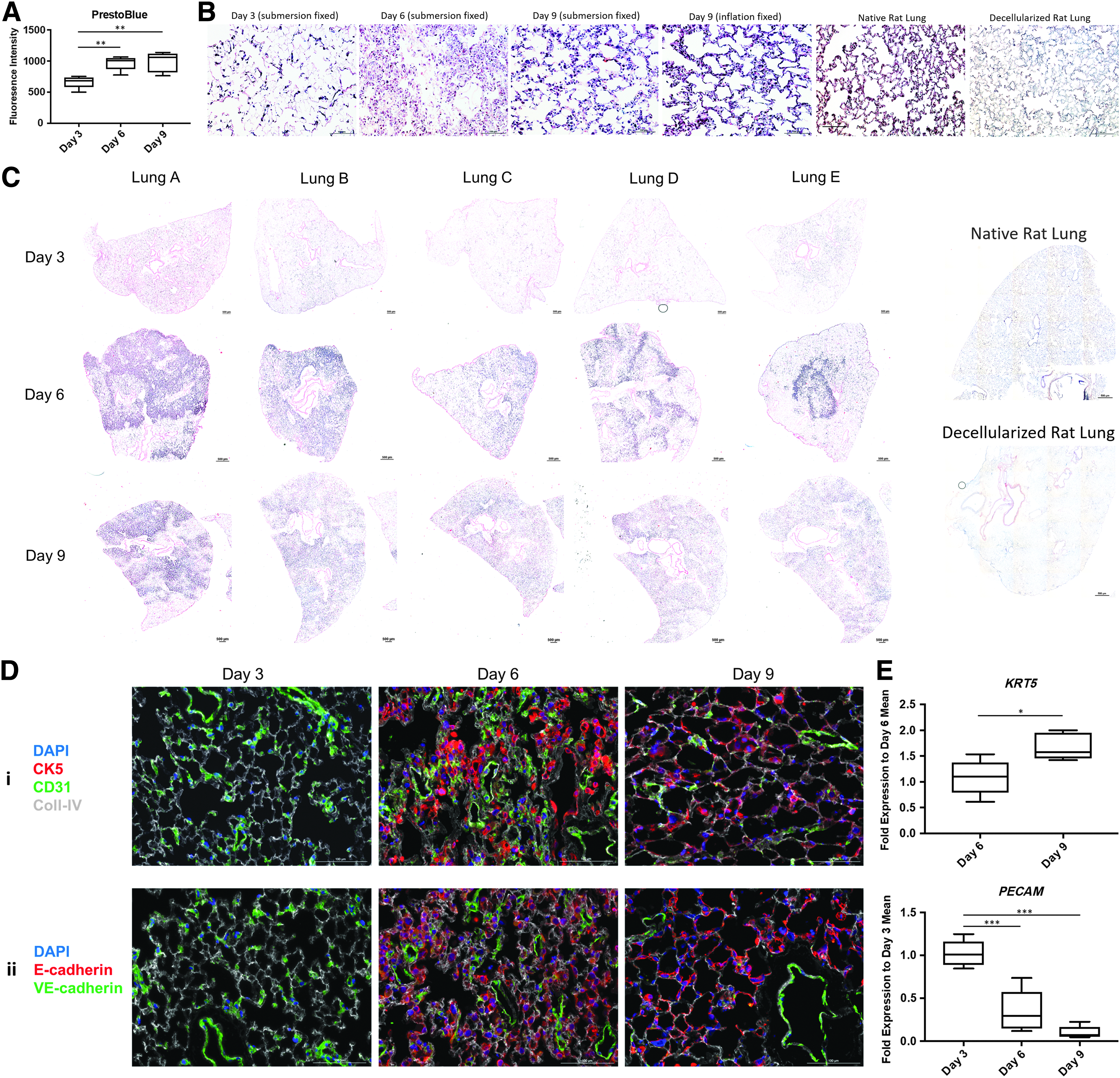
Analysis of cell coverage, tissue morphology, and endothelial/epithelial markers.
PrestoBlue assays across each time point revealed a significant increase in total cellularity of 0.45 ± 0.12-fold from days 3 to 6 and 0.49 ± 0.12-fold from days 3 to 9, but no significant change between days 6 and 9 (Fig. 3A). It should be noted that the day 3 PrestoBlue data also factored in cellularity of the right accessory lobe, which was excised before the days 6 and 9 data points. In addition to enhanced scaffold coverage and total cellularity, higher magnification images revealed a transition of distal lung tissue to a more native morphology after ventilation at day 9 (Fig. 3B). Immunostaining of tissue at each time point further demonstrated a consistent relative level of CD31+ PAECs in the days 3 and 6 time points, with a slight decrease on day 9 (Fig. 3D). Levels of Cytokeratin 5+ BEAS2B epithelial cells were consistent between the days 6 and 9 time points across lungs (Fig. 3D).
Gene expression levels were also analyzed using quantitative PCR. Cytokeratin 5 and CD31, epithelial and endothelial markers respectively, were analyzed against the housekeeping gene Beta Actin. Cytokeratin 5 expression levels, normalized to the day 6 average, showed a significant increase between days 6 and 9. CD31 expression levels, normalized to the day 3 average, showed significant decrease from day 3 to both days 6 and 9 (Fig. 3E).
Additional staining was performed for E-cadherin and VE-cadherin, and cell adhesion molecules in epithelial and endothelial cells, respectively. A similar pattern was found for the cell junctional markers, with a slight decrease in VE-cadherin from days 3 and 6 to day 9 and relative consistency in E-cadherin expression on days 6 and 9 (Fig. 3D).
Immunostaining for Ki67 revealed widespread and consistent expression of nuclear Ki67 on both days 6 and 9 of lung culture (Fig. 4A). Quantification of the percent Ki67-positive cells, using n = 3 sections of each lung at each time point, found a trend toward increased percent Ki67-positive cells on day 9 relative to day 6. Additional qPCR gene expression analysis for Ki67 found a significant increase in Ki67 expression on day 9 (Fig. 4C).

Analysis of cell proliferation and apoptosis.
TUNEL staining for apoptotic cells revealed a low level of TUNEL-positive cells on days 6 and 9 (Fig. 4D). The percentage of TUNEL-positive cells (n = 3 sections analyzed per time point) was not significantly different between days 6 and 9 (Fig. 4E).
Discussion
Our results detail the use of an automated multichannel bioreactor system to achieve reproducible tissue regeneration in up to five rat lungs cultured in parallel with a multiphase biomimetic culture method. Building upon previous research, we developed a 9-day protocol that incorporates three specific culture phases and allows for the analysis of multiple endpoints to assess cell viability, fate, and tissue regeneration. This system allows for each lung to receive controlled biomimetic conditions throughout culture, which results in consistent tissue recellularization across multiple lungs.
Tissue regeneration is a multifactorial process that involves cell–cell and cell–matrix interactions, as well as biochemical and biomechanical cues. 12 The regulation of these specific parameters to recreate the reparative tissue niche represents important factors in ex vivo organ regeneration. To this point, recellularization of decellularized lung scaffolds using standard vascular perfusion as well as certain methods of air and fluid ventilation has achieved substantial yet incomplete tissue regeneration.2,6 Our system allows for control of this critical microenvironment by specifically defining the culture conditions, including vascular perfusion and airway ventilation, which provide important signals to the regenerating tissue.13,14 By testing and then combining targeted culture parameters, we aim to drive the key responses that govern cellular function and tissue maturation within our ex vivo system and improve overall tissue regeneration in recellularized lungs.15–18
We describe a biomimetic culture method that transitions from a low vascular flow rate (1 mL/min) by perfusion through both the PA and PV to a higher flow (4 mL/min) through only the PA. Air traps are incorporated into each individual bioreactor's tubing line, but must currently be manually deaired on a daily basis to avoid air perfusion into the vasculature. To support ventilation, a single dose of supplemental porcine surfactant 19 can be delivered to the airways and a controlled respiratory rate (5 bpm) with low tidal volume (0.5 mL) applied. These parameters aim to recapitulate the biophysical environment of the native organ in vivo, but in a protective fashion so as not to damage the lung during regeneration. 9 Roller pump-based ventilation in this system can be controlled by both volume and pressure and works similarly to standard ventilators, but should also be monitored on a daily basis as recellularized lungs occasionally require extra recruitment breaths. Our system provides continuous pressure monitoring, which demonstrated consistent vascular perfusion and airway pressures across five parallel lungs. We aim to further optimize these conditions in future experiments and utilize the high-throughput design of our system to define the methods for specific, reproducible tissue regeneration.
We present several useful evaluation methods to analyze the regenerating tissue during ex vivo culture. Altogether, these endpoints demonstrate that both airway and vascular recellularization can be achieved with low lung-to-lung variability. Several of these analyses allow for repeated noninvasive testing and monitoring of the lungs during culture, which is an important consideration for clinically scale, patient-specific organ regeneration. 20 The molecular concentration of glucose and lactate, as measured by iSTAT, can be used to estimate the kinetics of cell growth in culture, 21 while the resazurin metabolic assay provides a correlate metric to further detail the overall cell number during culture. 10 These methods allow for real-time adjustments to the organ culture, without the need to terminate the experiment for analysis.
Additional histological and gene expression analysis of the tissue can be used to further validate the lung tissue before further functional testing and transplantation. Functional testing of regenerated lungs is an important endpoint to examine before transplantation. Vascular barrier function is crucial for in vivo function, and this can be determined by performing a dextran perfusion and bronchioalveolar lavage assay. 6 Additionally, quantification of gas exchange by comparing O2 and CO2 levels at the arterial and venous sides of the lung is a useful measurement to assess higher-level organ function. While metabolic assays are very useful as a correlate for total cell number and activity, confirmation of full scaffold coverage is still most accurately achieved by full organ histological analysis.
With the automated nature of this system, experiments can be easily scaled to facilitate the culture of 20 or more cultured lungs in parallel. This provides the power to test multiple variables in a reproducible manner. We have described methods to aid in the analysis of the lung within our system and have proven the ability to maintain consistency of these analytics. To increase cellularity and functional tissue regeneration, parameters, including cell source, culture medium components, extracellular matrix modification, and targeted biomechanical culture conditions, must be individually optimized. This system will now be integral to the further testing and optimization of isolated lung recellularization parameters such as these, with the ultimate aim to upscale these methods for clinical-scale lung regeneration.
Footnotes
Acknowledgments
The authors would like to thank Charles Klassen for help with building the multichannel bioreactor system and the Massachusetts General Hospital (MGH) Center for Skeletal Research Core (NIH P30AR066261) for histological processing.
Disclosure Statement
No competing financial interests exist. H.C.O. is founder and stockholder of IVIVA Medical, Inc.; this relationship did not affect the content or conclusions reported in this article.