
Abstract
Magnetic delivery system of mesenchymal stem cells (MSCs) has been developed for cartilage repair. It provides an effective and minimally invasive method for MSC transplantation. In this study, we evaluated the safety and quality of magnetically labeled human MSCs for practical applications. The safety of magnetically labeled MSCs was assessed using karyotyping, colony-forming assay using soft agar, and cell proliferation in a long-term culture. Magnetic labeling did not affect the karyotype, and MSCs retained their ability to grow and proliferate in an anchorage-independent manner even after exposure to magnetic force. The quality of the magnetically labeled MSCs was assessed by chondrocyte differentiation and reactivity toward magnetic forces. Magnetic labeling inhibited the chondrogenic differentiation of MSCs at higher densities of magnetic particles. MSCs labeled with ferucarbotran nanoparticles were retained by magnetic forces in a dose-dependent manner. The magnetization of MSCs with an appropriate density of magnetic particles maintained both qualities in MSCs. However, the uptake quantity of iron into MSCs varied across donors, even for the same density of magnetic particles. Therefore, the appropriate density of magnetic particles for use in MSC delivery for cartilage repair should be examined for every donor before treatment.
Impact Statement
This study is very important for a preclinical assessment of the safety and quality of magnetically labeled mesenchymal stem cells (MSCs) for use in cartilage repair. The findings of this study show that magnetic labeling with an appropriate density of magnetic particles has no harmful effects on the safety and quality of MSCs.
Introduction
Mesenchymal stem cells (MSCs) have been widely used in regenerative medicine from animal models to clinical trials. A particularly useful application of MSCs has been cartilage repair, as MSCs possess a chondrocyte differentiation capacity.1,2 A magnetic delivery system for MSCs, termed magnetic targeting, has been previously developed for the treatment of articular cartilage defects. 3 In this cell delivery system, superparamagnetic iron oxide nanoparticles (ferucarbotran) are incorporated into the cytoplasm of MSCs for magnetic cell labeling. Magnetically labeled MSCs are then injected into a joint, where they can be controlled and accumulated in a cartilage lesion by the use of magnetic forces. The efficacy and safety of this treatment for articular cartilage repair have been reported in a miniature swine model as well as a clinical trial.4,5 The safety and quality assessments of bone marrow MSCs for articular cartilage repair have also been reported.6,7 However, for magnetically labeled human MSCs such an assessment has never been reported. Therefore, a safety and quality evaluation of magnetically labeled human MSCs is highly warranted before realizing the practical application of magnetic targeting. The purpose of this study is to clarify the effect of magnetic labeling on the safety and quality of human bone marrow MSCs, for use in cartilage regeneration therapies.
Materials and Methods
Human bone marrow MSCs
In this study, commercially available human bone marrow MSCs were purchased (PT-2501; Lonza, Walkersville, MD). MSCs from eight donors were used in this study (Lot number: 1F4287 from a 22-year-old male, 2F3478 from a 43-year-old male, 296578 from a 45-year-old male, 310956 from a 24-year-old female, 318006 from a 27-year-old male, 326162 from a 38-year-old male, 429365 from a 30-year-old male, and 451491 from a 25-year-old male). All these MSCs passed the quality inspection conducted by Lonza company using cell viability (>75%), adipogenic and osteogenic differentiation ability (Oil Red O Staining and Calcium Deposition Staining), and flow cytometric analysis of cell surface markers (>90% were positive for CD29, CD44, CD105, and CD166, and <10% were positive for CD14, CD34, and CD45). Six of these lots (Lot numbers: 1F4287, 2F3478, 296578, 310956, 318006, 326162) were used for safety assessments, including karyotype analysis, proliferation capacity in long-term culture, colony-forming assay in soft agar, and quantification of magnetic labeling. On the contrary, three of these lots (1F4287, 429365, and 451491) were used for quality assessments, including viability, content of iron, the reactivity for magnetic attractive force, and chondrogenic differentiation capacity of MSCs.
Culture of MSCs
Human bone marrow MSCs at passage 2 (P2) were centrifuged at 1200 rpm for 5 min and resuspended in culture medium, containing Dulbecco's modified Eagle's medium (DMEM; Thermo Fisher Scientific, Waltham, MA), 15% fetal bovine serum (FBS; Sigma-Aldrich, St. Louis, MO), 20 mmol/mL 4-(2-hydroxyethyl)-1-piperazineethanesulfonic acid (HEPES; Thermo Fisher Scientific), 50 μg/mL gentamycin (Gentacin®; MSD, Kenilworth, NJ), and 0.25 μg/mL amphotericin (Fungizon®; Bristol-Myers Squibb, New York, NY). The MSCs were seeded at a density of 3500–5000 cells/cm2 onto 10-cm culture dishes and cultured at 37°C with 5% CO2. The culture medium was changed every 4 days. On reaching subconfluence, the cells were harvested with trypsin (TrypLE™ select; Thermo Fisher Scientific) and reseeded. The proliferation capacity of MSCs was examined by long-term culture. The MSCs were cultured until their number decreased for two successive passages during long-term culture.
Magnetic labeling of MSCs
On reaching 70% confluence, the MSCs were magnetically labeled with ferucarbotran (Rizovist®; FUJIFILM RI Pharma, Tokyo, Japan). The MSCs were incubated in culture medium containing ferucarbotran at a concentration of 97.6 μg iron (Fe)/mL for 12 h in a standard method (Fe × 1, 12 h). As a severe condition, MSCs were magnetically labeled under two types of conditions, including 195 μg Fe/mL labeling medium for 12 h (Fe × 2, 12 h) and 97.6 μg Fe/mL labeling medium for 36 h (Fe × 1, 36 h) for long-term culture. For the quality assessment of MSCs, ferucarbotran was added to the MSC culture at P3. The MSCs were incubated in the culture medium containing ferucarbotran at six different concentrations for 12 h. The concentration of 97.6 μg Fe/mL was defined as the standard dose (standard group), whereas the other concentrations were defined as follows: control group (0 μg Fe/mL), one-quarter group (24.4 μg Fe/mL), one-half group (48.8 μg Fe/mL), three-quarters group (73.2 μg Fe/mL), and double-dose group (195 μg Fe/mL). After incubation for magnetic labeling, all the cells were washed thrice with sterile phosphate-buffered saline (PBS) and used for assays.
Exposure of MSCs to magnetic field
A portion of the standard-dose MSCs were exposed to a magnetic field for the safety assessments. A superconducting magnetic device was used for this purpose 4 (Fig. 1A). Magnetically labeled MSCs were suspended in 3 mL of culture medium in a 50-mL polypropylene tube. The tube was placed at the center of the magnetic field generated from the device for 10 min. The exposure to the labeled MSCs as 0.55–5.48 tesla (T), considering the size of the tube and magnetic flux distribution (Fig. 1B). The labeled MSCs were aggregated in the tube because of the magnetic force (Fig. 1C). They were resuspended by gentle pipetting and used for safety assessments, including karyotype analysis, soft agar assay, and long-term culture.

Karyotype analysis
The karyotypes of the labeled MSCs were analyzed by the G-banding technique. For each sample, 20 or 50 metaphase cells were analyzed. The chromosomes were analyzed by visual inspection. In the assessment before the exposure to the magnetic field, the MSCs were labeled at P2 and at P5 and analyzed at P5 (n = 20 cells). In the assessment after the exposure to the magnetic field, the MSCs were labeled at P5, and analyzed at P5 and P10 (n = 50 cells).
Culture in soft agar
Before and after the exposure to the magnetic field, 1 × 10⁴ MSCs (labeled at P5 and analyzed at P5) were mixed with the culture medium containing 0.33% agar and overlaid on 0.5% agar in a 60-mm culture dish. The cells were cultured for 21 days. HeLa-S3 cells and MRC-5 cells were used as positive and negative control, respectively. The viability of the cells was confirmed by observing their ability to form a colony within 14 days. The assay was performed in triplicate.
Quantification of magnetic labeling
Quantification of the magnetic labeling of MSCs was performed on cytospin slides. The cell suspension was transferred to disposable cytofunnels with attached cytospin slides and spun at 1000 rpm for 5 min in a Cytospin4 Cytocentrifuge (Thermo Fisher Scientific). The slides were then fixed in 4% paraformaldehyde for 20 min and washed with phosphate buffer. The cells on the slides were stained with Berlin blue (FUJIFILM Wako Pure Chemical Corporation, Osaka, Japan) and counterstained with Kernechtrot (Muto Pure Chemicals, Tokyo, Japan). The labeling index was calculated by dividing the total blue intensity in the cell area in 10 pictures by the cumulative cell number. The blue intensity in pictures was estimated by computerized measurement (WinROOF, version 7.0; Mitani Co., Tokyo, Japan). The labeling index of each passage was normalized with the values obtained immediately after magnetic labeling and displayed as a relative value.
Cell viability
After the magnetic labeling, MSCs at P4 were collected after trypsinization and suspended in 1 mL of PBS. A 10 μL volume of this solution was mixed with 0.4% (w/v) Trypan blue solution (FUJIFILM Wako Pure Chemical Corporation), and the viability of MSCs labeled with different magnetic concentrations was evaluated using cell counter (TC10™ Automated Cell Counter; BIO-Rad Laboratories, CA).
Iron content in MSCs
Lysates were made from magnetically labeled MSCs from three different donors at six different concentrations of ferucarbotran for 12 h. The iron content of the lysate was measured using Metallo Assay kit (Metallo Assay; Metallogenics, Chiba, Japan). The MSCs in each experimental group were first counted, after which, they were homogenized in cell lysate buffer using ultrasonic sonicator. The crude lysate thus obtained was then mixed with HCl (0.01 M final concentration) and incubated at 20°C for 30 min. The lysate was then centrifuged at 4°C for 15 min, and the supernatant was quantified according to the manufacturer's instructions. The iron content was expressed as pg/cell.
Assessment of chondrogenic differentiation capacity
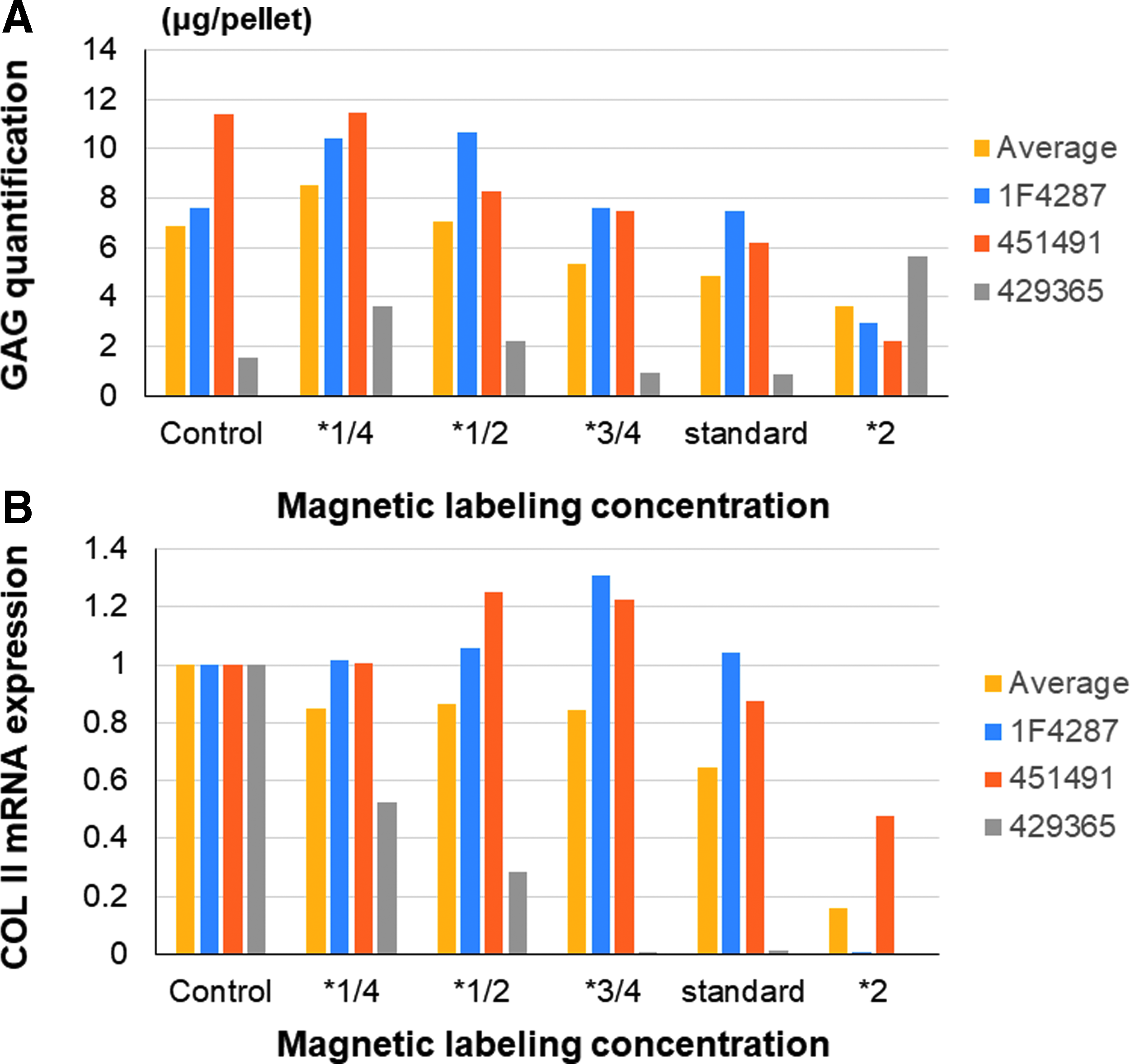
Chondrogenic differentiation ability of the MSCs from each donor was evaluated using pellet culture, according to Sekiya's method.8–10 About 2.5 × 10⁵ of MSCs at P4 was centrifuged at 450 g for 10 min using 15-mL polyethylene terephthalate tube. The pellet was cultured at 37°C with 5% CO2 in 500 μL of chondrogenic medium containing 500 ng/mL bone morphogenetic protein (BMP)-6 (R&D Systems, Minneapolis, MN) in addition to high-glucose DMEM supplemented with 10 ng/mL TGF-β3 (R&D Systems), 10−7 M dexamethasone, 50 μg/mL ascorbate-2-phosphate, 40 μg/mL proline, 100 μg/mL pyruvate (Sigma-Aldrich), and 50 mg/mL ITS+ Premix (6.25 μg/mL insulin, 6.25 μg/mL transferrin, 6.25 ng/mL selenous acid, 1.25 mg/mL bovine serum albumin, and 5.35 mg/mL linoleic acid; Becton Dickinson, Franklin Lakes, NJ). The medium was replaced every 3 to 4 days for 21 days. For each concentration of the m-MSCs (magnetic MSCs), the long diameter of the spheroids was measured. For histological evaluation under microscopy, the pellets were embedded in paraffin, cut into 5-μm sections, and stained with 0.05% toluidine blue solution and safranin-O/Fast green.
For the glycosaminoglycan (GAG) quantification, the pellets were digested in 3000 U/mL collagenase (Merck, Darmstadt, Germany) and 0.25% trypsin (Thermo Fisher Scientific). After 30 min of digestion, the homogenate was centrifuged at 12,000 rpm for 10 min. The GAG content of the supernatant was measured using a Blyscan Glycosaminoglycan Assay kit (Funakoshi, Tokyo, Japan) according to the manufacturer's instructions.
In addition, mRNA expression levels of collagen type II (COL II, COL2A1) were evaluated. Total RNA was isolated from pellets using a Qiagen RNeasy Micro Kit (Qiagen, Valencia, CA). cDNA was synthesized from the RNA using Super Script VILO Master Mix (Thermo Fisher Scientific). As a control, total RNA was isolated from normal knee cartilage dissected from skeletally matured cadaveric donors (Articular Engineering, Northbrook, IL). Quantitative polymerase chain reaction was performed using Power SYBR Green Master Mix (Thermo Fisher Scientific). cDNA samples were analyzed for both COL2A1 and the reference gene (glyceraldehyde-3-phosphate dehydrogenase [GAPDH]). The assays were performed according to the manufacturer's instructions. The mRNA expression of COL2A1 was normalized to that of GAPDH.
Reaction of MSCs to magnetic attractive force
Reactivity of MSCs toward magnetic attractive force under the six treatments (0, 24.4, 48.8, 73.2, 97.6, and 195 μg Fe/mL) was measured using a method described in the MACS manual separator (Miltenyi Biotec, Cologne, Germany). 11 Microfluidics chip (Sumitomo Bakelite Co, Tokyo, Japan) and four different magnetic flux densities of the neodymium magnet (20, 100, 250, and 500 mT) were used. The neodymium magnet was placed at the center of the column. The cell density was maintained at 3.0 × 10⁵ cells/mL, and 2 μL of cell solution was injected into one hole. The flow of the cell solution was measured under the influence of magnetic force, as well as without the magnetic force (Fig. 2).
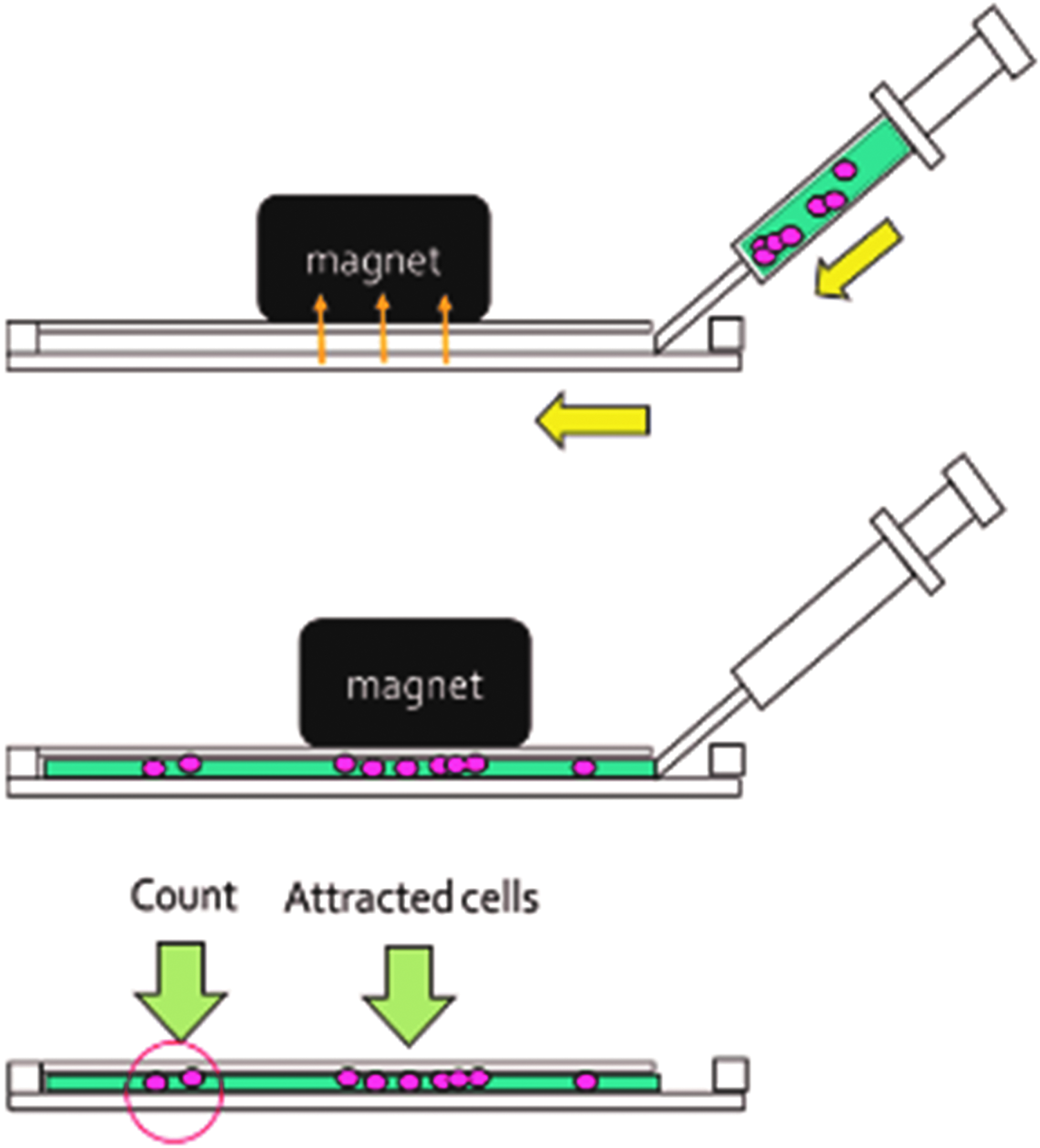
The assessment of reactivity of magnetically labeled MSCs for magnetic attractive force. The eternal magnet is put on the center of the column in the microfluidics chip. The cell solution is injected into one hole. The MSCs passed under the magnet are counted.
Statistical analysis
All the results were compared between the examination groups and the donor groups. Statistical analysis was performed using software Statcel4; a statistical program for performing statistics in MS Excel (Statcel4: Nebula Company, Tokyo, Japan). The results are represented as mean value ± standard deviation. Multiple comparisons were performed for all experiments using a single-factor ANOVA and Bartlett's test. When the p-value was significant, the Turkey–Kramer method was used to find the pairwise differences among groups.
Results
Karyotype of MSCs
Three lots of human MSCs were used for karyotype analysis (1F4287, 2F3478, and 296578). The karyotype was analyzed in P2 and P5 MSCs without labeling, P5 MSCs labeled at P2, and P5 MSCs labeled at P5. No abnormalities were seen in 2F3478 and 296578 in any of the conditions. Although P2 and P5 MSCs without labeling and P5 MSCs labeled at P2 were normal in 1F4287, 2 of the 20 P5 MSCs labeled at P5 in 1F4287 had an abnormality, balanced reciprocal translocation between chromosome 13 and chromosome 10 (Fig. 3A–C). To verify whether this abnormality in 1F4287 was caused by labeling, the karyotype of MSCs in 1F4287 was reanalyzed. P5 MSCs labeled at P5 (n = 50) and P10 MSCs labeled at P5 (n = 50) were analyzed, and no abnormalities were observed (Fig. 3D, E).
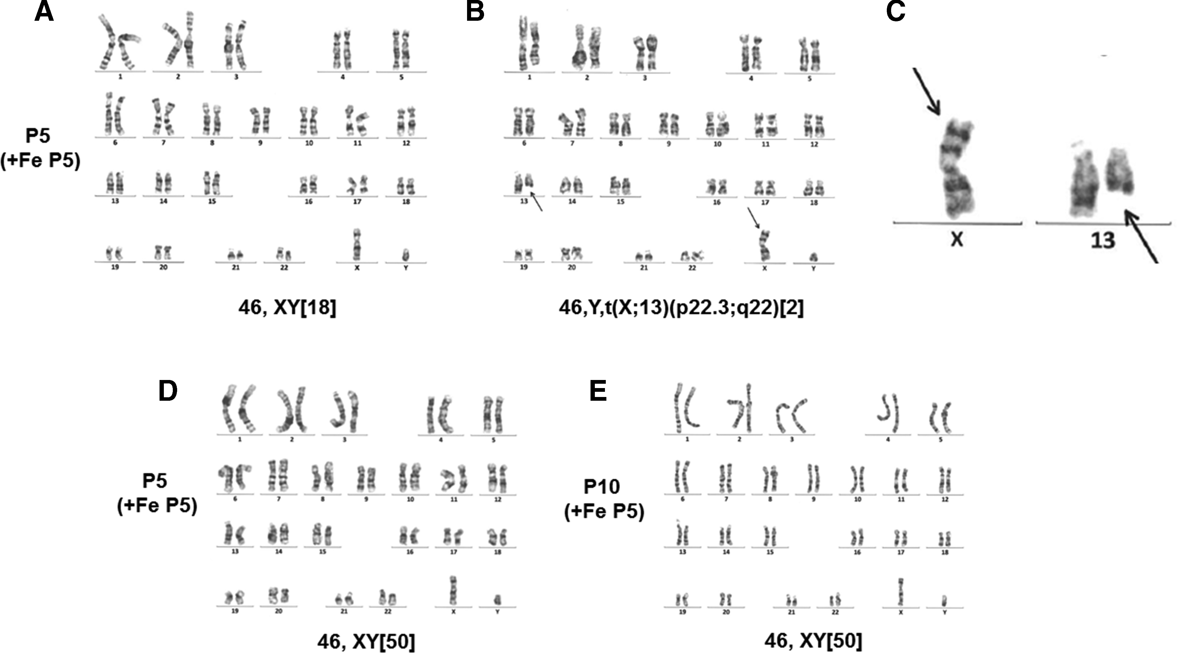
The karyotype analysis in magnetically labeled MSCs from 1F4287. Although 18 of 20 P5 MSCs labeled at P5 were normal
Proliferation of magnetically labeled MSCs
Three lots of human MSCs were used for proliferation assessments (1F4287, 2F3478, and 296578). Proliferation capacity was examined in long-term culture of MSCs during the four labeling treatments (control MSCs without labeling, Fe × 1, 12 h; MSCs labeled with 97.6 μg Fe/mL labeling medium for 12 h, Fe × 2, 12 h; MSCs labeled with 195 μg Fe/mL labeling medium for 12 h, Fe × 2, 12 h; and MSCs labeled with 97.6 μg Fe/mL labeling medium for 36 h). Initially, magnetic labeling promoted a slight proliferation of MSCs in all lots. Especially, MSCs of donor 0000296578 have 5–10 more population doublings after prolonged or 2 × concentration incubation with ferucarbotran. However, MSCs of all treatments finally lost their proliferation capacity in all lots (Fig. 4).
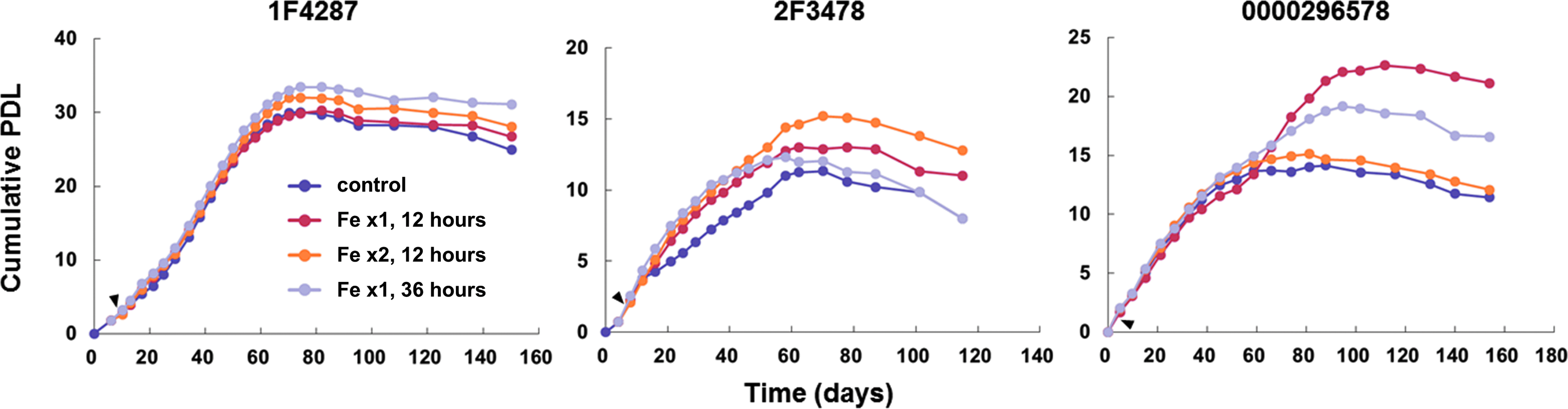
Plots of PDL during long-term culture of labeled MSCs (1F4287, 2F3478, and 296578). MSCs were labeled at P3 (arrowhead). “Fe × 1” and “Fe × 2” mean that cells were labeled by 97.6 μg/mL Fe and 195 μg/mL Fe, respectively. “12 hours” and “36 hours” mean that cells were labeled for 12 and 36 h, respectively. The data represent the average of duplicate values. PDL, population doublings level.
To evaluate the anchorage-independent growth of MSCs, P5 MSCs labeled at P5 were cultured in soft agar. While the positive control cells (HeLa-S3) formed colonies in soft agar, the negative control cells (MRC-5) and MSCs from any of the donors did not form colonies (data not shown).
Effect of exposure to magnetic field on the safety of MSCs
All the safety examinations were performed after a 10-min exposure of MSCs to magnetic field for using the same three lots of MSCs as above (1F4287, 2F3478, and 296578) as well as an additional three lots (310956, 318006, and 326162). The karyotypes of 50 P5 MSCs labeled at P5 (1F4287, 2F3478, and 296578) were examined. No abnormalities were observed in the magnetically labeled MSCs (Fig. 5A). In addition, the P5 MSCs labeled at P5 (1F4287, 2F3478, and 296578) did not form any colony when cultured in soft agar (data not shown).

Further, the proliferation capacity of MSCs (310956, 318006, and 326162) exposed to magnetic field in a long-term culture under four types of conditions (−Fe, −Mg: MSCs without magnetic labeling and exposure to magnetic field; −Fe, +Mg: MSCs without magnetic labeling exposed to magnetic field; +Fe, −Mg: magnetically labeled MSCs without exposure to magnetic field; and +Fe, +Mg: magnetically labeled MSCs exposed to magnetic field) was determined. MSCs of donor 0000326162 have three more population doublings upon incubation with ferucarbotran than without ferucarbotran. However, the proliferation of all these MSCs ceased within 100 days (Fig. 5B).
These findings indicated that exposure to magnetic field does not cause malignant transformation in magnetically labeled MSCs.
Release of iron particles from MSCs
Three lots of MSCs (1F4287, 2F3478, and 296578) were magnetically labeled under three types of conditions. After the magnetic labeling, the percentage of magnetically labeled MSCs was measured during culture using Berlin blue staining for all three types of labeling conditions (Fe × 1, 12 h; MSCs labeled with 97.6 μg Fe/mL labeling medium for 12 h, Fe × 2, 12 h; MSCs labeled with 195 μg Fe/mL labeling medium for 12 h, Fe × 2, 12 h; and MSCs labeled with 97.6 μg Fe/mL labeling medium for 36 h). Hundred percentage of MSCs were magnetically labeled at the start of culture. However, the percentage of the magnetically labeled MSCs decreased with time (Fig. 6A), and they became rare after culturing for >2 or 3 weeks in all lots under all conditions (Fig. 6B).

Viability of MSCs
Three lots of MSCs (1F4287, 429365, and 451491) were used for the quality assessments, including viability, iron content, the reactivity toward magnetic attractive force, and chondrogenic differentiation capacity of MSCs. For the quality assessments, the magnetic labeling conditions were defined as follows: control group (0 μg Fe/mL), one-quarter group (24.4 μg Fe/mL), one-half group (48.8 μg Fe/mL), three-quarters group (73.2 μg Fe/mL), standard group (97.6 μg Fe/mL), and double-dose group (195 μg Fe/mL).
The average viability of MSCs was 90.1% ± 6.6%. There were no significant differences in cell viability among the donors under any of the conditions (data not shown).
Iron content in MSCs
The iron content increased gradually, with increasing concentration of ferucarbotran in the MSCs from all donors (Fig. 7). On an average, in three donors, the iron content increased proportionally to the concentration of ferucarbotran in the medium to half the standard concentration, and saturated at three-quarters of the standard concentration or higher. However, there were differences in iron contents among the magnetically labeled MSCs from different donors. These findings suggest that the capacity for ferucarbotran uptake into MSCs varies with the donor.
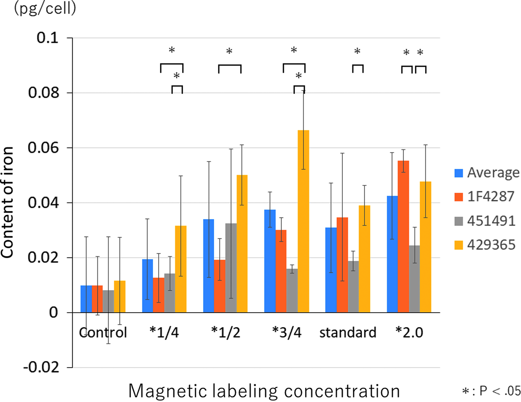
The content of iron in magnetically labeled MSCs. There were differences of iron contents in the magnetically labeled MSCs among donors (n = 9). On an average in three donors (1F4287, 429365, and 451491), the content of iron increased proportionally to the concentration of ferucarbotran in the medium. The results are represented as mean value ± SD. SD, standard deviation.
Chondrogenic differentiation of MSCs
The chondrogenic differentiation capacity of MSCs was assessed using pellet culture. The long diameter of spheroids from donor 429365 was especially smaller than the others (Fig. 8). Histologically, metachromasia was observed at every concentration of iron, but the toluidine blue and safranin-O stain abilities of the pellets from all donors in the double-dose group were lower than those of the other groups (Fig. 8). For the quantitative assessments of chondrogenic differentiation, the expressions of GAG and COL II mRNA were measured. The GAG expression in the MSCs from donor 429365 was decreased in the three-quarters group and the standard group (n = 1). However, GAG quantification increased in the double-dose group (Fig. 9A). Very high concentration of iron in the double-dose group might have induced an error in the absorbance measurement of GAG. The mRNA expression level of COL II from donor 429365 decreased in the three-quarters group and the higher concentration groups (Fig. 9B). On the contrary, the expressions of GAG and COL II mRNA from donors 1F4287 and 451491 decreased only in the double-dose group (Fig. 9A, B). These findings showed that the chondrogenic differentiation capacity of MSCs was maintained at the standard concentration of iron for donors 1F4287 and 451491, and at one-half concentration of iron for donor 429365.
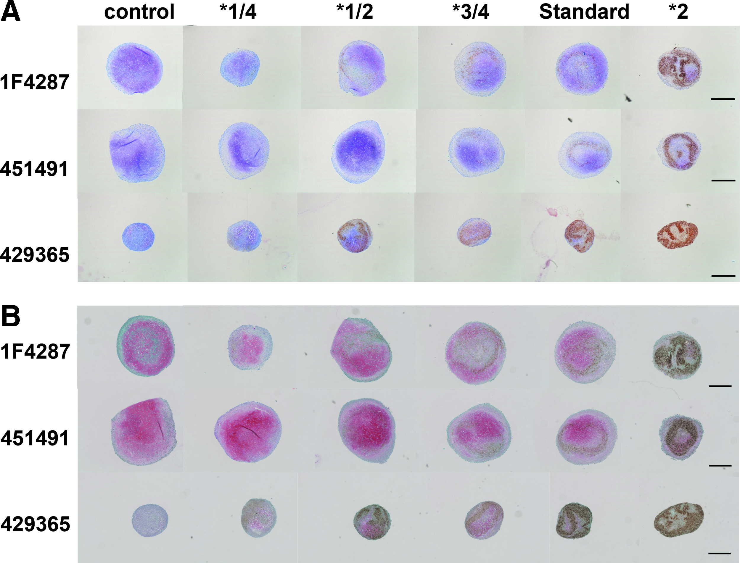
Histological assessments of pellets using the toluidine blue
The quantitative assessments of chondrogenic differentiation of magnetically labeled MSCs (n = 3; 1F4287, 429365, 451491) using the measurement of GAG
Reactivity of the MSCs toward magnetic force
In the control group (0 mT), there were no differences in the cell number across concentrations, but as the flux density of the magnet increased, the number of cells not attracted to the magnet decreased. The cell numbers decreased depending on the density of magnetic flux and the iron concentration. A magnetic flux density of ≥100 mT was required to decrease the cell number in all donors. At a magnetic density of 100 mT, one-half concentration of iron was enough to decrease the cell number in donor 429365. However, the standard concentration of iron was required to decrease the cell number in donors 1F4287 and 451491 (Fig. 10).
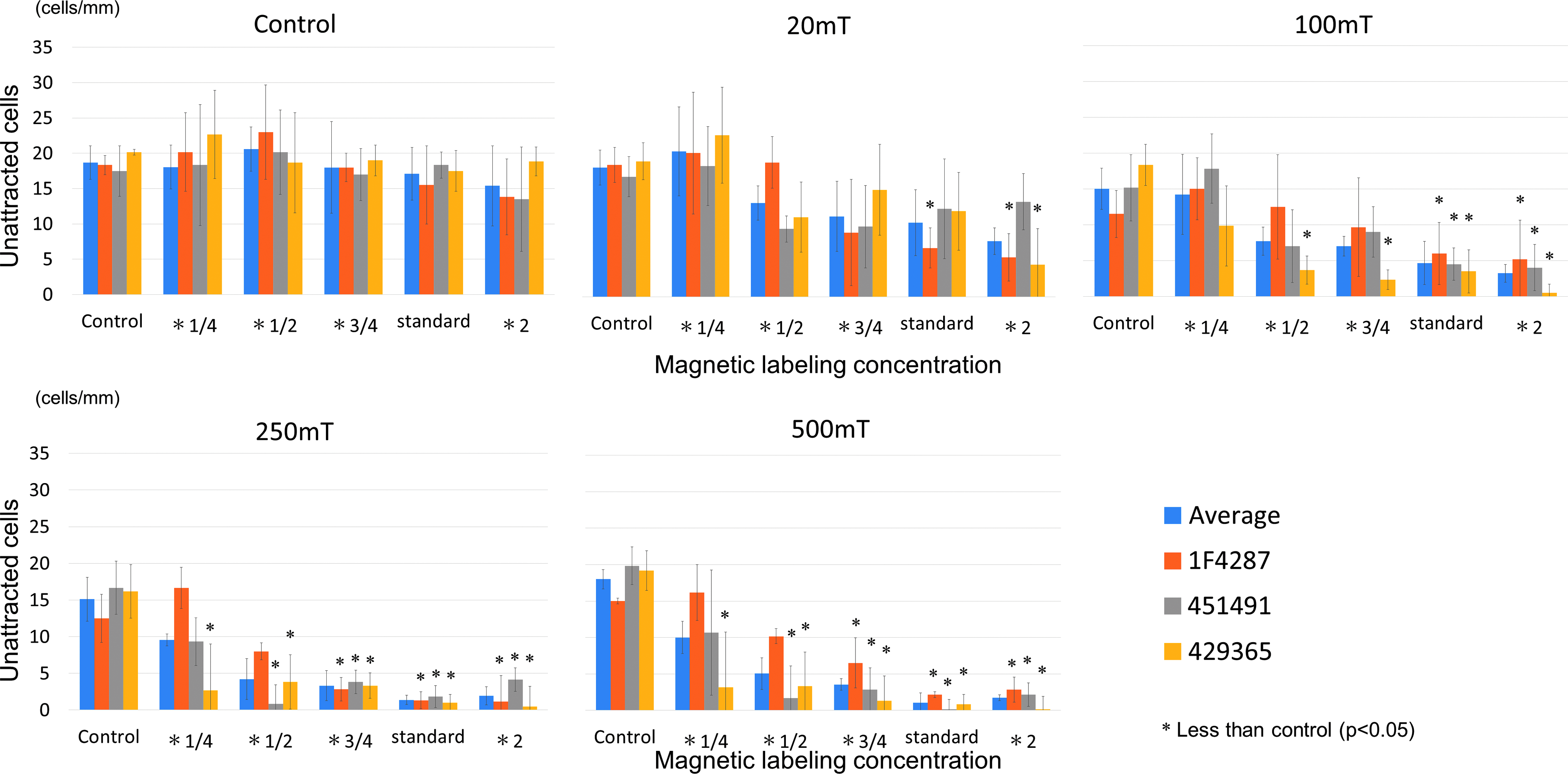
The reactivity of MSCs labeled in six different conditions (0, 24.4, 48.8, 73.2, 97.6, and 195 μg Fe/mL) for the magnetic field (n = 6; 0 mT control, 20, 100, 250, and 500 mT). A decrease in cell number indicates the attraction of MSCs to the magnet. The results are represented as mean value ± SD. Statistical significant differences are shown only compared with control in the figure.
Discussion
This study demonstrated that magnetic labeling did not impair the safety of MSCs as assessed by karyotyping, colony-forming assay in soft agar, and cell proliferation in long-term culture. Although magnetic labeling inhibited the chondrogenic capacity of MSCs, an appropriate concentration of iron was able to maintain their chondrogenic differentiation capacity as well as their reactivity toward magnetic force. The appropriate concentration of iron for the magnetic labeling of MSCs varied according to the donor.
For 1 of the 3 donors (1F4287), the karyotype analysis showed an abnormality in 2 of the 20 P5 MSCs labeled at P5. However, the second karyotype analysis of MSCs for 1F4287 (P5 MSCs labeled at P5 and P10 MSCs labeled at P5) failed to show a single abnormality among 50 cells. It is known that chromosomal instability of MSCs occurs in normal culture. 12 Therefore, the abnormality observed in lot 1F4287 was considered a rare, occasional event that occurred independent of the labeling. Magnetic labeling promoted the proliferation of MSCs in the long-term culture. A previous study also showed that the proliferation activity of the magnetically labeled MSCs was slightly higher than that of the nonlabeled MSCs 13 ; however, the magnetically labeled MSCs finally lost their proliferation capacity similar to the nonlabeled MSCs as observed in this study. In addition, the magnetically labeled MSCs never formed colonies in soft agar. These findings suggest that magnetic labeling does not cause malignant transformation of MSCs. Even a 10-min exposure of magnetically labeled MSCs from the same three donors to a magnetic field did not show any abnormality in karyotyping, colony-forming assay using soft agar, or cell proliferation in a long-term culture. Therefore, we concluded that the magnetic labeling and exposure to magnetic field did not adversely affect the safety of MSCs.
In the assessment of the percentage of magnetically labeled MSCs during culture using Berlin blue staining, almost all MSCs were labeled just after labeling. However, the percentage of magnetically labeled MSCs markedly decreased within 2 or 3 weeks. This property might be advantageous for ensuring safety after transplantation, because iron particles will not permanently remain at the transplanted site. However, the in vivo kinetics of ferucarbotran after transplantation should be clarified.
During the quality assessment, magnetic labeling did not affect the viability of MSCs regardless of the ferucarbotran concentration. However, magnetic labeling using high-density ferucarbotran inhibited the chondrogenic differentiation of MSCs. A previous study reported that magnetic labeling at a concentration of 100 μg Fe/mL does not affect the chondrogenic differentiation of MSCs. 14 Kamei et al. showed, using a minipig model, that the magnetic delivery system with a standard dose of ferucarbotran was better than the control for the regeneration of hyaline cartilage in cartilage defects. 4 On the contrary, Henning and Bulte showed that chondrogenic differentiation of m-MSCs in cartilage defects was inhibited by iron in a concentration-dependent manner.15,16 Iron oxide nanoparticles have been widely reported to produce highly reactive hydroxyl radicals. 17 These radicals and the subsequent oxidative stress are considered biotoxic and have the ability to inhibit chondrogenic differentiation.18–21 In this study, the concentration of ferucarbotran necessary to inhibit the chondrogenic differentiation of MSCs varied across donors, probably due to the variation in the amount of intracellularly incorporated iron among the donors. However, the exposure of magnetically labeled MSCs to the magnetic field (1.5 T, 10 min) did not affect the chondrogenic differentiation capacity of magnetic MSCs in our previous report. 4
To our knowledge, no previous study has investigated the relationship between the iron concentration used for magnetic labeling and MSC reactivity toward magnetic attractive force. In our study, magnetic flux density of ≥100 mT was required for inducing the reactivity of MSCs to the magnetic attractive force. The concentration of ferucarbotran necessary to show the reactivity of MSCs to the magnetic attractive force varied across donors. For donor 1F4287, magnetic labeling at the density of 48.8 μg Fe/mL maintained the chondrogenic differentiation capacity of MSCs as well as the reactivity of MSCs to the magnetic attractive force. On the contrary, magnetic labeling at the density of 97.6 μg Fe/mL maintained the chondrogenic differentiation capacity of MSCs and the reactivity of MSCs to magnetic attractive force in donors 429365 and 451491. These were considered appropriate conditions for the magnetic labeling of MSCs for use in cartilage repair. A previous report showed that the labeling of MSCs with iron protamine sulfate is not toxic and does not affect their ability to differentiate. 22 Hence, iron protamine sulfate might be helpful for the magnetic delivery system. However, protamine sulfate was not used in this study because it has not been approved clinically for intra-articular administration in Japan.
In vivo kinetics of iron particles after the transplantation of magnetically labeled MSCs has been assessed by MRI and Berlin blue staining of tissue sections. 23 These iron particles decreased in number at 4 weeks and disappeared at 12 weeks after the transplantation.
Footnotes
Acknowledgments
This work was supported by the Highway Program for Realization of Regenerative Medicine (16bm0504004h0005) to M.O. and the Research Project for Practical Applications of Regenerative Medicine (18bk0104010h0001) to N.K. from Japan Agency for Medical Research and Development.
Disclosure Statement
No competing financial interests exist.