
Abstract
The objective of this study was to investigate the utility of gelatin hydrogel nonwoven fabrics (GHNFs) as a carrier membrane in preparing multilayered cell sheets. The GHNFs were fabricated by a blow method of gelatin solution. When the cell sheet of human mesenchymal stromal cells was piled up to formulate three-layered cell sheets, the GHNFs were used to allow the cell sheet to detach from the cell culture dish and transfer. The cell sheet harvest and transfer processes were performed simpler and faster than those without using the GHNF. The lactate/glucose ratio of a metabolic activity was significantly lower, and the ATP production was higher for the three-layered cell sheets formulated with the GHNF than that obtained without the GHNF. During the culture, the cell migration from cell sheets into the GHNF was observed. The GHNF was promising to simply and fast fabricate three-layered cell sheets with the activity remaining.
Impact Statement
This study introduces the utility of gelatin hydrogel nonwoven fabrics (GHNFs) for cell sheet engineering. The GHNF had the mechanical property strong enough to hold by forceps even in the swollen condition. The cell sheet harvest and transfer processes were performed simpler and faster than those without using the GHNF. The GHNF facilitates the metabolic activity of three-layered cell sheets, and the cell migration from cell sheets into the GHNF was observed. The GHNF is a promising material used to support cell sheets during the process of assemble formulation and contributes to the improved biological functions of tissue-like cell constructs.
Introduction
Cell sheet engineering has been extensively used for cell-based therapies, drug discovery, regenerative medicine, and tissue engineering.1–6 The multilayering of cell sheets is one of the promising technologies to obtain three-dimensional (3D) cell constructs. Various 3D cell constructs were fabricated by the layering technique of cell sheets. However, since a single-layer cell sheet is quite fragile, it is easy to tear and be crumbed in harvesting the cell sheets from the culture dish with forceps. In general, the rate of success largely depends on the skill and experience of researchers/technicians. Therefore, several methods to manipulate cell sheets have been tried to develop, such as pipettes, 7 plunger-like devices,8,9 and other processes. 10 Among them, a membrane is used as a support material to harvest and transfer the cell sheet.11,12 The support membrane was placed onto the cell sheet, and combined cell sheet and support membrane were harvested together as a uniform sheet. The membrane and cell sheet harvested are subsequently transferred to another dish, and then the support membrane is peeled. By repeating this process, multilayered cell sheets can be fabricated. On the contrary, the fabric of poly(lactic-co-glycolic acid) (PLGA) electrospun is used as a support membrane. 13 Multilayered cell sheets are fabricated by layering the sheets of human mesenchymal stromal cells (hMSCs) with the PLGA fabrics. In this system, the fabric is not peeled off but remains in the multilayered cell sheets. Another issue of multilayered cell sheets fabrication is long time period is taken to complete the adhesion between cell sheets in the medium. As one trial to shorten the time period of adhesion, centrifugation is tried. 14 However, it is still necessary to further develop technologies to reduce the incubation time of cell sheets adhesion.
When the thickness of multilayered cell sheets becomes large, the cells present inside layered cell sheets often weaken or die due to the lack of oxygen and nutrient supplies. 15 Several strategies are reported to improve the cell viability, such as the combination of vascular endothelial cells and the use of porous membrane.16,17 In addition, it is demonstrated that the incorporation of gelatin hydrogel microspheres (GHMs) between cardiovascular cell sheets improved the cell viability. 18
To tackle the problem of multilayered cell sheets fabrication, the sheet handling, the incubation time of cell sheets adhesion, and the cell viability inside the multilayered cell sheets, we have designed a nonwoven fabric of gelatin hydrogels. This study is undertaken to evaluate the utility of gelatin hydrogel nonwoven fabrics (GHNFs) for the fabrication of multilayered cell sheets. During the fabrication process of cell sheets formulated with the GHNF, the simplicity of cell sheets manipulation and the incubation time of cell sheets adhesion were evaluated in comparison with those of cell sheets fabrication without GHNF. We examine the glucose consumption, the lactate production, and the ATP production of three-layered cell sheets formulated with the GHNF.
Materials and Methods
Preparation of GHNF
The GHNFs were prepared by the solution blow spinning method. 19 The spinning apparatus used in this study consisted of a nozzle through which an aqueous solution was injected into a stream of accelerated gas. The setup consisted of a source of compressed air equipped with a pressure regulator, a syringe pump to control the injection rate of the aqueous solutions, and a collector. The collector was positioned at a fixed working distance from the nozzle. An aqueous solution of 37.5 wt% gelatin (isoelectric point 5.0; Nitta Gelatin, Inc., Osaka, Japan) was preheated at 60°C. The spinning parameters were an air pressure of 0.375 MPa, the internal diameter of syringe pump nozzle of 250 μm, and the working distance of 50 cm to collect fibers. The solution was pumped through the nozzle with the application of high-velocity air. The solution was collected in the state of fiber as the GHNF at the collector. Then, the GHNFs prepared were air-dried at room temperature and freeze-dried. The noncrosslinked and dried GHNF was treated in a vacuum oven at 140°C and 1 × 10⁻⁵ MPa for the dehydrothermal crosslinking of gelatin for 48 h according to the method previously reported. 20 The crosslinked GHNF was punched out into 6-mm-diameter disks. The disks prepared were sterilized using ethylene oxide gas. The pictures of GHNF in the dried and swollen states were taken with a light microscope (BX-X710; Keyence Corp., Osaka, Japan). The diameter of 100 fibers was measured using a computer program (PhotoRuler, Hyogo, Japan), and the mean diameter was calculated. The thickness without a load was measured using a rheometer (RE2-33005C; Yamaden, Tokyo, Japan). The weight was measured in the dried and swollen states. In the swollen state, the excess water of swollen GHNF was removed with Kimwipe (Nippon Paper Crecia, Tokyo, Japan). The thickness and the weight of five samples were measured, and the density of GHNF was calculated from the diameter, thickness, and weight measured.
Preparation of hMSC sheets
hMSCs immortalized by introducing human telomerase reverse transcriptase (hMSC) were provided by Dr. Toguchida. 21 hMSCs (1 × 10⁵ cells) were seeded onto a 12-well dish in minimal essential medium alpha (Thermo, Inc., Waltham), supplemented with 10 vol% fetal bovine serum (Thermo, Inc.), 1 vol % penicillin and streptomycin (Nacalai Tesque, Kyoto, Japan), and 100 μM of ascorbic acid phosphate (AA) (Wako-Fujifilm, Osaka) for 5 days at 37°C in a humidified incubator with 5% CO2. The AA was used to promote the cell sheet formation. 22 The medium with AA was exchanged every 2 days. After the culture, the confluent cells as a monolayer sheet were detached from the 12-well dish by flushing several times with the culture medium using a 1000-μL micropipette.
Transfer of cell sheet by the GHNF
One GHNF swollen with 10 μL of culture medium was placed onto one cell sheet. The cell sheet associated with the GHNF was picked up using forceps and transferred to another culture dish. The conventional method used a 5-mL pipette to transfer a cell sheet. 7 Each transfer time was recorded. Experiments were performed independently five times. In addition, a coefficient of variation (CV) of transfer time was calculated from the standard deviation divided by the mean.
Assessment of cell sheet attachment
Attachment between the cell sheet and the GHNF was assessed as described previously. 23 In brief, one cell sheet with the culture medium was gently aspirated into the tip of a 5-mL pipette, transferred onto a new six-well culture dish, and the remaining culture medium was removed. In the GHNF-used method, one GHNF in the swollen state with 10 μL of culture medium was placed onto one cell sheet. The cell sheet associated with the GHNF was picked up using forceps and transferred to another six-well dish. After incubation, 2 mL of culture medium was added, and then the dish was shaken at 90 rpm for 120 s on an orbital shaker (NR-80; Taitek, Saitama, Japan), and the attachment or detachment of the cell sheets was observed. If detachment between the cell sheet and the GHNF was not observed for 120 s, it was estimated as the successful attachment. Experiments were performed independently three times unless otherwise mentioned.
Layering cell sheets without or with the GHNF
In the conventional method, layering cell sheets without the GHNF was carried out as reported previously. 7 A detached cell sheet was collected by flushing with the medium. One cell sheet with the culture medium was gently aspirated into the tip of a 5 mL-pipette and transferred onto a new six-well culture dish. After spreading the cell sheet without any folds by aspirating the medium, the dish was incubated for 60 min at 37°C to allow the cell sheet to adhere to the surface of dish. Next, another cell sheet was placed on top of the first one and attached without folds by aspirating the medium, and then the dish was incubated for 60 min at 37°C to allow the two cell sheets to adhere. By repeating the similar process, three-layered cell sheets were formulated on the six-well dishes.
Figure 1 shows schematic illustration of layering procedure of cell sheets formulated with the GHNF. The cell sheet was detached from the 12-well dishes, and the culture medium was removed from the dishes. The GHNF in the swollen state was placed onto the cell sheet. A combination of the GHNF and the cell sheet was harvested as a uniform sheet with forceps, and then transferred onto a new six-well dish. Similarly, a second cell sheet with the GHNF was placed on top of the first one to allow it to attach. By repeating the layering procedure, three-layered cell sheets formulated with quadruple-layered GHNF constructs were formulated on the six-well dish. The dish was incubated at 37°C for 60 min to allow the cell sheet to adhere to the GHNF.

Schematic illustration of layering procedure of cell sheets with the GHNF. GHNF, gelatin hydrogel nonwoven fabrics.
Histological analysis
Three-layered cell sheets formulated with the GHNF were fixed with 4 wt% paraformaldehyde solution. Specimens were embedded in an optimal cutting temperature compound (Sakura Finetek Japan Co. Ltd., Tokyo, Japan) and frozen. The frozen samples were sectioned using a cryotome (CM3050S; Leica Microsystems, Wetzlar, Germany), and then stained with hematoxylin and eosin. Prepared specimens were examined under a microscope (BZ-X710; Keyence Co. Ltd., Tokyo, Japan).
Cell sheets culture experiments
The three-layered cell sheets formulated without or with the GHNF were cultured upon preincubation for 12 h in a six-well dish with 4 mL of medium in the static condition to allow cell attachment between the cell sheets. Subsequently, 4 mL of fresh medium was exchanged, and the samples were cultured further for 24 h in a static or shaking condition before the following measurements. In the shaking condition, the dish was set on a reciprocal shaker (BC-730; Biocraft, Tokyo, Japan).
Measurement of glucose consumption, lactate production, lactate/glucose ratio, and ATP production of three-layered cell sheets formulated without or with the GHNF in the static or shaking culture condition
The amount of glucose consumed by three-layered cell sheets was determined by measuring the change in glucose concentration of the culture medium using Glucose C2 kit (Fujifilm Wako, Osaka, Japan). The amount of lactate produced by three-layered cell sheets was determined with a PicoProbe Lactate Fluorometric Assay Kit (BioVision, San Francisco). The lactate/glucose ratio was calculated as a measurement of aerobic condition. 24 To calculate the lactate/glucose ratio, the amount of glucose was determined by dividing the weight (mg) by the molecular weight (180 g/mol). The amount of ATP produced by three-layered cell sheets was determined using an ATP Kit for Aggregate (Toyo-b-net, Tokyo, Japan).
Measurement of glucose uptake and ATP production of cell sheet
Similarly, three-layered cell sheets formulated with the GHNF were prepared. After preincubation for 12 h, 2-deoxyglucose (2DG) was added according to the manufacturer's instructions. After incubation for 40 min in the static or shaking (90 rpm) condition, the cell sheets with the GHNF were destacked with forceps to obtain the upper, middle, and bottom sheets. The amount of glucose uptake by each cell sheet was determined by measuring the uptake of 2DG using a Glucose Cellular Uptake Measurement Kit (CosmoBio, Tokyo, Japan). In addition, the amount of ATP production by each cell sheet was determined by the ATP Kit for Aggregate (Toyo-b-net). Since the cell sheets formulated without using the GHNF could not be destacked, the uptake of 2DG and the amount of ATP production of three-layered cell sheets were measured to compare with the total uptake and amount of upper, middle, and bottom cell layers.
Statistical analysis
All the data were statistically analyzed and expressed as mean ± standard error of the mean. The data were analyzed by Student's t-test to determine the statistically significant difference while the significance was accepted at p < 0.05.
Results
Characterization of GHNF
Table 1 summarizes the structural parameters of GHNF. Figure 2 shows appearance of GHNF in the swollen condition and microscopic pictures of GHNF surface in the dried and swollen conditions. The GHNFs had the mechanical property strong enough to hold by forceps even in the swollen condition. The GHNFs were composed of fibers with homogeneous size and had a homogeneous porous structure. When the GHNFs were swollen by water, the intrastructure did not change, although the diameter of fibers became large.

Structural Parameters of Gelatin Hydrogel Nonwoven Fabrics in the Dried and Swollen Conditions
The values are reported in mean ± standard deviation.
Layering time to fabricate three-layered cell sheets formulated with the GHNF
Figure 3 shows the comparison in the layering time of cell sheets formulated with or without GHNF. The layering time was significantly shorter for the GHNF-used method than for the conventional method. In addition, the CV of the present method (CV = 0.15) was smaller than that of the conventional method (CV = 0.48).
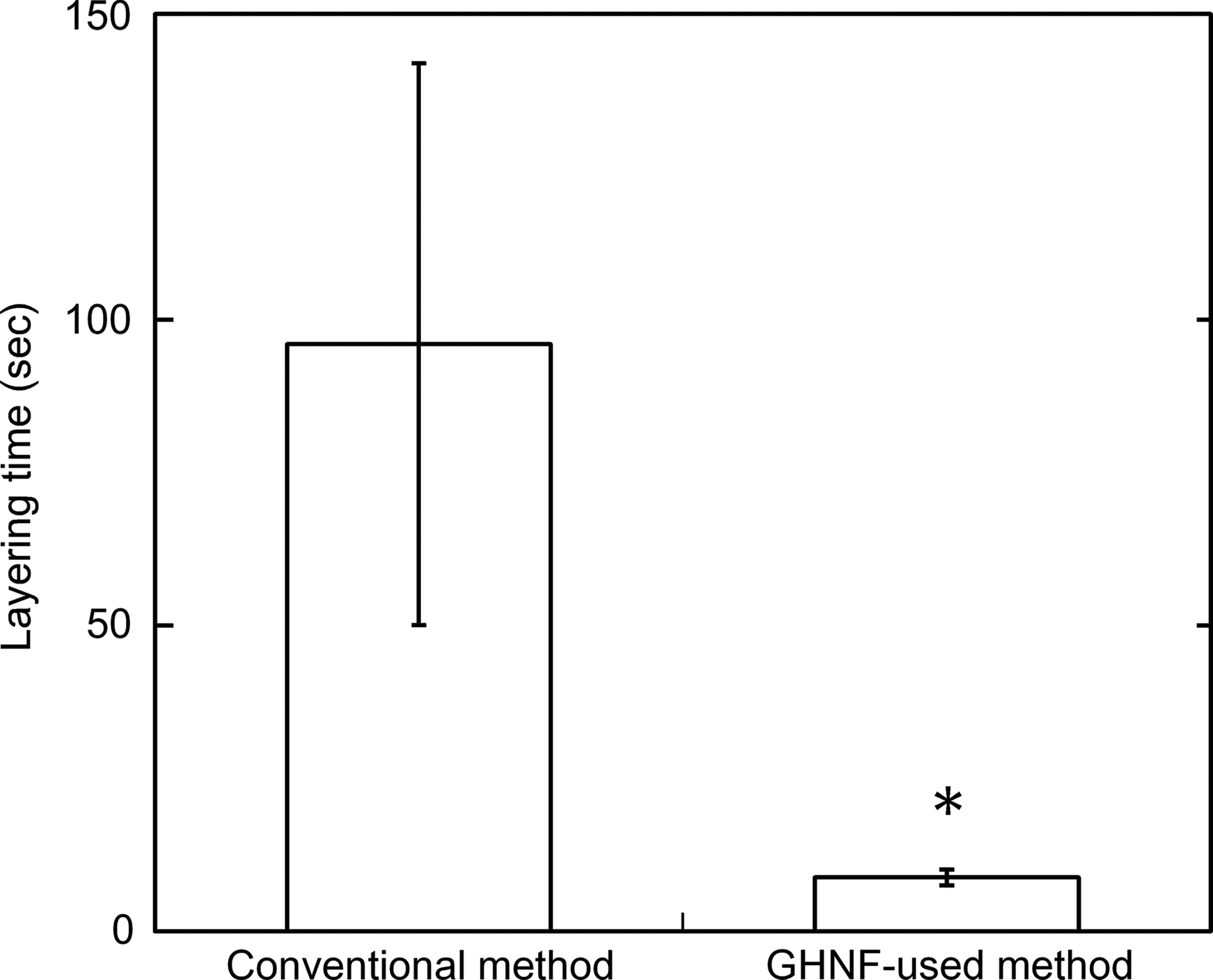
Comparison of the layering time of cell sheets between the conventional and the GHNF-used methods. *p < 0.05; significant difference between the two groups.
Attachment between the cell sheet and the GHNF
Table 2 shows the influence of incubation time on the detachment between the cell sheets and the GHNF. No layered cell sheets were detached when the incubation time was 10, 20, and 30 min. The incubation time of GHNF-used method was shorter than that of conventional method. The following experiments were incubated for 60 min. Figure 4 shows a light microscopic picture of cross-section of three-layered cell sheets formulated with GNHF 12 h after incubation in the static condition and 6 days after incubation in the shaking condition. With the incubation time, the cell migration from the cell sheets into the GHNF was observed.
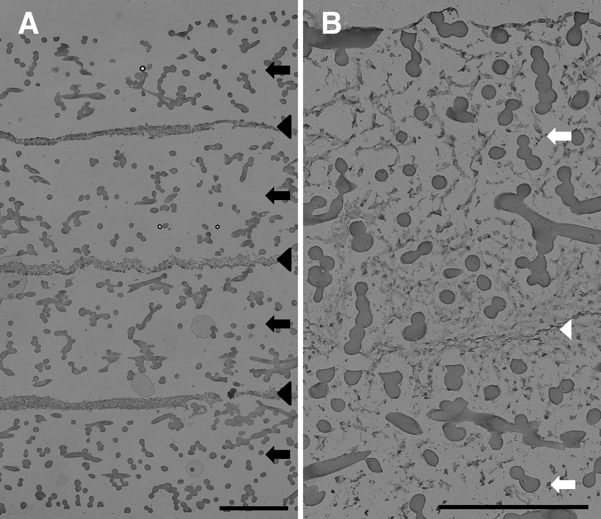
A light microscopic picture of cross-section of three-layered cell sheets formulated with four GHNFs.
Influence of Incubation Time on the Detachment Between Cell Sheets and Gelatin Hydrogel Nonwoven Fabrics
Cell activity of three-layered cell sheets formulated without or with the GHNF in the static or shaking culture condition
Figure 5 shows the glucose consumption, the lactate production, the lactate/glucose ratio, and the ATP production of three-layered cell sheets without or with the GHNF 24 h after incubation in the static condition. A significantly higher glucose consumption, lower lactate/glucose ratio, and higher ATP production were observed for three-layered cell sheets formulated with the GHNF than those observed without GHNF, prepared by the conventional method.
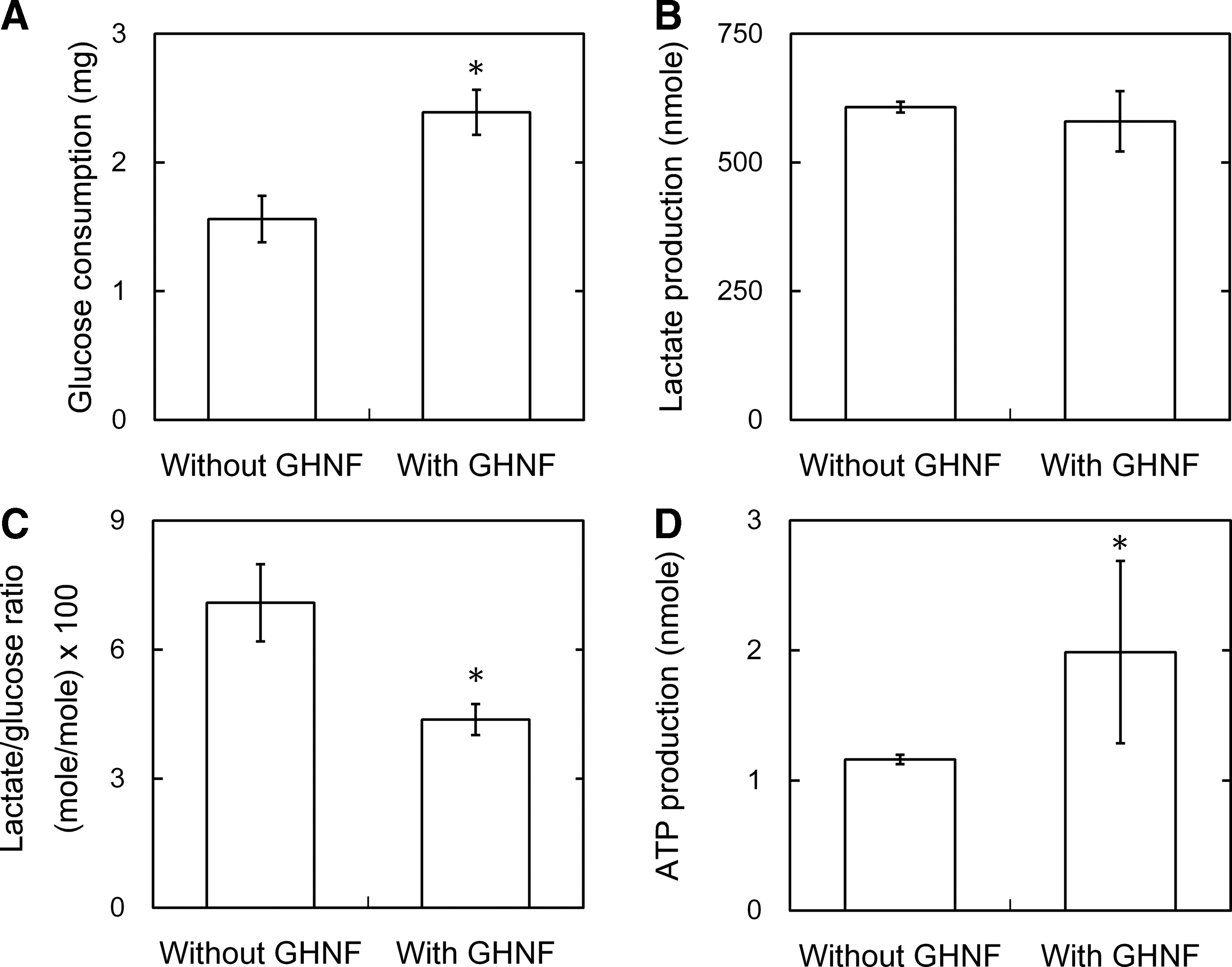
Cell activity of three-layered cell sheets formulated with or without GHNF 24 h after culture in the static condition:
Figure 6 shows the influence of shaking speed on glucose consumption of three-layered cell sheets formulated with the GHNF. The glucose consumption increased to a significant extent with an increase of the shaking speed. The shaking speed was fixed at 90 rpm for the following experiments.

Influence of shaking speed on the glucose consumption of three-layered cell sheets formulated with GHNF for 24 h in the shaking condition. *p < 0.05; significant difference between the two groups.
Cell activity of three-layered cell sheets formulated with the GHNF in the static or shaking culture condition
Figure 7 shows the glucose consumption, the lactate production, the lactate/glucose ratio, and the ATP production of three-layered cell sheets with the GHNF 24 h after incubation in the static or shaking condition. A significantly higher glucose consumption, lower lactate production, lower lactate/glucose ratio, and higher ATP production were observed in the shaking condition than in the static condition.
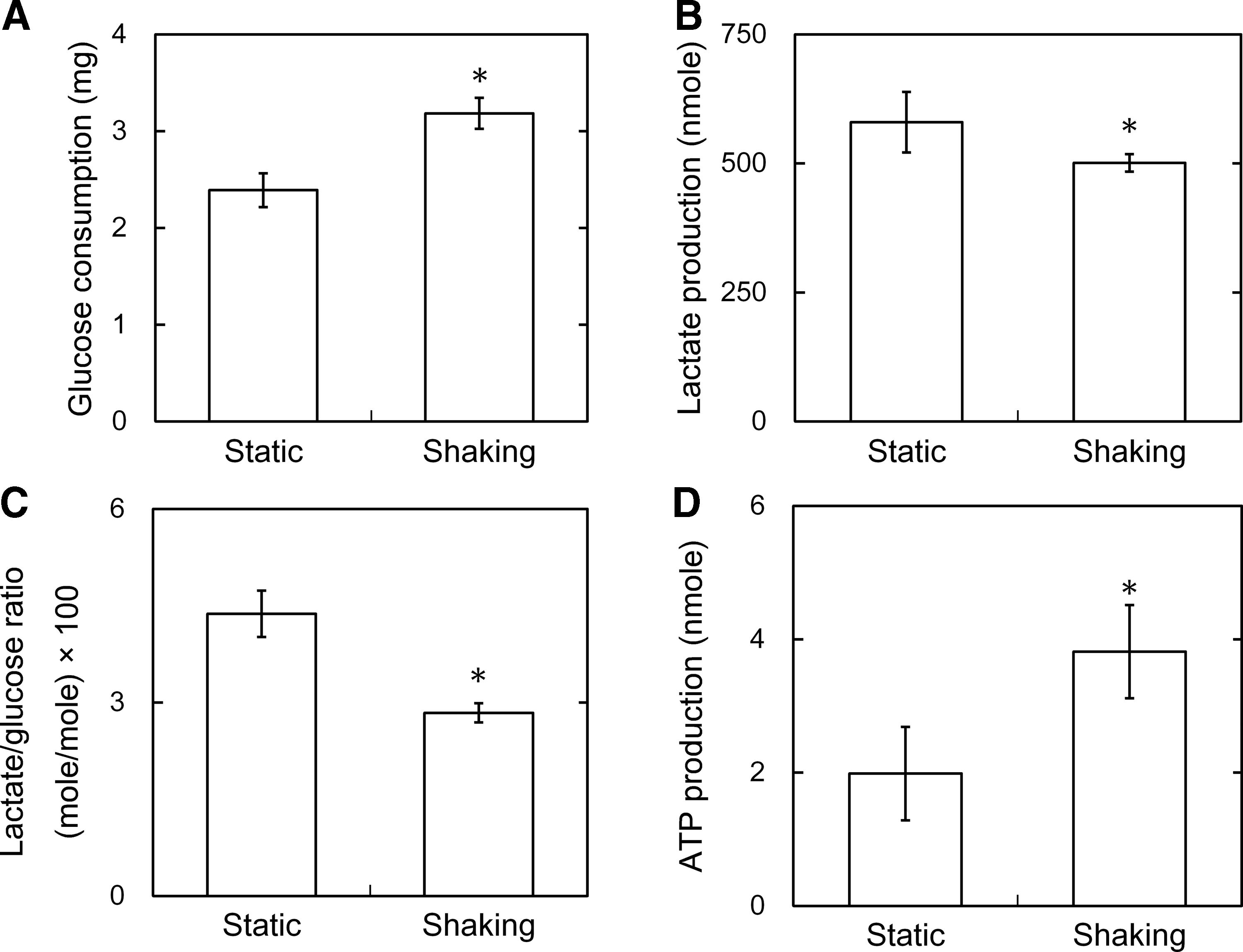
Cell activity of three-layered cell sheets formulated with GHNF 24 h after culture in the static or shaking (90 rpm) condition:
2DG uptake of respective cell layers destacked from three-layered cell sheets formulated with the GHNF
Figure 8 shows the 2DG uptake of the upper, middle, and bottom cell layers destacked from three-layered cell sheets formulated with the GHNF in the static or shaking culture condition. For both the static and shaking culture, the 2DG uptake of respective layers was in a similar level. The total amount of three-layer 2DG uptake was larger than that of three-layered cell sheets without the GHNF.

2DG uptake of three-layered cell sheets formulated with GHNF 40 min after incubation in the static or shaking (90 rpm) condition. Glucose cellular uptake of respective layers destacked from three-layered cell sheets formulated with GHNF. *p < 0.05; significant difference between the two groups.
ATP production of respective cell layers destacked from three-layered cell sheets formulated with the GHNF
Figure 9 shows the ATP production of the upper, middle, and bottom cell layers destacked from three-layered cell sheets formulated with GHNF in the static or shaking culture condition. For both the static and shaking culture, a similar ATP production was observed for respective layers. In the shaking condition, the total ATP production of upper, middle, and bottom layers of cell sheets formulated with the GHNF was significantly high compared with that of cell sheets without the GHNF.
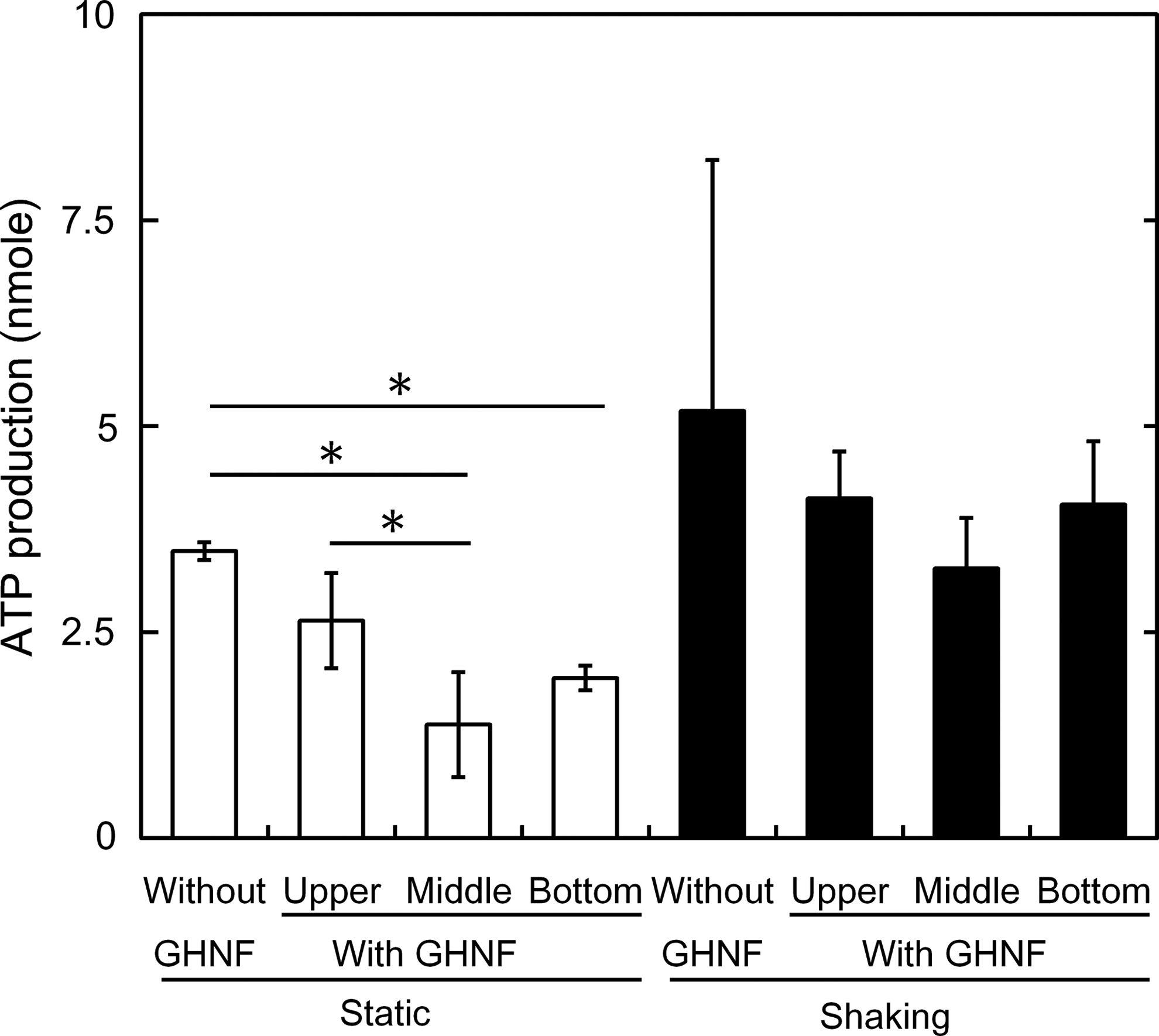
ATP production of each layer destacked from three-layered cell sheets with or without GHNF 24 h after incubation in the static culture or shaking (90 rpm) condition. ATP production of respective layers destacked from three-layered cell sheets formulated with GHNF.*p < 0.05; significant difference between the two groups.
Discussion
GHNFs were fabricated by a solution blow spinning method. In this method, gelatin fibers are accumulated on the collector in a wet state, and the fibers of GHNF are connected at the cross-point of fibers. Even when swollen, the GHNF showed a mechanical property strong enough to hold by forceps (Fig. 2A). In addition, the density of GHNF (ρ = 0.26 ± 0.02) in the dried condition was higher than that of gelatin sponges commercially available (ρ = 0.01 g/cm3, Gelfoam; Pfizer, Tokyo, Japan). It is likely that the presence of fiber cross-points in the GHNF prevents the slipping between the gelatin fibers and functions as the strong framework, leading to no-deformed property even in the wet condition.
It is apparent from Figure 3 that the layering time was significantly shorter for cell sheets fabricated with the GHNF than that observed for the conventional method. In addition, the CV of layering time was smaller for cell sheets with the GHNF than that of the conventional method. The GHNF-used layering method was superior to the conventional method in terms of layering time and the procedure fluctuation of layering; because, in the conventional method, the cell sheet must be detached from the culture dish by flushing with the medium in every layering process. On the contrary in the GHNF-used method, one-time incubation is sufficient to formulate layered cell sheets. In addition, it is apparent from Table 2 that the incubation time required for the attachment of cell sheets layering was short in the GHNF-used method compared with that in the conventional method. It has been reported that spaces present between the cell sheet and the culture dish prevent their adhesion. 23 In the GHNF-used method, liquid present on the surface of cell sheet would be absorbed by the GHNF, and consequently spaces between the cell sheet and the GHNF would become smaller. As a result, the whole fabrication time of layered cell sheets would be shortened. In the case of three layers fabrication, the time necessary for sheet layering was reduced to approximately one-third compared with that observed for the conventional method. After the incubation, a layer-by-layer structure of cell sheets and the GHNF was observed (Fig. 4A). Cell migration into the GHNF after 6 days (Fig. 4B) indicates that cells in the three-layered cell sheets are biologically active, and the GHNFs were of good cytocompatibility.
The ratio of lactic acid production to glucose consumption is a general measure of aerobic metabolism. 24 The lower the ratio, the higher the aerobic metabolism of cells. In addition, it is recognized that the ATP production is higher in the aerobic condition than in the anaerobic condition. The glucose consumption of three-layered cell sheets increased using the GHNF (Fig. 5A). In addition, a decrease of the lactate/glucose ratio and an increase of the ATP production suggest that the cell condition was more aerobic (Fig. 5C, D). It is reported that the diffusion limits of nutrients and oxygen are reduced by creating a structure of blood vessels or inserting GHMs between layered cell sheets.18,25 In the GHNF-used method, it is likely that the GHNF functions as a reductant of diffusion limits, resulting in improved cell activity of three-layered cell sheets incorporating GHNF. However, it should be noted that the interaction between the layers of cell sheets is poor compared with the methods described above. An increase of glucose consumption, the decrease of lactate/glucose ratio, and the increase of ATP production by the shaking culture will be due to the promotion of oxygen and nutrients diffusion through the GHNF present between cell sheets (Figs. 6 and 7). The GHNF had a fibrous and porous structure (Fig. 2C). In addition, since the cell sheet is sandwiched between two GHNFs on both sides, the multilayered cell sheets did not adhere to the bottom of culture dish. Therefore, even in the shaking culture, the multilayered cell sheets were in a floating state in the medium. It is highly conceivable that glucose is better supplied to top and bottom sides of multilayered cell sheets.
To investigate the distribution of glucose uptake inside the three-layered cell sheets formulated with the GHNF, the 2DG assay was performed. 26 As a result, the amount of 2DG uptake was similar for respective layers of three-layered cell sheets with the GHNF (Fig. 8). As compared with the uptake amount of whole three-layered cell sheets, the amount was larger in the shaking culture than in the static one. We can say with certainty that the shaking culture increases medium flow, resulting in an enhanced exchange of oxygen and nutrients of cells even inside layered cell sheets. To investigate the distribution of ATP production inside three-layered cell sheets formulated with the GHNF, the ATP assay was performed. The ATP production of the middle layer became significantly larger by the use of the GHNF in the shaking culture. This indicates a better metabolic condition of cells present inside three-layered cell sheets. Insertion of materials between cell layers has been reported in several studies for improved condition of cell sheets. For this purpose, PLGA-based membranes and GHMs are used.13,18 Gelatin hydrogel is a biodegradable biomaterial that is of good cytocompatibility as a cell culture substrate. As the water content of gelatin hydrogel is >95%, 27 the culture medium and body fluids containing oxygen and nutrients can be easily impregnated into gelatin hydrogels.28–30 The GHMs will be more suitable for promoting diffusion of nutrients and oxygen inside multilayered cell sheets than the hydrophobic material of PLGA. The GHM has such an excellent property, but is not always useful as a carrier material to support and transfer cell sheets because GHMs are dispersed hydrogel. In addition, GHMs are very small to readily manipulate with forceps like the GHNF. On the contrary, since gelatin sponges commercially available have poor mechanical property in the swollen state, it is practically hard to handle them in the swollen condition. Based on the property, it may be unsuitable as a carrier to support cell sheets. 31 Several studies have been reported to design carrier materials for cell sheets transplantation.11,12 For this end, the materials should be simply used to support and transfer cell sheets, and must be degraded and disappear after implantation into the body. From this viewpoint, the GHNF is a promising material, which can be applied for cell sheet technology.
In the three-layered cell sheets formulated with the GHNF, the GHNF portion is much thicker than the cell sheet. Cell sheet technology is often applied to cell-dense tissues, such as heart and liver, without the use of scaffold. 32 From this viewpoint, the GHNF-used method is limited to apply to the cell-dense tissues. However, cell infiltration, spreading, and proliferation from cell sheets to porous polymer sheets are reported. 33 Therefore, cell-sparse tissues such as cartilage or bone may be suitable for the GHNF technology.34,35 On the contrary, vessel-like structures containing cells and fiber scaffolds prepared by a cell electrospinning are demonstrated to show a high cell activity.36,37 The electrospinning technology is promising to design cell-scaffold constructs with the remaining biological activity. The present GHNF formulation of fiber structure is a promising material to support and transfer cell sheets during the process of assemble formulation, and contributes to the improved biological functions of tissue-like cell constructs.
Conclusion
When the cell sheet of hMSCs was piled up to formulate three-layered cell sheets, the GHNFs were used to allow the cell sheet to detach from the cell culture dish and transfer. The cell sheet harvest and transfer processes were performed simpler and faster than those without using the GHNF. The lactate/glucose ratio of metabolic activity was significantly lower, and the ATP production was higher for the three-layered cell sheets formulated with the GHNF than that without the GHNF. It is concluded that the GHNF is a promising tool used to simply and fast fabricate three-layered cell sheets with the remaining activity.
Footnotes
Disclosure Statement
No competing financial interests exist.