




Abstract
Human mesenchymal stem cells (hMSCs) have wide applications in regenerative medicine but their clinical translation is largely hindered by limited production capacity of current cell expansion regime. This study utilizes a novel dispersible and dissolvable porous microcarrier tablet, 3D TableTrix™, in stirred bioreactor to demonstrate a scalable expansion protocol for industrial manufacturing of hMSCs. The 3D TableTrix is a ready-to-use tablet that disperses into 10s of 1000s porous microcarriers upon contact with culture media, eliminating the need to prepare microcarriers before cell seeding, hence simplifying operation process. We demonstrate over 500 times expansion of adipose-derived hMSCs using serum-free culture medium in 11 days with bead-to-bead transfer for a partial scale-up from laboratory-scale spinner flasks to a 1-L bioreactor system. A final yield of 1.05 ± 0.11 × 10⁹ hMSCs was achieved, and yield of over 3 × 10⁹ with an overall expansion factor of 1530 could theoretically be realized with full scale-up. Cells were harvested by dissolving microcarriers with 98.6% ± 0.1% recovery rate. Cells retained their immunophenotypic characteristics, trilineage differentiation potential, and genome stability with low indications of senescence phenotype. This study illuminates the potential of industrializing clinical-grade hMSC production using 3D TableTrix microcarrier tablets and stirred tank bioreactors.
Impact statement
The 3D TableTrix™ is a newly available microcarrier ingeniously designed as dispersible and dissolvable porous microcarrier tablets for human mesenchymal stem cell (hMSC) expansion. This eliminates the need of tedious preparation work usually required for microcarriers and its dissolvable nature allows for high cell recovery rate of 98.6% ± 0.1%. Over 500 times expansion of adipose-derived mesenchymal stem cells in serum-free culture media using a 1-L bioreactor system demonstrates its tremendous potential for industrial production of hMSCs.
Introduction
Human mesenchymal stem cells (hMSCs) are one of the most preferred adult stem cells in clinical applications, demonstrating their great value and potential for cell therapy and tissue engineering. 1 However, challenges remain for successful clinical translation of hMSCs therapies, and one critical bottleneck issue is scalable and controllable expansion of MSCs. 2 It is anticipated that production of 1012 cells per batch would be necessary for industrialization and commercialization of hMSCs, 3 hence there is a strong drive to develop robust bioprocesses for large-scale cell production. A major challenge for stem cell therapy companies is scaling up of their manufacturing process 4 and stirred-tank bioreactors are one of the most cost-effective bioprocessing system available.5,6
Bioreactors are common large-scale manufacturing systems used in production of biologics, such as protein drugs and vaccines, with potential to scale from small vessels (a few 100 mL) to large vessels (1000 L).6,7 As hMSCs are adherent cells, identifying a suitable supporting matrix is key to successful expansion of hMSCs in stirred tank bioreactors.4,6–9 Researches have been conducted on commercially available microcarriers and bioreactor parameters to optimize hMSCs yield and quality, which have been reviewed elsewhere.5,10 However, regardless of the characteristics and nature of microcarriers investigated in published literatures, a common feature among all microcarriers is the need for long preparation work before cell inoculation. The process includes weighing out the microcarriers and rehydrating them for hours before sterilizing and then balancing with culture medium, with some microcarriers needing additional protein coating procedures to enable cell adhesion. Such preparation is not only troublesome but also time consuming, especially when scale-up to vessels of 100 to 1000-L capacity is anticipated for commercial manufacturing of hMSCs.
In this article, application of dissolvable macroporous gelatin-based microcarriers in a novel and proprietary dispersible tablet formulation, 3D TableTrix™, for scalable expansion of hMSCs is demonstrated. The 3D TableTrix is a macroporous, highly water-adsorbent, and elastic gelatin-based microcarriers packed into weight-defined sterile tablets which, upon contact with aqueous solution, readily absorb liquid, and disperse into individual microcarriers again. Thus, no weighing, rehydration, coating, or sterilization are required prior cell inoculation. Furthermore, by using complimentary microcarrier dissolution reagent to fully dissolve these microcarriers, cells can be gently harvested with superior recovery rate, hence integrating expansion and cell harvesting in the same vessel, further simplifying bioprocesses of large-scale hMSCs production.
Around 29.8 ± 3.0- and 5.47 ± 0.93-fold expansions were achieved for adipose-derived hMSCs (AD-MSCs) and umbilical cord-derived hMSCs (UC-MSCs) after 4 days of culture in a spinner flask with serum-free medium (SFM), respectively. With bead-to-bead transfer for a partial scale-up from 125-mL spinner flask to a 1-L bioreactor system, high yield of 1.06 × 10⁹ cells in 11 days from 110 tablets (1.2 g microcarriers) were obtained. High vitality and quality cells were harvested by dissolving all microcarriers and their phenotypic markers and differentiation potentials were not affected. This study thus indicates the potential of using 3D TableTrix microcarrier tablets and stirred tank bioreactors to industrialize clinical-grade hMSC production.
Materials and Methods
Characterization of 3D TableTrix
The 3D TableTrix tablets were obtained from Beijing CytoNiche Biotechnology Co., Ltd. To analyze particle size, each 3D TableTrix tablet was hydrated in 3 mL phosphate-buffered saline (PBS) for 10 min and analyzed with a particle size analyzer (SCF-105B; Zhuhai OMEC Instruments, China) according to the manufacturer's instruction. The 3D TableTrix tablets were dispersed in DI water, frozen at −20°C for 2 h, and lyophilized for 5–6 h to obtain dry microcarriers in powder form for scanning electron microscopy. Microcarriers were coated with gold for 90 s and then imaged with a scanning electron microscope (FEI Quanta 200; Thermo Scientific). Pore sizes were analyzed from these scanning electron micrographs using ImageJ software (National Institutes of Health).
Atomic force microscope (AFM) was used to measure the local Young's Modulus of microcarriers that were ground into small pieces according to a method detailed elsewhere. 11 Particle density (number of microcarriers per mg) was analyzed by counting the number of microcarriers adhering to a piece of 3 cm by 3 cm transparent tape under the microscope and divided by the weight of microcarriers that adhered to this tape.
Monolayer culture of human mesenchymal stem cells
AD-MSCs were isolated as reported elsewhere. 12 Around 1 × 10⁶ of AD-MSCs or UC-MSCs (Nuwacell Ltd., China) were inoculated to T75 for monolayer culture with the SF hMSC culture medium (RP02010; Nuwacell Ltd., China) to 80–90% confluence and harvested with 0.25% Trypsin-EDTA (325-043-EL, Wisent, Canada). P4–P6 cells were used for subsequent experiments.
Cell inoculation and expansion on 3D TableTrix in spinner flask
Spinner flask (SF125; CytoNiche Biotech, China) with magnetic impeller adjusted to appropriate height, according to the manufacturer's instructions, were autoclaved and oven dried. These spinner flasks were equipped with vented caps for gas exchange. The 3D FloTrix™ miniSPIN system (M2; CytoNiche Biotech, China) was set up inside a 37°C, 5% CO2 incubator (CCL1708-8; ESCO, Singapore). The miniSPIN M2 has a stirrer system to sit four flasks and two controllers to independently control two flasks each (Fig. 5A). The controller allowed for programmable agitation protocols.
Five to ten tablets of 3D TableTrix (F01; CytoNiche Biotech, China) were introduced to a sterile 125-mL spinner flask with 10 mL cell culture medium (SFM hMSC culture medium (RP02010; Nuwacell Ltd., China), chemically defined (CDM) hMSC culture medium (NC0103; Yocon, China) and serum-containing (MSCM) medium (M001; Viral Therapy Technologies, China) through the side arm and fully dispersed by gentle agitation. A total of 1–10 × 106 AD-MSCs were then added and cell culture medium was topped up to a final volume of 40 mL (for five tablets) or 60 mL (for 10 tablets) immediately.
Spinner flasks were then placed on a 3D FloTrix miniSPIN system (Fig. 5A) inside the 37°C, 5% CO2 incubator and different agitation protocols were programmed. Agitation speeds could be set to 0 rpm for “Delayed” protocol, 60rpm for “Constant” protocol, and 58 cycles of 60 rpm × 5 min and 0 rpm × 20 min for “Intermittent” protocol. Agitation was set to 60 rpm after a 24-h inoculation period. Cell growth was monitored by taking 1 mL samples from the side arm with a pipette and cells were enumerated. To ensure uniform sampling, constant agitation of 60 rpm was used while aliquoting samples of microcarriers. Attachment efficiency was evaluated by determining the number of cells in supernatant of the samples at 2, 6, and 24 h after inoculation. 13 All cells were cultured for 4 days under this method.
Bead-to-bead transfer
2 × 10⁶ AD-MSCs (P4) were cultured on 10 tablets of 3D TableTrix in 60 mL SFM with a 125-mL spinner flask for 4 days according to method described in section cell inoculation and expansion on 3D TableTrix in spinner flask, under constant agitation of 60 rpm. Microcarriers laden with 2 × 10⁶ cells were aliquoted from the spinner flask (or cryopreserved microtissues were thawed and resuspended in medium) and added to a new 125-mL spinner flask containing fresh 10 tablets of 3D TableTrix and topped up with SFM to a final volume of 60 mL. Cells were cultured for 4 days and samples were taken according to method described in section cell inoculation and expansion on 3D TableTrix in spinner flask.
Cryopreservation and thawing of microtissues
One tablet worth of microcarriers laden with cells were resuspended in 1 mL of cryoprotectant containing 10% dimethyl sulfoxide and 90% culture medium and then frozen slowly at −1°C/min to −80°C for 24 h. They were then transferred to liquid nitrogen for longer storage.
To thaw, microtissues in cryopreservation vials were removed from liquid nitrogen and immediately submerged in 37°C water bath for 2 min with constant swirling to facilitate thawing. When the microtissues were fully thawed, they were transferred to a 15-mL centrifugal tube and 10 mL of 37°C culture medium was slowly added. Centrifugation at 1000 rpm for 5 min were performed to sediment the microtissues and supernatant was removed. Cells were harvested from thawed microtissues and assayed for viability according to protocol in the section, cell harvesting & enumeration. Thawed microtissues were then resuspended to appropriate cell density in culture medium and inoculated to new microcarriers according to bead-to-bead transfer protocol in the section, bead-to-bead transfer.
Scale-up to 1-L bioreactor
Around 2 × 10⁶ AD-MSCs were cultured on 10 tablets in 60 mL SFM with a 125-mL spinner flask for 4 days according to the method described in the section, cell inoculation and expansion on 3D TableTrix in spinner flask, under constant agitation of 60 rpm. Then, microcarriers laden with 2 × 10⁷ cells were aliquoted from the spinner flask for bead-to-bead transfer to a 1-L bioreactor (Fig. 5B). Fifty tablets of fresh 3D TableTrix (F01; CytoNiche Biotech, China), cultured microcarriers laden with 2 × 10⁷ cells, and 500 mL SF hMSC culture medium (RP02010; Nuwacell Ltd., China) were added to a sterilized 1-L culture vessel of 3D FloTrix vivaSPIN bioreactor system (V1; CytoNiche Biotech, China, Fig. 5B) through the vessel's side arm.
After setting up the culture vessel and medium feeding tubing according to the manufacturer's instructions, a 7-day cell culture protocol was set to: temperature: 37°C, gas inlet: 20 ccm for 5% CO2 mixed with 95% air, agitation: 60 rpm. Additional 500 mL fresh medium was fed into the system at 24 h. After which, 50% medium was aspirated and equivalent amount of fresh medium was fed every 48 h. Thirty minutes settling time (no agitation) was set to allow microcarriers to settle before medium aspiration and agitation was automatically resumed to 60 rpm after aspiration was completed. All medium flow was set to 100 mL/min. Cell growth was monitored by taking 1 mL samples through the sampling port, which was connected to a dip tube at a height corresponding to 50% of the vessel's working volume.
Cell harvesting & enumeration
Cell-laden microcarriers were allowed to sediment to the bottom of the sampling tube/EP tube or culture vessel, and medium was aspirated carefully to ensure no microcarriers were removed. A 3D FloTrix Digest (CNR001–500; CytoNiche Biotech, China) was then added at a ratio of 0.15 mL/mg microcarrier and incubated at 37°C for 30 min. Agitation of 60 rpm was used when harvested in culture vessels while gentle pipetting was performed for sampling tubes/EP tubes every 10 min to assist dissolution of microcarriers and dissociating cells. Cell number and viability were counted by Trypan Blue exclusion assay using an automatic cell counter (Countstar BioTech, ALIT Life Science, China).
Fluorescence staining of cells on microcarriers
Cell-laden microcarriers were allowed to sediment to the bottom of the wells of 96-well microplate, and medium was aspirated carefully to ensure no microcarriers were removed. They were then stained with Calcein AM and Propidium Iodide (PI) (Wako, Japan), diluted in PBS according to the manufacturers' instruction, at 37°C for 15 min. Dye solution was aspirated and PBS was added to the wells before imaging with a fluorescence microscope. Confocal images were maximum projections of Z-stack images taken by Zeiss LSM710 confocal microscope at 10 μm step intervals for a total of 150 μm.
Differentiation of adipose-derived human mesenchymal stem cells
Adipogenic (HUXMD-90031), osteogenic (HUXMD-90021), and chondrogenic (HUXMD-9004) differentiation and characterization kits were purchased from Cyagen. AD-MSCs harvested from monolayer culture (inoculated P5 cells at 1 × 10⁶ in T75 and cultured for one passage, i.e., harvested as P6) or 3D TableTrix expansion (from section scale-up to 1-L bioreactor) were plated, cultured, differentiated, and characterized for adipogenic, osteogenic, and chondrogenic lineages using respective culture and characterization kits. All protocols were performed according to the manufacturer's instructions.
Flow cytometry
AD-MSCs from monolayer culture (inoculated P5 cells at 1 × 10⁶ in T75 and cultured for one passage, i.e., harvested as P6) or 3D TableTrix expansion (from section scale-up to 1-L bioreactor) were harvested according to respective protocols and then stained directly for the positive markers, CD73-FITC, CD90-FITC, and CD105-FITC as well as negative markers CD45-PE, CD34-FITC, CD14-FITC, CD19-PE, HLA-DR-FITC, and CD11b-PE. With the exception for CD11b-PE (561001; BD Biosciences), all antibodies were purchased from BioLegend and were preconjugated. More than 5 × 10⁴ total events were acquired on an Aria SORP FACS (BD Biosciences) and analyzed using FlowJo Ver. 10.1 (BD Biosciences).
SA-β-gal staining
AD-MSCs harvested from monolayer culture (inoculated P5 cells at 1 × 10⁶ in T75 and cultured for one passage, i.e., harvested as P6) or 3D TableTrix expansion (from section scale-up to 1-L bioreactor) were plated at 1 × 10⁴ cells/cm2 in six-well microplates and cultured for 3 days before staining with SA-β-gal Staining Kit (G1580; Solarbio, China) according to the manufacturer's instructions.
Karyotype analysis
AD-MSCs harvested from monolayer culture (inoculated P5 cells at 1 × 10⁶ in T75 and cultured for one passage, i.e., harvested as P6) or 3D TableTrix expansion (from section scale-up to 1-L bioreactor) were cryopreserved and sent to a certified third-party laboratory (KingMed Diagnostics, China) for standard chromosome analysis at 550-band resolution according to standard procedures.
Statistical analysis
Data are presented as mean ± standard deviation. Analysis of variance was used for statistical analysis and specific p-values are given. p value >0.05 is considered not statistically significant and hence not reported.
Results
3D TableTrix, a novel dispersible porous microcarrier tablet
Unlike most microcarriers, which are in powder form, 3D TableTrix (Cytoniche, China) is a novel and proprietary formulation of microcarriers. These microcarriers are provided as weight-defined tablets, at 1.58 ± 0.01 mm thick and 7.98 ± 0.06 mm diameter (Fig. 1A, iii-iv show a bottle of 3D TableTrix tablets and its enlarged image). These tablets readily disperse into individual microcarriers upon contact with aqueous solution (Fig. 1B, Supplementary Movie S1), and with agitation, microcarriers can be uniformly dispersed within a minute.
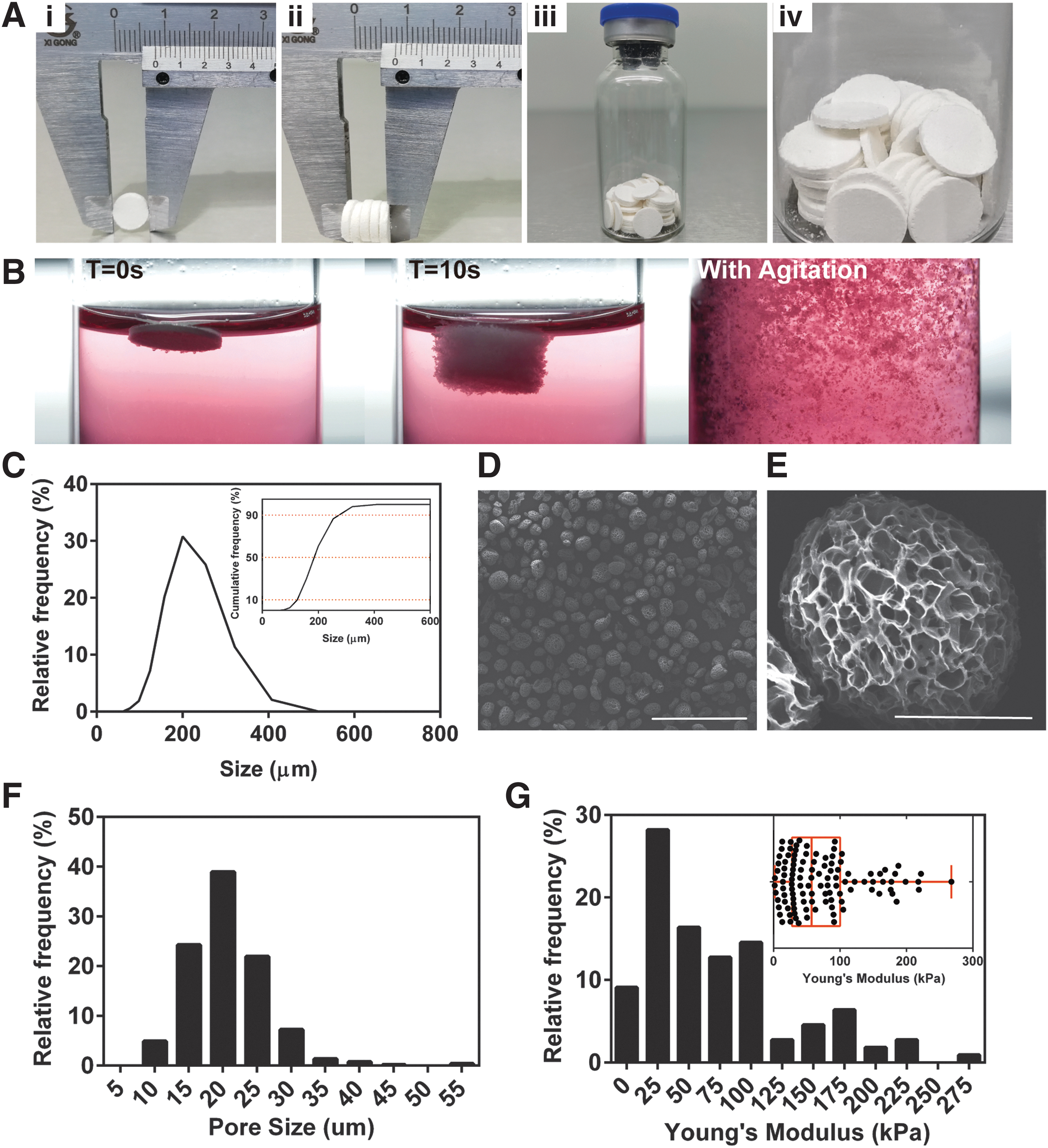
Characterization of 3D TableTrix™.
At a density of 4893 ± 288 microcarriers/mg and a D10–D90 size distribution between 125–273 μm (hydrated, Fig. 1C and insert), 3D TableTrix pack over 9 × 10⁴ microcarriers into each 20 mg tablet, offering at least 9000 cm2/g surface area, calculated based on the modest assumption that these microcarriers are perfectly round nonporous solid spheres. Microcarriers packed in 3D TableTrix, however, are in fact highly porous (Fig. 1D, E) with pore sizes of 20.6 ± 5.7 μm (Fig. 1F), hence it is highly possible that much more surface area than theoretical value is available for high cell density culture to realize efficient cell production. The median local Young's Modulus of these microcarriers (i.e., Young's Modulus sensed by cells attached to walls in these porous microcarriers) were tested to be 57.23 kPa (Fig. 1G insert) as measured with AFM, with a majority of elasticity of modulus falling between 25 and 100 kPa (Fig. 1G).
Inoculation protocol for ready-to-use 3D TableTrix in stirred culture system
Agitation played a significant role in affecting cell attachment to microcarriers. In this work, laboratory-scale stirred systems, that is, spinner flasks, were used to investigate effects of three different agitation modes, namely delayed, constant, and intermittent, on cell attachment within the first 24 h of inoculation. Significantly lower attachment rate (80.2% ± 1.2%) was observed for constant agitation compared with delayed or intermittent inoculation protocol (94.6% ± 1.2% and 96.6% ± 0.5%, respectively) at 2 h postinoculation (Fig. 2A), as attested by fluorescence staining of cells, which showed several cells not attached to microcarriers for constant agitation (Fig. 2B).
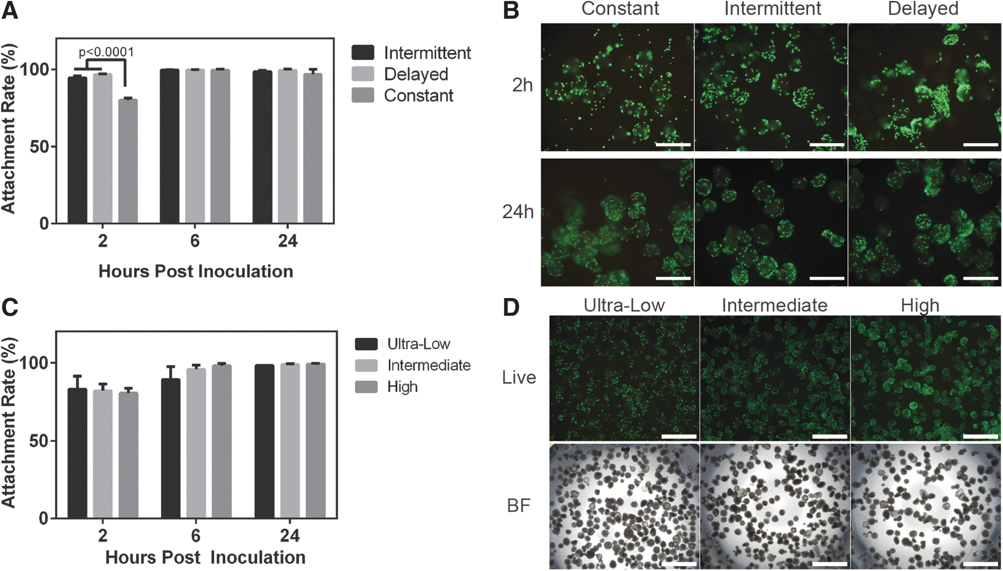
Inoculation protocol for 3D TableTrix.
Another parameter affecting cell attachment rate was the ratio between cells and microcarriers. Around 1 × 10⁶ (ultra-low), 5 × 10⁶ (intermediate), and 10 × 106 (high) AD-MSCs were inoculated to five microcarrier tablets (0.1 g) in 40 mL medium for this investigation. Even at a constant agitation of 60 rpm, more than 80% attachment rates were observed for all inoculation densities at 2 and 6 h, more than 98% of all cells were attached to microcarriers at 24 h postinoculation (Fig. 2C, D).
Dynamic expansion of hMSCs cultured on 3D TableTrix in spinner flasks
Results in Figure 2 demonstrated that cell attachment to 3D TableTrix was highly efficient. Comparison of cell growth for different inoculation densities was then analyzed. It was evident from cell enumeration that after the initial cell attachment phase, the log phase expansion began from day 2 (Fig. 3A). Total cell yield for ultra-low, intermediate, and high inoculation density after 4 days of culture were 2.98 ± 0.30 × 10⁷, 8.48 ± 1.75 × 10⁷, and 9.92 ± 0.63 × 10⁷, respectively (Fig. 3A). These corresponded to expansion factors of 29.8 ± 3.0, 17.0 ± 3.5, and 9.92 ± 0.63, where expansion factors for ultra-low density became significantly higher than those of intermediate and high seeding density from day 3 (Fig. 3B). As there had been reports that found seeding MSCs at low densities (e.g., 10–1000 cells/cm2) promoted cell proliferation rate and maintained better trilineage potential, 14 seeding density of 2 cells/microcarrier (equivalent to 2 × 10⁶ cells for 10 tablets) was used for subsequent experiments.

Expansion of hMSCs on 3D TableTrix in spinner flask.
As it had been reported that medium compositions, such as serum or growth factor concentrations, may affect cell attachment and expansion on microcarriers,15,16 we then investigated if medium composition would affect 3D TableTrix performance in dynamic expansion of AD-MSCs, and specifically we tested three different culture mediums, that is, CDM, SFM, and MSCM. AD-MSCs were able to proliferate well on 3D TableTrix in all three cell culture mediums, although a slightly lower final yield on day 4 when cultured with CDM (Fig. 3C). Nonetheless, cell viability of higher than 90% is assured in these three culture mediums (Fig. 3D), suggesting that successful culture of AD-MSCs on 3D TableTrix was not restricted by medium composition, further affirming the potential application of 3D TableTrix in large-scale production of clinical-grade hMSCs in which Good Manufacturing Practice (GMP)-compliant SFM or CDM are preferred.
To demonstrate the compatibility of 3D TableTrix to various sources of hMSCs, dynamic expansion of UC-MSCs is also verified. Total yield of 1.09 ± 0.19 × 10⁷ cells (5.47 ± 0.93-fold expansion) were harvested from 10 tablets in 120 mL working volume after 4 days of SFM culture, with 92.7% ± 6.5% cell viability (Fig. 3E). Live/dead staining of cells on microcarriers attested the growth of UC-MSCs on microcarriers (Fig. 3F).
Bead-to-bead transfer enables continuous expansion
Cells would eventually stop proliferation when the surface area available for growth become limited. To continue expanding cells for higher yield, they would have to be provided with more surface area, which in typical monolayer cultures would mean harvesting them from the culture flasks and inoculating them to more new flasks to increase surface areas. For microcarriers, we could realize continuous expansion without having to harvest cells and reinoculating, but to supply new microcarriers at adequate time points of the culture process so that sufficient surface areas were available for the expanding population of cells. This expansion strategy would require bead-to-bead transfer technology, which was a process where cells migrate from microcarriers, during culture and cell division, to fresh microcarriers.6,17
The effect of agitation protocols on effective bead-to-bead transfer was first examined in spinner flasks by inoculating microcarriers laden with 3 × 10⁶ cells (“microtissues,” Fig. 4A) to 10 tablets. Agitation was set to intermittent or constant (agitation protocols investigated for free cell inoculation) for the first 24 h and then constant agitation at 60 rpm for the rest of the culture period. Confocal imaging attested that cells could migrate from one microcarrier to another as bridges of cells could be seen between microcarriers after fresh microcarriers were inoculated (Fig. 4B). Fluorescence staining of live cells further confirmed that cells could successfully transfer to new microcarriers (Fig. 4C). No significant difference in final cell yield or cell viability were observed in both protocols (Fig. 4D).
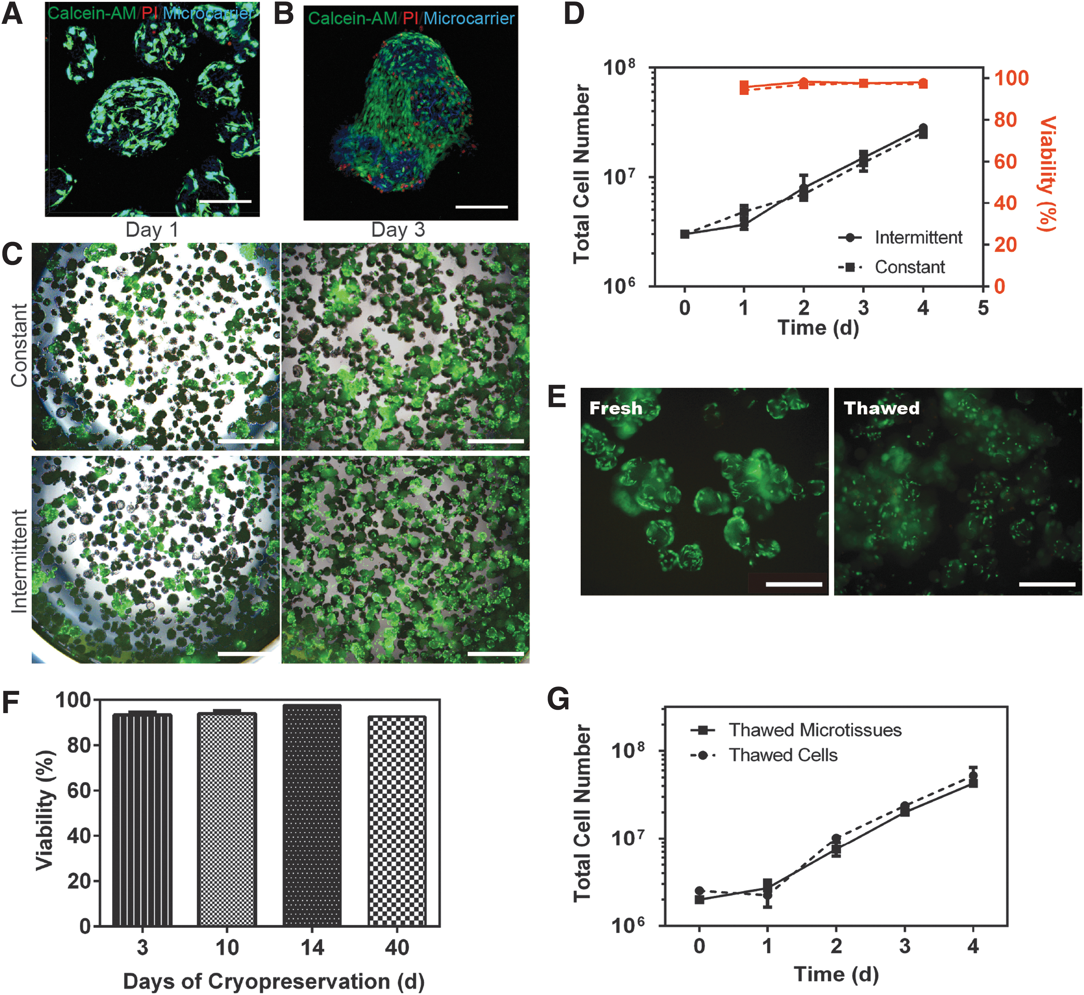
Bead-to-bead transfer with microtissues.
A unique feature of 3D TableTrix is its compatibility with conventional cryopreservation technology, cells remained attached to microcarriers (Fig. 4E) and retained more than 90% viability upon thawing after cryopreservation for 3, 10, 14, and 40 days (Fig. 4F). Cell expansion was also successful using thawed cell-laden microcarriers (microtissues), which was not significantly different from using thawed cells as starting seeds for expansion (Fig. 4G), thus demonstrating the feasibility of storing microtissues as seeds for bead-to-bead transfer in subsequent batches of culture.
Scalable production of hMSCs on 3D TableTrix in stirred tank bioreactor
With the feasibility of bead-to-bead transfer on 3D TableTrix verified, scale-up to larger culture volume was performed. Stirred tank bioreactors are scalable, robust, and well-controlled bioprocessing systems that facilitate microcarrier-based suspension culture for large-scale production of adherent cells.6,18 Hence, a stirred tank bioreactor with a 1-L glass vessel with temperature and gas control (Fig. 5B) was used to illustrate scalability of hMSC production using 3D TableTrix microcarriers using microtissues obtained from laboratory-scale spinner flasks (Fig. 5A).
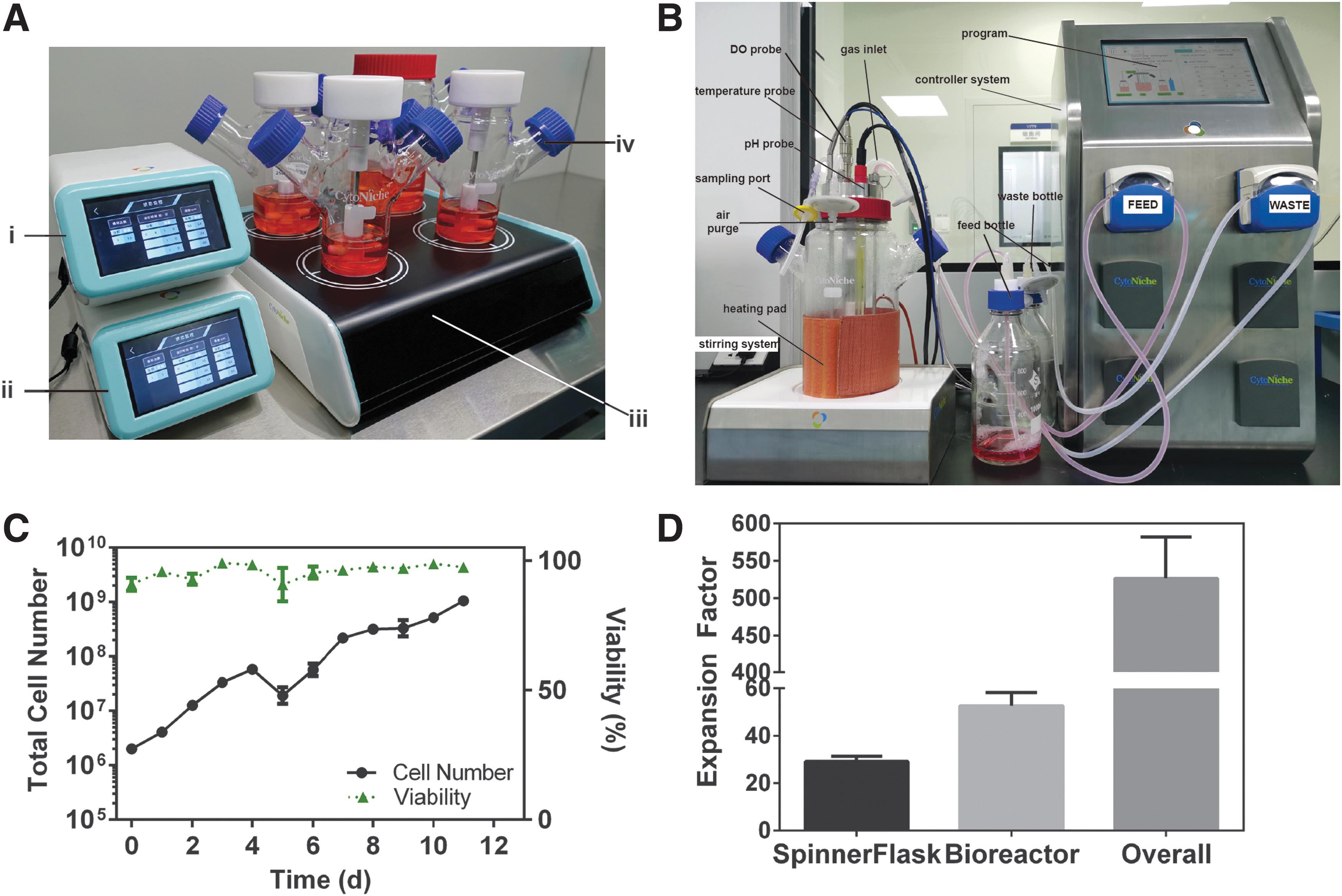
Scale-up of AD-MSC expansion to 1-L bioreactor.
Starting with 2 × 10⁶ cells with 10 tablets in a 125-mL spinner flask, 5.82 ± 0.45 × 10⁷ AD-MSCs (29.1 ± 2.2-fold expansion) were yielded after 4 days of culture (Fig. 5C, D). Microtissues (microcarriers laden with cells) containing 2.0 × 10⁷ cells from the spinner flaks were aliquoted and inoculated to 1 g (50 tablets) microcarriers in a 1-L bioreactor and cultured for another 7 days. Total final yield achieved was 1.05 ± 0.11 × 10⁹ cells (97.5% ± 0.8% viability) from these microcarriers (Fig. 5C). Thus, more than 500-folds of expansion was realized in 11 days (Fig. 5D).
Cell recovery and cell characterization
The 3D TableTrix was a dissolvable microcarrier and hence all cell enumeration and harvesting in this article were accomplished by fully dissolving 3D TableTrix microcarriers with its specific dissolution reagent, 3D FloTrix Digest (CytoNiche, China). The 3D TableTrix microcarriers could be fully dissolved within 30 min under gentle agitation as observed under microscope (Fig. 6A). A 98.6% ± 0.1% cell recovery rate was accomplished and cell viability was maintained at above 90% (Fig. 6B). Harvested cells were then characterized for their immunophenotype, trilineage differentiation ability, and karyotype stability. 3D TableTrix-expanded AD-MSCs retained their phenotypic surface markers where expression was >98% and <1% for positive and negative markers (Fig. 6C), meeting the criteria set by International Society of Cell Therapy (ISCT). 1

Cell recovery and quality assessment.
Cells also maintained their ability to differentiate into adipogeneic, osteogenic, and chondrogenic lineages (Fig. 6D). In comparison with 2D counterparts (population doubling were 2.47 in one passage, 3–4 days culture, harvested at 80–90% confluence), cells harvested from 3D TableTrix expanded in the 1-L bioreactor scale-up process had a total population doubling of 10.6 in 11 days (4.86 in 4-day spinner flask culture and additional 5.71 in subsequent 7-day bioreactor), thus concerns with excessive expansion leading to genome instability and senescence were raised. Despite the higher population doubling, karyotyping affirmed genome stability (Fig. 6E) of microcarrier-expanded cells and these AD-MSCs did not show increased β-galactosidase activity as characterized with SA-β-gal staining in comparison to their 2D counterparts (Fig. 6F).
Discussion
Microcarrier selection is one of the most important choice to make for successful large-scale expansion of hMSCs in stirred tank bioreactors.4,18,19 Efficient cell seeding protocol is essential as cell attachment to microcarriers is critical to the success of expansion in stirred tank bioreactors. High percentage and uniform attachment of cells to initiate cell expansion is desired. Important parameters affecting cell attachment include surface chemistry of microcarriers, agitation protocols, and cell and microcarrier concentrations, among others. 18 As 3D TableTrix microcarriers are made of gelatin, it is presumed that cell adhesion motifs were inherently available20,21 and thus no precoating is performed for these microcarriers for cell attachment. Also, since these microcarrier tablets are sterile and readily absorbed liquid to disperse into individual hydrated microcarriers, they can be introduced directly into culture medium together with cell suspensions without weighing, rehydrating, autoclaving, or other prior preparations.
Agitation mode is another important parameter investigated. Literatures had recommended a 20-h delay of agitation after cell inoculation to aid cell attachment, 22 or intermittent agitation between high and low speed (or intervals of agitated and non-agitated periods) to maximize cell/microcarrier interaction with homogenous distributions facilitated by agitation and cell anchorage to microcarriers during low or no agitation periods.6,13,18,23,24 While no agitation achieved the best efficiency of 99.7% ± 0.1% and 99.4% ± 0.8% at 6 and 24 h postinoculation respectively, our study did not find significant differences in attachment rate and uniformity for different agitation protocols. Cell inoculation density for microcarriers ranging from 0.6 to 30 × 103/cm2 (corresponding to less than 1 cell/microcarrier to over a 100 cells/microcarrier ratio) have been reported,4,8,9,13,17,19,23–28 with 6000 cells/cm2 being a common choice of density in a number of studies. As inoculation density varied largely in literature, we investigated densities that cover the range of densities reported. By inoculating 1 × 10⁶, 5 × 10⁶, and 1 × 10⁷ cells to five tablets of 3D TableTrix (0.1 g which gave a total of at least 900 cm2 surface area and nearly 5 × 10⁵ microcarriers), we were able to get inoculation densities of at most 1111 cells/cm2 (ultra-low), 5555 cells/cm2 (intermediate), and 11,111 cells/cm2 (high) or about 2, 11, 22 cells per microcarrier, respectively. Our study found that these three densities were all suitable cell/microcarrier ratio for efficient inoculation on 3D TableTrix.
In comparison to most microcarriers investigated thus far, performance of 3D TableTrix is unprecedent. In our experiments, we have demonstrated that 3D TableTrix could achieve a yield of up to 2.48 × 10⁶ cells/mL in 4 days from 40 mL culture volume in 125-mL spinner flask. Most commercial microcarriers investigated thus far were nonporous spheres made of plastic or dextran (e.g., Cytodex, Hillex, Plastic, Synthemax II), which typically yielded 1–3 × 10⁵/mL for AD-MSCs13,26,29 and 1–8 × 10⁵/mL for other hMSCs.13,30,31
Theoretically, use of macroporous microcarriers would provide higher surface area per unit volume to achieve higher cell density, however, investigations found that macroporous CultiSpher did not outperform nonporous microcarriers in cell yield. In one study, only 2.5 × 10⁴/mL bone marrow-derived (BM) MSCs were grown on CultiSpher after 144-h culture, while Plastic or collagen-coated plastic microcarriers more than triple the yield. 8 Other study on fetal MSCs also demonstrated that CultiSpher performance was poorest among the microcarriers investigated, with Cytodex-3 and Plastic being the best, yielding 6.8 × 10⁵ cells/mL. 31 While a study showed that Cultispher had higher cell adhesion rate for AD-MSCs than Plastic (88% vs. 69%), the eventual yield was not significantly different for these two microcarriers, expanding about 14 times in 7 days to reach an eventual 1.4 ± 0.5 × 10⁵ cells/mL. 13 Another study also demonstrated that CultiSpher only yielded 4.2 × 10⁵/mL bone marrow-derived MSCs. 16
These data (2.5 × 10⁴–4.2 × 10⁵/mL) for CultiSpher are mainly obtained from cells cultured in 100-mL spinner flasks for 6–8 days (with culture volume ranging from 50 to 100 mL), and in comparison, 3D TableTrix yielding 7.45 × 10⁵–2.48 × 10⁶ cells/mL in 125-mL spinner flask (with 40 mL culture volume, and depending on inoculation density) within 4 days show evidently superior performance. As these two microcarriers are of similar chemical composition and macrostructure, the difference in performance between these two microcarriers would require more systematic studies to identify the performance-determining differentiation factor. Nonetheless, our study on 3D TableTrix provides some evidence to support the theory that porous microcarriers could yield more cells than nonporous microcarriers, which potentially offer more advanced expansion configuration for stem cell manufacture.
We also further demonstrate that with bead-to-bead transfer technology, scaling up hMSCs expansion on 3D TableTrix could be realized without enzymatic dissociation of cells from microcarriers. We demonstrate over 500 times expansion of AD-MSCs in SF culture media, starting with 2 × 10⁶ cells and yielding 1.05 × 10⁹ cells in 11 days with bead-to-bead transfer from 125-mL spinner flask to a 1-L bioreactor. As only 34.3% of the cells from the spinner flask was used as starting materials during the 1-L bioreactor scale-up, we anticipate that yield of over 3 × 10⁹ cells with an overall 1530 times expansion could theoretically be realized with full scale-up.
With bead-to-bead transfer, it was found that cells could migrate from one microcarrier to another microcarrier during bead-to-bead transfer to populate fresh microcarriers added to the culture system, greatly simplifying the process of passaging cells in a scaled-up system. The ability for cells to form cell bridges between microcarriers as they migrate during bead-to-bead transfer raise concerns that large aggregations of microcarriers would form during long-term culture and thus hindering mass transfer and further expansion of cells. We did observe that loose clusters would form as cell population on microcarriers increased, however, these clusters could easily break up with gentle pipetting and agitation. No increase of cell deaths were found in these clusters too, as observed by live/dead staining. As these microcarriers are highly porous, we believe that gas and nutrient could still be adequately supplied to cells even when these loose clusters formed. Regardless, as reported by a literature that investigated means to limit aggregation during MSC expansion on microcarriers, 25 optimizing the time for fresh microcarrier addition could prevent large clusters from forming, as the increasing cell population would be suitably distributed to more surface areas provided by the new microcarriers.
Besides expansion efficiency, effectively harvesting cells from microcarriers is equally important for bioprocesses that need cells as final products. Currently, cell harvesting process is one of the most challenging step in ensuring hMSC cell quantity and quality. 19 Most commercially available microcarriers are nonbiodegradable and require additional filtration steps to separate and harvest cells from microcarriers,23,32 which cost loss of cells. Hence, some studies have favored biodegradable microcarriers, such as gelatin-based CultiSpher, so that cells could be released by fully dissolving microcarriers, which simplify downstream purification processes without having to physically remove microcarriers from final desired living cell products.9,16 Other than CultiSpher, reports of commercially-available biodegradable microcarriers are few, of which Corning dissolvable microcarriers are noteworthy and have been studied for scalable iPSCs expansion. 33 In this study, we not only achieved high expansion efficiency with 3D TableTrix, high cell recovery rate of 98.6% ± 0.1% was also realized as 3D TableTrix could be fully dissolved using respective dissolution reagent. This eliminates complex downstream processing (i.e., filtration and separation) to ensure higher cell yield as compared with nonbiodegradable microcarriers. Cells harvested were of high vitality and retained critical immunophenotypes and functionality.
In addition to using microcarriers for cell expansion, we find that there is increased interest in developing biodegradable microcarriers for both in vitro expansion and in vivo therapy. Some of these promising microcarriers are made of biodegradable polymers, such as polycaprolactone (PCL) and poly(lactic-co-glycolic acid) (PLGA). Lam et al. fabricated porous PCL microcarriers coated with extracellular matrix proteins for expansion of hMSCs, and they successfully expanded MSCs from bone marrow, cord blood, fetal, and Wharton's jelly, with a yield of 3.6–4.4 × 10⁵/mL in either 100-mL spinner flask or 1-L bioreactor in 5 days, which was comparable to commercial microcarrier Cytodex-3. 34
Besides using these microcarriers for expansion, this research team further explored the potential of these microcarriers as cell delivery tools for tissue engineering purposes. The team was able to induce bone formation when MSC-laden PCL microcarriers were implanted in a mouse model. 35 Another team working on PLGA microcarriers was able to expand 3.2-folds of MSCs in 12 days and then induced these MSCs into smooth-muscle-like cells for potential transplantation into smooth muscle tissues. 36 Other than synthetic microcarriers, natural biomaterial-based carriers had also been used to deliver cells. Li et al. have developed gelatin-based porous microscaffolds as injectable microniches for efficient MSC delivery, thus enabling low-dosage treatment of lower limb ischemia in mice. 37 Commercial gelatin-based microcarriers such as CultiSpher and Spheramine had also been reported as cell delivery agents for tissue repair.38–40 The advantage of such biodegradable microcarriers is that cells do not have to be detached from their adhesion surface after expansion for transplantation, which would otherwise be detrimental and result in poor clinical outcomes. 36 Other microcarriers that could serve as cell delivery tools for tissue engineering are reviewed extensively elsewhere.10,41–43 In this retrospective, gelatin-based 3D TableTrix, with its possibility of been stored as cryopreserved microtissues (cell-laden microcarriers), has high potential for use as part of cell therapy strategies to deliver cells for enhanced therapeutic effects.
In conclusion, 3D TableTrix as a newly available microcarrier option on the market has displayed its tremendous potential for industrial production of hMSCs with its results reported in this study. The ingenious design of 3D TableTrix as dispersible and dissolvable porous microcarrier tablets eliminates the need of tedious preparation work usually required for microcarriers, and with bead-to-bead transfer eliminating the need for enzymatic dissociation during passaging, bioprocesses involved in scaling up hMSCs expansion could be greatly simplified. Not only so, cells could be cryopreserved in situ on 3D TableTrix microcarriers and be successfully thawed with high vitality and retain the 3D macrostructure, this providing an option to store cell-laden microcarriers (microtissues) as seeds for bead-to-bead transfer in subsequent batches of culture, as well as for use as implantable carriers to aid cell therapy.
While our results supersede microcarriers currently reported in literature, more theoretical and practical considerations1,22,44 need to be investigated to realize the full potential of 3D TableTrix microcarriers for hMSCs production, including potential risks of aggregation, ease of removing any microcarrier-associated chemical residues in the final cellular products, and more comprehensive analysis of cell quality. 1 Nonetheless, with this high-performance microcarrier, it is expected that cost-effective, industrial-scale production of quality hMSCs or MSC-derived products, such as exosomes, under a controllable process compliant with GMP practices5,6,10,19,44 will soon be possible.
Footnotes
Acknowledgments
The authors would like to acknowledge the help from Mr. Kun Hou and Mr. Le Li for the help with microcarrier characterizations. They thank Mr. Yuyang Chen from Prof. Yanan Du's laboratory for isolating and providing AD-MSCs. They thank Mr. Fan Gao and Mr. Zheng Wang for support on videos and animations. They also thank Prof. Yan Shi and Dr. Tie Xia from School of Medicine, Tsinghua University for technical support for AFM.
Disclosure Statement
Y.D. is scientific advisor and X.Y., K.Z., Y.Y., D.D., H.X., and W.L. are employees of Beijing CytoNiche Biotechnology Co. Ltd. The other authors have no commercial, proprietary, or financial interest in the products or companies described in this article.
Funding Information
This work is financially supported by Beijing Municipal Science & Technology Commission (Z181100001818005), National Key R&D Program of China (2017YFA0104901), and R&D program (RD03) of Beijing CytoNiche Biotechnology Co. Ltd.

References
Supplementary Material
Please find the following supplemental material available below.
For Open Access articles published under a Creative Commons License, all supplemental material carries the same license as the article it is associated with.
For non-Open Access articles published, all supplemental material carries a non-exclusive license, and permission requests for re-use of supplemental material or any part of supplemental material shall be sent directly to the copyright owner as specified in the copyright notice associated with the article.
