
Abstract
Hydrogel microspheres have been widely used as cell carriers and three-dimensional cell culture matrices. However, these microspheres are associated with several unfavorable properties for bone tissue engineering applications, for example, their surface is too smooth to attach cells and they do not contain inorganic materials. This article presents a new method to overcome these disadvantages by depositing CaCO3 crystals on the hydrogel microsphere surface. Specifically, we used a nonplanar flow-focusing microfluidic device to produce gelatin methacrylate (GelMA)-/Na2CO3-based microspheres. We subsequently obtained CaCO3 crystals by a chemical reaction between Na2CO3 and CaCl2. The efficacy of this method was demonstrated by in vitro experiments with human umbilical vein endothelial cells (HUVEC) and immortalized mouse embryonic fibroblasts (iMEF). Cell culture on GelMA/CaCO3 microspheres showed that cells can easily attach and adhere to GelMA/CaCO3 microspheres and maintain high viability. Alkaline phosphatase (ALP) expression was increased as well. These results suggest that this novel microsphere has a high potential for bone tissue engineering applications.
Impact statement
Microspheres as cell culture substrates have attracted a great deal of attention. The combination of organic and inorganic materials offers the unique merits in bone tissue engineering. In this study, there are two contributions. First, the organic and inorganic material of gelatin methacrylate (GelMA) and CaCO3 were successfully combined, especially, CaCO3 was formed as crystals to enhance cell attachment. Second, microspheres were successfully fabricated with one-step process: that is, the microfluidic technique was coupled with the CaCO3 precipitation in situ. Cell culture shows that the GelMA/CaCO3 microspheres proposed in this study have a high potential for bone tissue engineering applications.
Introduction
Microspheres with diameters varying from 1 to 1000 μm have wide application, such as in chemical analysis, biology research, pharmacy, diagnostics, and food industries.1–8 In literature, several conventional approaches have been reported to produce microspheres, such as agitation, sonication, and so on.9,10 Agitation promotes the emulsification of a polymer solution in an aqueous solution through mechanical stirring, which usually results in the quick and bulk formation of microspheres. 11 Sonication can form small microspheres even at nanoscale, which improves the dispersion of microspheres by separating possible microsphere aggregations. 12 However, both methods have poor control over the size and uniformity of microspheres. 13 In contrast, microfluidics is a promising approach to produce microspheres due to its high yield capability and great monodispersity.14,15 Microfluidics takes full advantage of single and double emulsion techniques,16,17 the latter of which utilizes two immiscible fluids to form particles through the breakup of fluid flow in a flow-focusing or T-junction confluence.18–20 Recent studies have shown that the microfluidics approach can produce porous structures, in particular, two-dimensional (2D) membranes and 3D scaffolds with a well-defined geometry, such as honeycomb-like scaffolds, by one-step simple setup at low cost.21,22
Gelatin methacrylate (GelMA) is a widely recognized photo-cross-linkable biocompatible material that has been studied in literature.23–25 GelMA microspheres, also referred to as GelMA microgels, have many biological applications, including drug delivery, 26 3D cell culture,27,28 and tissue engineering. 29 In particular, for stem cell expansion application, microspheres can ensure the quality of stem cells being cultured due to their uniform size distribution and essential active factor encapsulating abilities within the microspheres, as prepared by microfluidics.30–32 However, microspheres made of hydrogels, including GelMA microgels, have a smooth surface and lack essential properties, such as osteoconductivity, and mechanical strength. Bones are composed of living cells embedded in a dense framework of organic matters such as collagen and saturated with the minerals calcium and so on. GelMA microspheres are only organic matters; the absence of inorganic compounds in these microspheres is a disadvantage for their application in bone tissue engineering.27,28
Different methods have been proposed to modify the microspheres to overcome the above-mentioned limitations. Choi et al. 33 fabricated graphene-attached poly (methyl methacrylate [MA]) microspheres to improve their thermal conductivity. Hwang et al. 34 decorated the surface of microspheres with silver nanoparticles through electroless deposition for molecular detection by surface-enhanced Raman scattering. Lu et al. 35 synthesized amphiphilic microspheres based on hydrophilic, pH-responsive acrylic acid (AA) and hydrophobic, nonionic n-butyl acrylate (BuA), which were shown to be able to encapsulate and deliver both hydrophobic and hydrophilic moieties. Cha et al. 30 demonstrated a facile and efficient approach for in vitro cell culture and injectable tissue engineering by culturing cardiac side population cells on GelMA microsphere surfaces. Baki et al. 36 used oxygen plasma treatment to modify the surface of injectable PLGA/GelMA microspheres, which can also enhance the proliferation rate of attached cells. Ghorbani et al. 37 cultured L929 cells on microspheres and studied their adhesion, spreading, and proliferation process. The cells were able to attach and spread on the constructs with many pseudopodia, and the cell viability was more than 90% after 2 weeks of incubation, which confirmed the potential of gelatin-siloxane microspheres for bioregeneration application. Zhuang et al. 38 fabricated GelMA hydrogels containing glial cell line-derived neurotrophic factor (GDNF). They observed that axonal regeneration and functional recovery in the GDNF-loaded microspheres group were similar to the autograft group. They concluded that GDNF-loaded gelatin microspheres combined with GelMA hydrogels can serve as a new biodegradable artificial nerve guide for nerve tissue engineering.
A disadvantage of the above-mentioned studies is that they do not allow the production of sufficiently regulated nanoscale structures on the surface of microspheres. Recent developments in microfluidics have shown that the T-junction microfluidics combined with self-assembly technique can produce nanoparticle modifications on the surface of microspheres in a highly ordered manner in a one-step process, which makes the microspheres applicable for a wide range of purposes.21,22,39 For example, Gultekinoglu et al. 39 produced poly(lactic-co-glycolic acid)-block-poly(ethyleneglycol) (PLGA-b-PEG) microspheres with nanoparticle modifications on their surface by a T-junction microfluidic device combined with a self-assembly process.
In bone regeneration, the microsphere and nanosphere structures are used to reduce the side effects and immune rejection of implants and scaffolds. 37 In addition, microspheres and nanospheres can be used to manufacture injectable scaffolds to be used in minimally invasive procedures. In view of this, Gong et al. 40 produced CaCO3 microspheres loaded with bone morphogenic protein-2 (BMP-2). The use of CaCO3 has the advantage that CaCO3 possesses osteoconductivity.41–43 Although the BMP2-loaded CaCO3 microspheres were found to induce osteogenic differentiation of bone marrow stromal cells and to stimulate bone healing, a disadvantage was that the size of the microspheres was in the range of a few micrometers with wide size distributions. 40 In addition, the size of the microspheres was too small to use them as an effective carrier of stem cells in cell-based bone regeneration procedures. 27 Also, growth factors are difficult to incorporate in microspheres produced with agitation. 30
In this article, a novel method is reported to produce GelMA/Ca2CO3 microspheres by using a nonplanar flow-focusing microfluidic device. The potential of these microspheres for 3D cell culture in bone regeneration application was explored in human umbilical vein endothelial cells (HUVECs) and immortalized mouse embryonic fibroblast (iMEF) cell cultures.
Methods
A flow-focusing microfluidic device was fabricated using polydimethylsiloxane (PDMS) replication molding as instructed by Eteshola and Leckband 44 and Bodas's and Khan-Malek 45 approach. Sylgard 184 PDMS (Dow Corning, Midland, MI, USA), mixed with the cross-linker at a ratio of 10:1, was degassed for 30 min in a vacuum freeze dryer (BiLon, Shanghai, China), and then poured onto the silicon wafer engraved with microchannel structures (Fig. 1a–c). The microfluidic structures were cured in a drying baker (Fuma Instruments, Shanghai, China) at 80°C for about 2 h ensuring the membrane was completely compliant. Finally, the PDMS replica was peeled off from the wafer with the pattern, and then activated on both surfaces through oxygen plasma treatment machine (Mingheng Corp., Chengdu, China); the replica was bonded to another one that had been bonded to a clean glass slide. The microchannel had two inlets with different heights of 50 and 150 μm, respectively (Fig. 1d).

Nonplanar flow-focusing microfluidic device.
Subsequently, GelMA was synthesized by following Nichol's 46 instruction. In brief, 10% (wt/v) of type A porcine skin gelatin was mixed into Dulbecco's phosphate-buffered saline (PBS) and constantly stirred at 55°C until fully dissolved. 10% (v/v) MA was subsequently added to the gelatin solution under stirred conditions at 55°C and allowed to react for 3 h. The fraction of lysine groups reacted was modified by varying the amount of MA present in the initial reaction mixture. Following a 2 × dilution with additional warm (37°C) PBS to stop the reaction, the mixture was dialyzed against distilled water using 12–14 kDa cutoff dialysis tubing for 1 week at 37°C to remove salts and methacrylic acid. The solution was lyophilized through a vacuum freeze dryer (FD-1A-50 Lyophilizer; Shanghai Bilon Instrument Co. Ltd., China) for 1 week and stored at −4°C until use to generate GelMA microspheres.
To determine the appropriate amount of Na2CO3 for preparation of GelMA/CaCO3 microspheres, preliminary assays were performed by varying the Na2CO3 concentration from 1% to 4%. The GelMa, PEGDMA, and Igracure 2959 were fixed at concentration of, 10% (wt/v), 2% (wt/v), and 0.2% (wt/v) respectively. GelMA/Na2CO3 films were fabricated with help of a polytetrafluoroethylene (PTEE) plate (Fig. 2a). A chemical reaction was triggered by soaking GelMA/Na2CO3 films in 3% (wt/v) CaCl2 solution. The transparency of the films decreased significantly after the chemical reaction, which was caused by the precipitation of CaCO3 crystal on the film surface (Fig. 2b). A scanning electron microscope (SEM) equipped with energy-dispersive X-ray spectroscopy (EDS) was used to quantify Ca on GelMA/CaCO3 films and to provide proof for the presence of CaCO3 crystals on the films. We found that a significant amount of Ca element (Fig. 2c) was obtained at GelMA/CaCO3 films, which were produced with 3% (wt/v) of GelMA/Na2CO3. The CaCO3 crystals obtained in the 3% (wt/v) Na2CO3 group were found to have a crystal size smaller than 5 μm (Fig. 2d). It was decided to chose a 3% (wt/v) Na2CO3 concentration for further experiments.
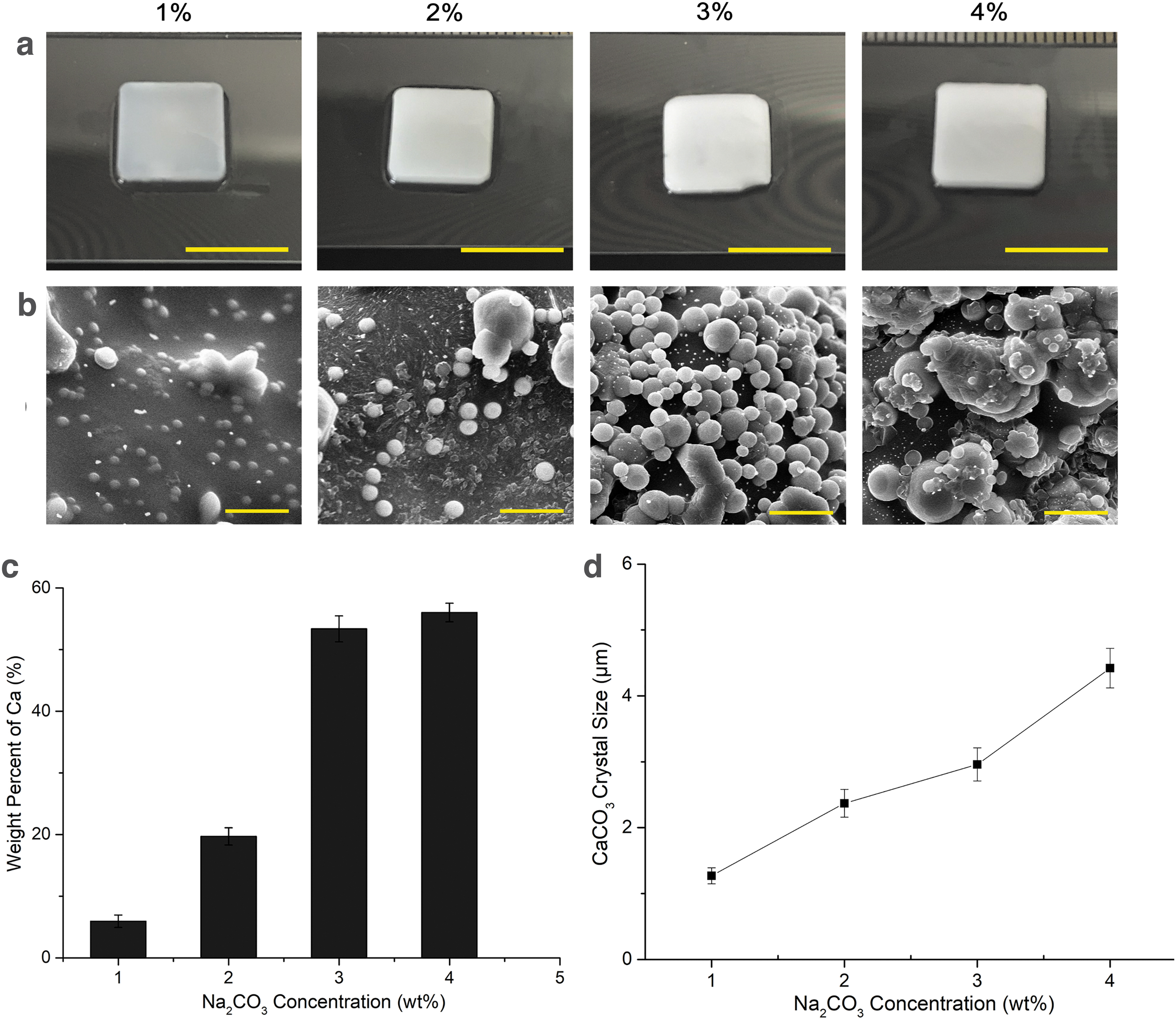
CaCO3 crystal formation at different concentrations of Na2CO3.
To further investigate CaCO3 crystal formation, the time effect on CaCO3 crystal morphology was studied (Fig. 3a) using GelMA/CaCO3 films, which were produced with 3% (wt/v) of Na2CO3. Briefly, GelMa/Na2CO3 films were fully immersed in 3% (wt/v) CaCl2 solution and allowed to react from 1 to 8 min. SEM analysis confirmed that CaCO3 crystals started to precipitate on film surfaces in 2 min and grew over time. The film surface was fully covered with CaCO3 crystals after 4 min of chemical reaction in the ambient environment. CaCO3 crystals changed into a nonspherical shape with a size larger than 5 μm after 5 min of reaction (Fig. 3b). The longer the reaction time, the larger the size. The amount of Ca followed the same trend and increased during time (Fig. 3c). On basis of these findings, a reaction time of 3 min was used in further assays.

CaCO3 crystal formation over time.
For microsphere preparation, the aqueous GelMA solution was dispersed in the continuous oil phase containing 20% (v/v) Span 80. Subsequently, this immiscible fluid was pumped into both inlets of the microfluidic device with Harvard syringe pumps (PHD Ultra, Harvard Apparatus, Holliston, MA). GelMA microspheres were generated and collected in a 5 mL centrifugal tube, and then subsequently exposed to ultraviolet (UV) light for 3 min to ensure full photopolymerization.
This method was also used to produce GelMA/Na2CO3 microspheres. Then, the aqueous phase contained 3% (wt/v) anhydrous Na2CO3, 10% (wt/v) synthesized GelMA, and 2% (wt/v) PEGDMA in PBS. Also, 0.2% (wt/v) Igracure 2959 was added into the aqueous solution as the photo-cross-linking agent. Subsequently, a chemical precipitation response between Na2CO3 and CaCl2 was applied to modify the microspheres with CaCO3 crystals (GelMA/CaCO3 microspheres). Briefly, 3% (wt/v) CaCl2 solution was added to GelMA/Na2CO3 microspheres and allowed to react for 3 min. The residual CaCl2 solution was removed from the microspheres by a syringe. Microspheres were then washed three times with deionized water to remove residual CaCl2. Modified microspheres were rinsed three times by PBS and dispersed again in PBS, and were stored in a refrigerator at −4°C after 24 h of UV sterilization. It is noticed that microspheres can easily be dispersed into a liquid phase. A centrifugal device (Cence L500, Hunan, China) was used during the process of oil removal. The relative centrifugal force was kept constant at 525 g and 3 min.
Using the microfluidic device, GelMA microspheres with high uniformity can be produced as shown in Figure 4a and b, with a mean diameter of 133 μm (Fig. 4c). The coefficient of variation (CV) is a common index used to evaluate the size distribution and monodispersity of double emulsions and microspheres. 47 CV is defined as the ratio of the standard deviation of size distribution to its arithmetic mean. Samples with a CV value of <5% were defined as monodisperse. The CV value of GelMA microspheres was found to be 2.4%, which indicated that highly monodispersed GelMA microspheres were fabricated. The mean size of GelMA/Na2CO3 microspheres was 312 μm with a CV value of 4.4% (Fig. 4d, e).
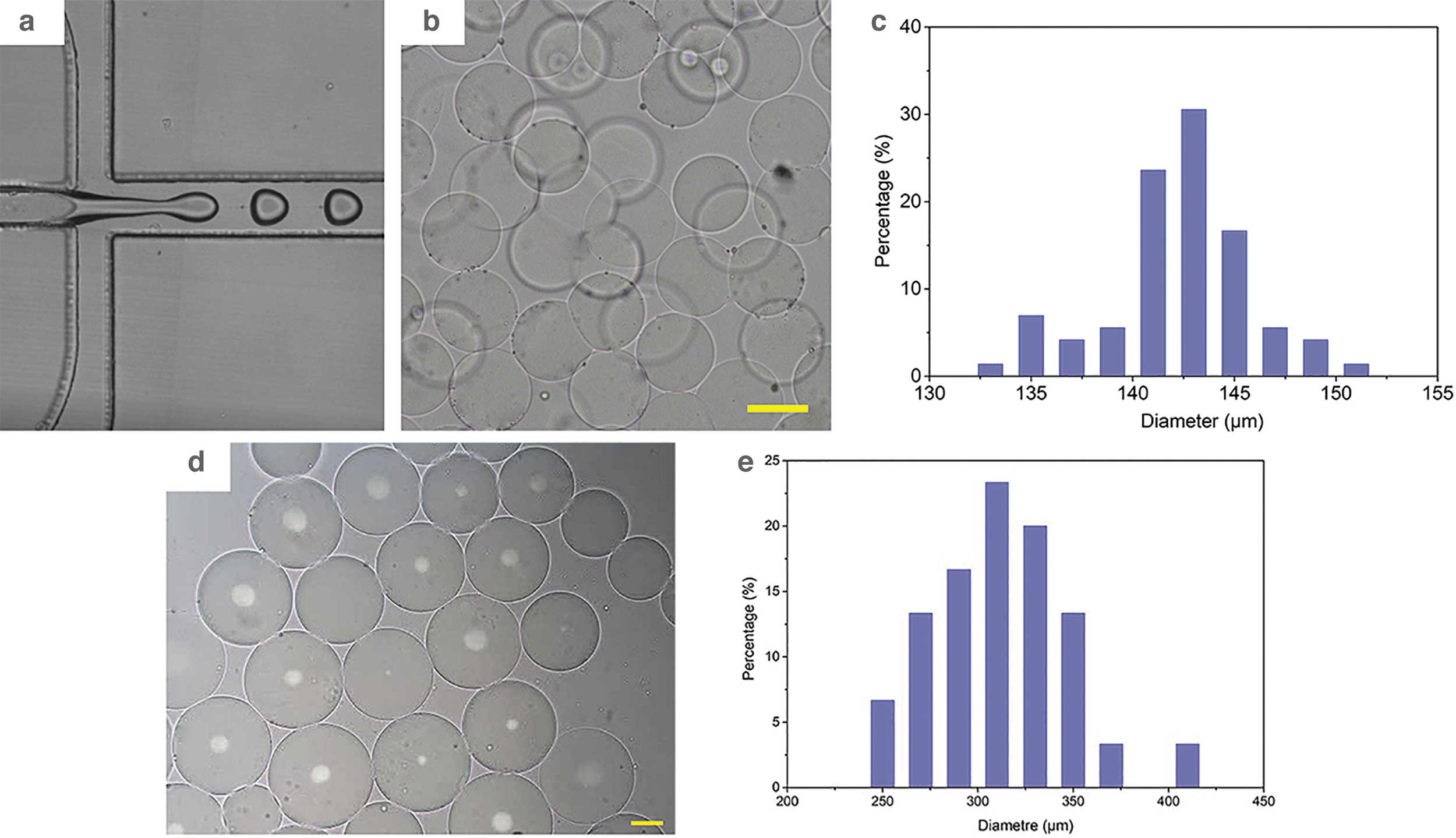
Microsphere formation.
Additional examination by optical microscopy revealed that unmodified microspheres presented a smooth and clear morphology (Fig. 5a). For the GelMA/CaCO3 microspheres, CaCO3 crystals were precipitated on the surface and within the microspheres (Fig. 5b), which resulted in a significant different morphology compared to the unmodified group. There was no sign of detachment of CaCO3 crystals from the microsphere surface after being dispersed in PBS for 1 week as shown in Figure 5c.
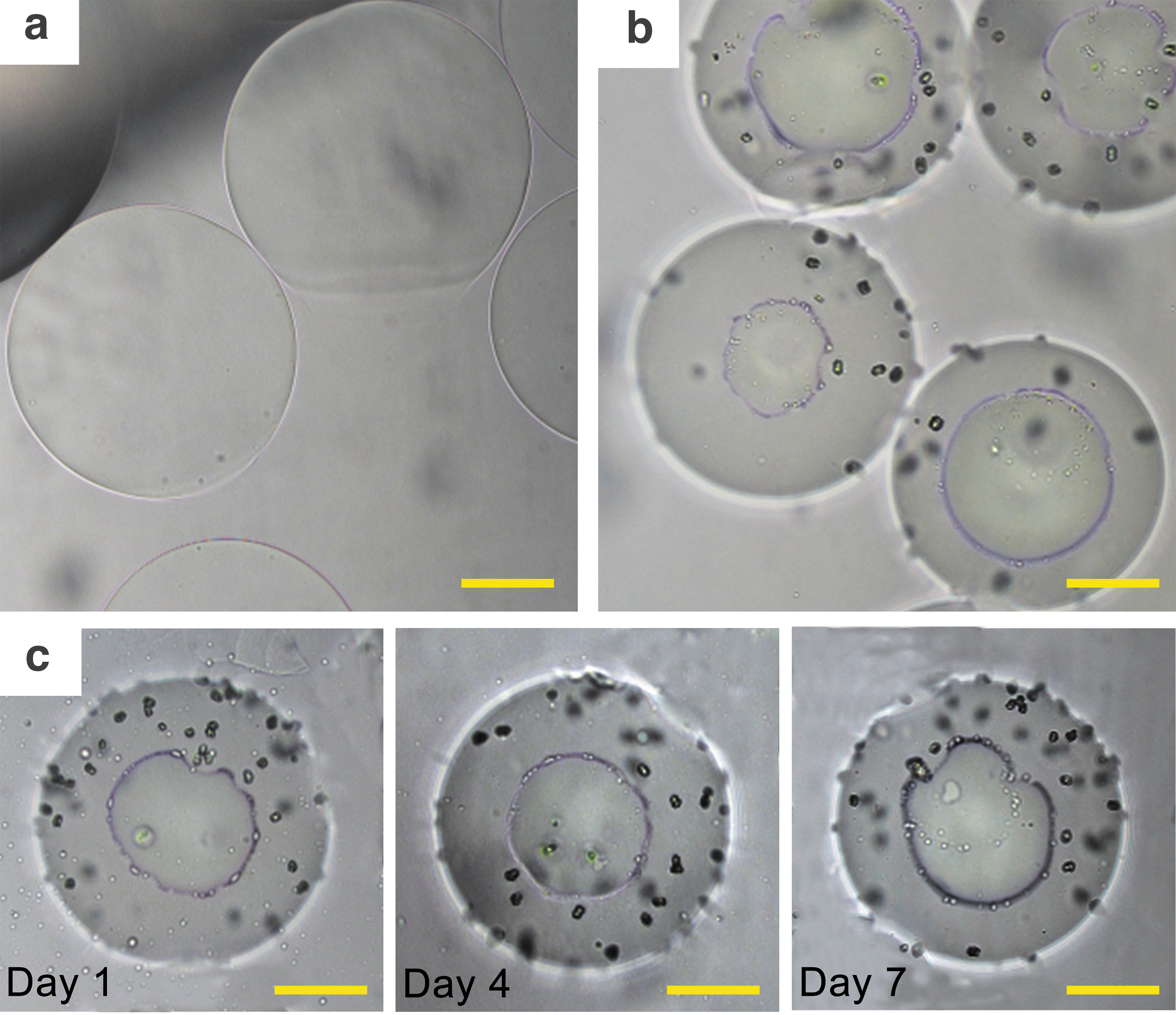
Modified and unmodified microspheres.
SEM examination was used to visualize the precipitated CaCO3 crystals on the unreacted GelMA/Na2CO3 microsphere surface (Fig. 6a, b). Higher magnification confirmed there was no sign of CaCO3 crystal deposition (Fig. 6c). In comparison, we found that the modified microspheres possessed small particles (Fig. 6d, e). At higher magnification (as shown in Fig. 6f), this was seen more clearly. EDS results indicated that the GelMA/Na2CO3 microspheres were composed of C, O, and Na in the control group, while there was no sign of the Ca element (Fig. 6g, h). CaCO3 crystals were successfully precipitated on the modified microspheres (Fig. 6i, j), as indicated by the EDS results depicted in the Figure 6i and j.
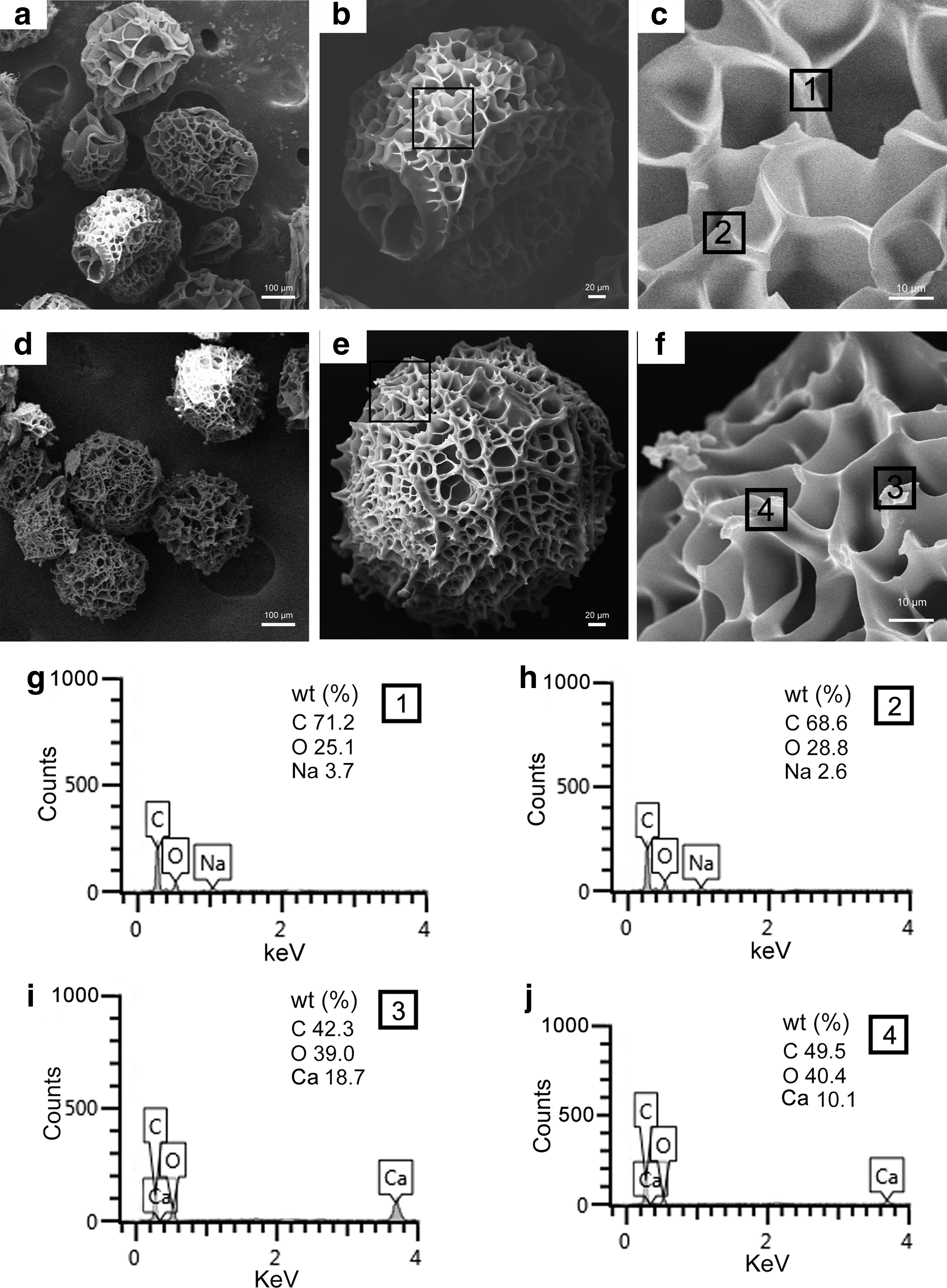
SEM image of GelMA/Na2CO3 and GelMA/CaCO3 microspheres.
Experiment
Experimental design
GelMA/CaCO3 microspheres and films
GelMA/CaCO3 microspheres were prepared in a one-step process using the described nonplanar microfluidic technique combined with a chemical precipitation process and the described manufacturing conditions. The GelMA/CaCO3 microspheres were used in cell culture assays. GelMA/Na2CO3 microspheres were used as control group. In addition to the microspheres, GelMA film samples of 10 mm in diameter precipitated with CaCO3 particles were prepared for a cell adhesion assay according to the method described by Braut-Boucher et al. 48
Cell culture
HUVECs were cultured in Dulbecco's modified Eagle's medium. The osteogenic medium contained 10 mM β-glycerophosphate, 50 μg/mL L-ascorbic acid, and 0.1 mM dexamethasone. Cells were incubated in an incubator (HERA cell 150, Thermo, Waltham, MA, USA) with 5% CO2 at 37°C.
HUVECs were cultured on the modified GelMA/CaCO3 microspheres. The unmodified GelMA/Na2CO3 microspheres were used as control group. HUVECs were detached from the cell culture dish with 0.05% trypsin (Life Technology, Grand Island, NY, USA) and were suspended at a cell density of 2 × 10⁶ cells/mL. Forty-eight-well plates were used for this cell culture, and an estimated number of 300 sterilized microspheres were placed into each well. HUVECs were subsequently added into the dish at a density of 1 × 10⁵ cells. The microspheres and cells were incubated in an incubator in an environment with 5% CO2 at 37°C, with medium changed every 3 days. The adhesion, spreading, and proliferation of HUVECs were investigated over 7 days of culturing.
Live/dead assay
Cell viability and apoptosis on the microsphere surface were measured using a live/dead assay by a calcein-AM (CAM) dye (Sigma-Aldrich) in conjunction with propidium iodide (PI; Sigma-Aldrich). The dye solution contained 2 μL CAM and 3 μL PI in 1 mL PBS, which was stored at −4°C away from light. Each sample was fixed in 4% paraformaldehyde (PFA) solution for 20 min and rinsed with PBS three times before fluorescent characterization of cells. Cells were stained by 500 μL CAM/PI dye solution for 20 min in a Φ35 mm culture dish. The dye solution was carefully removed using a 1 mL syringe. GelMA microspheres were again washed three times by PBS to remove residual dye solution. Apoptotic cells on the microspheres were examined under the inverted fluorescent microscope.
Cell viability and apoptosis on GelMA/Na2CO3 microspheres were also measured using 4′,6-diamidino-2-phenylindole (DAPI) and PI fluorescent staining. After removing the extra medium in the culture, cells were fixed with 4% PFA in PBS for 20 min and washed three times by PBS. To identify the cell viability, cells were incubated with 500 μL staining solution containing 5 μL DAPI and 1.5 μL PI in PBS. Cell viability was calculated from statistics generated from fluorescent images.
Cell proliferation assay
HUVECs grown in the Φ35 mm culture dish without microsphere interference were used as control group. The viability of the surrounding cells cultured in the dish together with microspheres was measured at day 1, 4, and 7 using a Cell Counting Kit-8 (CCK-8) assay (Kingmorn Life Science) in accordance with the manufacturer's instructions. Fresh cell culture medium DMEM containing 10% (v/v) CCK-8 solution was added into the culture dish. Subsequently, cells were incubated for 2 h in an incubator at 37°C. A 96-well microplate was used for the test, and the peripheral wells of the microplate were filled with 120 μL culture medium separately. Optical density (OD) was measured at a wavelength of 450 nm using a Multiskan Spectrum microplate reader (Thermo Fisher Scientific, South Africa).
Cell adhesion assay
iMEF cells were seeded in the culture dish 1 day before the test at 37°C and 5% CO2 in a humidified environment. Sterilized GelMA/CaCO3 films were put into each well of a 48-well plate with DMEM, also stored in the incubator for 90 min. iMEF cells were washed with PBS and incubated for 30 min at 37°C with 5 μM CAM in PBS. Then, the cells were detached with 0.25% trypsin-0.05% ethylenediaminetetraacetic acid, and 2 × 10⁵ cells were seeded into each well of a 48-well plate containing the film sample. The cells that did not attach to the film samples were removed by sucking out the culture medium. Fluorescence of total cells deposited (FT) and background fluorescence (FB) were read with a 490 nm excitation filter and a 530 nm emission filter using a Multiskan Spectrum microplate reader (BioTek). All wells were washed three times by PBS, and fluorescence of adherent cell (FA) could be read. All tests were performed in triplicate. The percentage of cell adhesion was calculated by FA, FT, and FB.
Alkaline phosphatase activity assay and protein assay
To evaluate the impact of CaCO3 crystals on iMEF cell differentiation, iMEF cells were cultured on GelMA/CaCO3 microspheres. GelMA/Na2CO3 microspheres were used as control group. An estimated number of 300 sterilized microspheres were placed into each well of the 48-well plates. iMEF cells were subsequently added into the well at a density of 3000 cells/well. After culturing for 7 days, the medium was replaced with the osteogenic medium. Subsequently, cells were cultured for 1and 7 days, and culture medium was refreshed every day. Alkaline phosphatase (ALP) activity was measured following the method by Lowry et al. 49 In brief, the medium contained 0.1 M of 2-amino-2-methyl-1-propanol, 1 mM of MgCl2, 8 mM of p-nitrophenyl phosphate disodium (Sigma), and cell supernatant. After 15 min of incubation, the reaction was stopped with 0.1 M NaOH, and OD for the enzyme products was measured at 405 nm. A standard curve was prepared with ρ-nitrophenol (Sigma). Cellular protein concentration was measured by the Bradford method with bovine serum albumin as the standard.
Statistical analysis
Quantitative data are presented as mean ± standard deviation, and the data were processed through Microsoft Excel and Origin software (Origin Lab).
Experimental Results
Cell adhesion test
Cell adhesion test was conducted using iMEF cells on GelMA/CaCO3 films. The GelMA/Na2CO3 films were used as control group. The result is shown in Figure 7. It can be seen from Figure 7a and b that there are a higher number of cells attached to CaCO3/GelMA films compared with GelMA/Na2CO3 even before washing with PBS. This suggests that cells can be easily attached to the modified films (Fig. 7a, b). After washing with PBS, there was a significant difference in cell adhesion between GelMA/CaCO3 and GelMA/Na2CO3 films (Fig. 7c). It can be concluded that CaCO3 crystals improve cell attachment and adhesion.

iMEF cell adhesion assays.
Cell viability and proliferation tests
HUVECs were cultured in conjunction with GelMA microspheres. The result is shown in Figures 8 and 9. The cells started to adhere on the GelMA microspheres within 24 h (Fig. 8a). Cells attached to microsphere surface formed a tightly compact layer in 7 days (Fig. 8a). The viability of the cells adhered on microspheres is shown in Figure 8b. The viability of cells was also measured using DAPI/PI double staining (Fig. 9). It can be seen from Figure 9 that the attached cells maintained high cell viability.
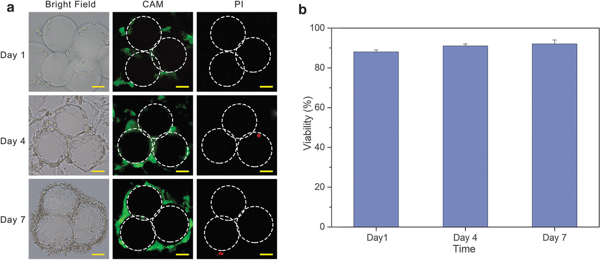
HUVEC viability test.
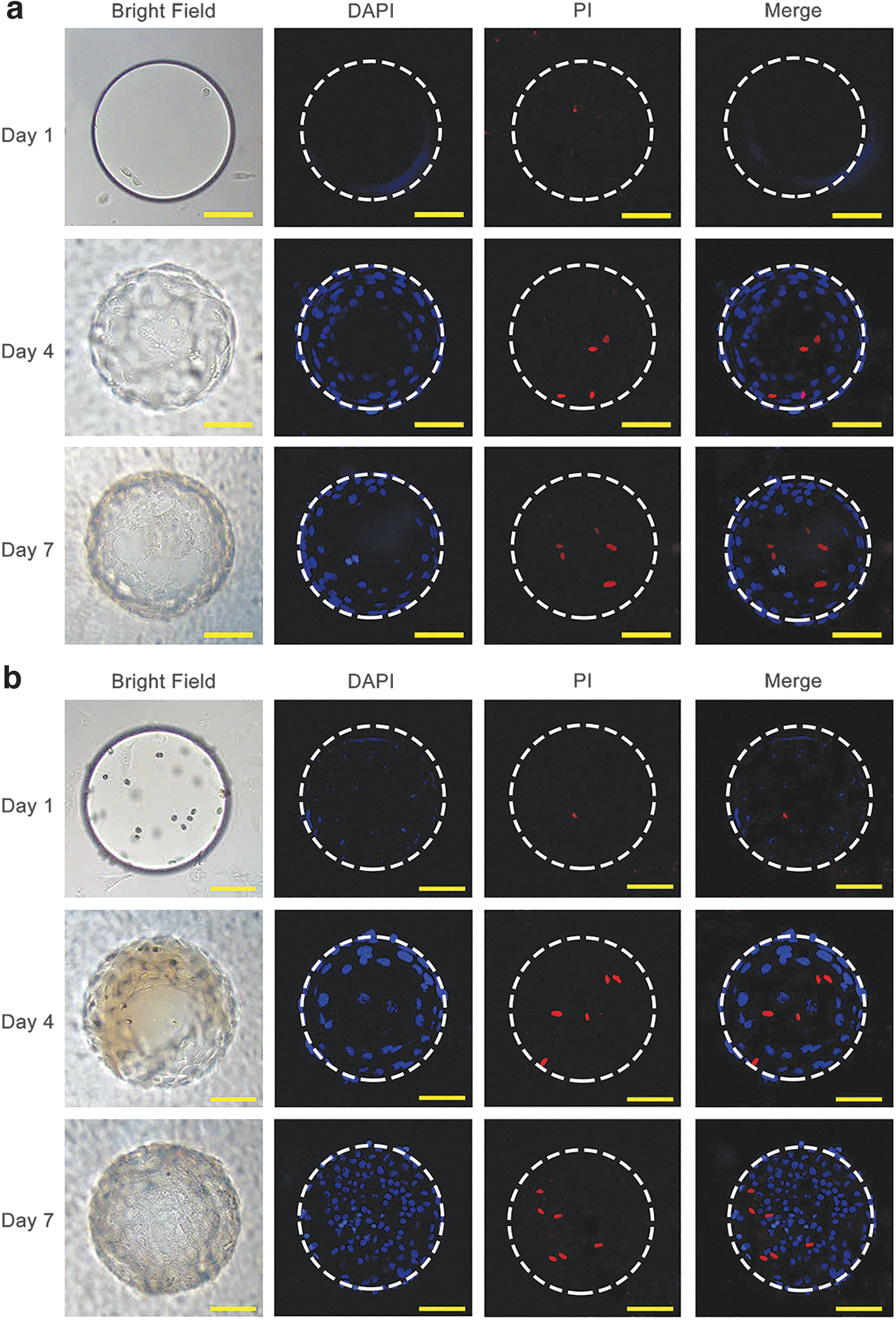
The viability of cells adhered on microspheres. The white contour circle is marked to highlight the microsphere position. HUVECs were stained with DAPI and PI and visualized by a confocal microscope.
Cell proliferation was visualized within 7 days in both unmodified and modified microsphere groups (Fig. 10a, b). The microspheres were almost fully covered with cells on day 3. The number of attached cells increased sharply on day 5. In contrast, the covering speed of cells on the unreacted microsphere was lower compared with the CaCO3 crystal-precipitated microspheres (Fig. 10c). The reason could be that the CaCO3 crystals increased the surface roughness, which is certainly in favor of cell attachments and adhesion.
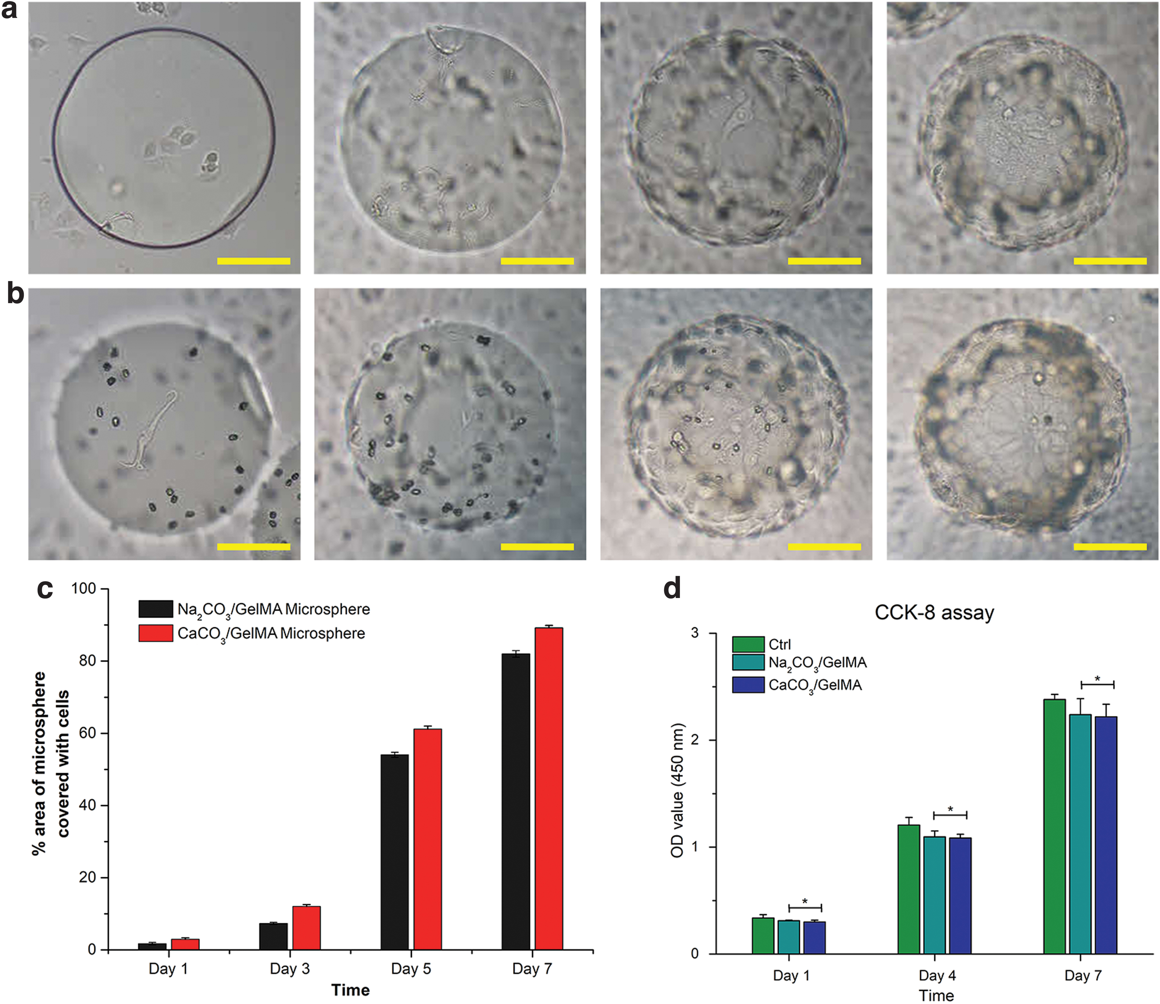
HUVEC proliferation test.
Both the modified and unmodified microspheres showed weakly acidic nature with a pH value of 6.5 ± 0.5. Cell proliferation rate increased sharply after 4 days, as shown in the CCk-8 result (Fig. 10d). However, there was a significant difference between the samples of the modified and unmodified microspheres. The cell proliferation rate of HUVECs was high in the unmodified group compared with the modified group.
Alkaline phosphatase tests
To test the bone tissue inducing capability of the GelMA/CaCO3 microspheres, osteogenic activity was conducted using iMEF cells, which were proven to be able to differentiate into bone cells under appropriate differentiating conditions. 50 iMEF cell proliferation was tested within 7 days in both the unmodified and modified microsphere groups (Fig. 11a, b). The CCK-8 results show that the proliferation rate of CaCO3 crystal-precipitated microspheres was higher compared with the unreacted microspheres (Fig. 11c). The ALP expression was significantly higher in the GelMA/CaCO3 microspheres compared with the GelMA/Na2CO3 microspheres on day 1 and 7 (Fig. 11d).

iMEF cell culture at a density of 3000 cells/well gradually covered the microsphere surfaces over time. Fluorescent images of represent live (green) and dead (red) iMEF cells stained by CAM/PI. The scale bar is 100 μm.
Discussion
Hydrogel microspheres are favorable candidates for cell culture and manipulation due to their 3D feature, cell friendly nature, and ease to use. However, hydrogel microspheres made of natural biomaterials, such as GelMA, collagen, and so on, have a smooth surface, which is unfavorable of cell adhesion. Several methods can be used to modify the microsphere surface, such as electroless deposition 34 and plasma discharge. 36 However, these methods are too complex as they include various steps before completion of the procedure and the quality of the surface treatment is not stable. Recently, Gultekinoglu et al. 39 proposed a process for the surface treatment of microspheres that can make two processes (microfluidics and self-assembly) to happen simultaneously, which is a so-called one-step process.
Our method is also a kind of one-step process, but differs 39 as we used the nonplanar microfluidic device combined with CaCO3 precipitation in situ. Nonplanar microfluidic devices are able to generate both W/O and O/W droplets and to produce microspheres over a broader size range, and with multiple spheres compared with the planar microfluidic device.51,52 Also, the use of the nonplanar microfluidic device avoids the need of modifying the surface of the channels for specific wetting characteristics, 52 which is one of the reasons that a nonplanar microfluidic device is more efficient than a planar microfluidic device.
In the one-step process proposed in our method, the inorganic compound of CaCO3 combines successfully with the organic compound of GelMA, which makes it favorable for bone tissue engineering applications.53,54 We found that different reaction times and concentrations of Na2CO3 are responsible for different sizes of CaCO3 crystals; specifically, the increase of the reaction time and the concentration of Na2CO3 will increase the size of CaCO3 crystals. In our approach, the reaction time was set at 3 min along with the concentration of Na2CO3 being 3% (wt/v) to make the size of CaCO3 crystals be about 3 μm, which favors cell compatibility. 54
The iMEF cell adhesion tests showed that there was a significant difference in the cell adhesion rate on the GelMA/CaCO3 and GelMA/Na2CO3 films, particularly that of the former being much higher compared with the latter. A similar result was obtained with HUVECs. The cells attached easily to the CaCO3 crystal-modified groups, because CaCO3 crystals increase the roughness of the microsphere surface. This observation corresponds with the study of Lampin et al. 55
The efficacy of the 3D microsphere structures as a bone tissue engineering scaffold was investigated by using two types of cells, that is, HUVECs and iMEF cells. Both cell types maintained a high viability, which suggests that the GelMA/CaCO3 microspheres are not toxic. It is interesting to notice that the proliferation rate of HUVECs on the GelMA/Na2CO3 microspheres did not significantly differ from HUVEC proliferation on the GelMA/CaCO3 microspheres. However, the proliferation rate of iMEF cells on the GelMA/CaCO3 microspheres was significantly higher than that on the GelMA/Na2CO3 microspheres. The explanation for this observation can be that HUVECs are not bone cells, and therefore are less affected by the scaffold properties in contrast to cells such as iMEF, which are close to bone cells. 56 In agreement with the proliferation results, the ALP expression for iMFF cells was significantly higher on the GelMA/CaCO3 microspheres than on the GelMA/Na2CO3 microspheres. All these results indicate that the GelMA/CaCO3 microspheres are unique 3D structures with a high potential for bone cell culture and tissue engineering applications.
Conclusion
This article presents a novel method to produce microspheres (GelMA/CaCO3). The microspheres were prepared in a one-step process using a nonplanar microfluidic technique combined with a chemical precipitation process; in particular, putting GelMA/Na2CO3 in CaCl2 solution resulted in the formation of CaCO3 crystals on the surface of the microsphere. The presence of CaCO3 crystals was confirmed by SEM and EDS. The organic compound of GelMA and the inorganic compound of CaCO3 were successfully integrated to favor bone tissue engineering, which agrees with the hybrid engineering principle. 57
The cell experiments confirmed that CaCO3 crystals significantly enhanced cell adhesion. The GelMA/CaCO3 microstructure also showed a high cell viability and ALP expression. This suggests that the GelMA/CaCO3 microspheres can be a promising 3D cell culture matrix for bone regeneration and tissue engineering applications.
In our future work, the nonplanar microfluidic technique will be further developed to produce hollow microspheres to encapsulate cells.
Ethics Approval
For the harvesting and use of HUVECs and iMEF cells, permission and approval were given by the Ethics committee of the Ninth People's Hospital, Shanghai, China. Protocol no. SH9H-2020-A603-1.
Footnotes
Disclosure Statement
None of the authors of this article has any competing interests to declare; therefore, no competing financial interests exist for any of the authors.
Funding Information
This work is financially supported by the Shanghai Science and Technology Innovation Program (No. 19441909600) and the Opening project of Shanghai Key Laboratory of Orthopedic Implant (KFKT2020001).