
Abstract
In vitro tissue-engineered cell culture models are an essential instrument to investigate physiological and pathophysiological wound healing mechanisms and to evaluate new beneficial wound dressing materials and therapeutics to identify possible drug targets and to improve regeneration processes in nonhealing and chronic wounds. In this study, the authors established an in vitro model for cutaneous wound healing, based on primary human dermal microvascular endothelial cells (HDMEC) and primary human dermal fibroblasts (HDF) to study wound healing-associated processes. Co-cultivation of HDMEC and HDF results in the formation of microvessel-like structures in long-term co-cultures. The proposed in vitro co-culture model can be easily modified by adding macrophages to simulate the process of inflammation, thus allowing in vitro investigation of pathophysiological wound healing processes present in nonhealing wounds. Furthermore, the beneficial in vitro wound healing model was used to evaluate a porous fiber-based drug delivery dressing material consisting of melt-spun porous fibers that were filled with a hydrogel carrier (gellan gum) containing vascular endothelial growth factor (VEGF). Angiogenic capability was chosen as functional parameter for improved wound healing, and release of deposited VEGF from the dressing material was evaluated up to 7 days of cultivation. The experiments demonstrated that the porous fiber-based drug delivery dressing material for dermal wound healing with incorporated VEGF strongly enhances the process of angiogenesis in the in vitro co-culture model through a release of VEGF over 7 days of cultivation. In conclusion, tissue-engineered human skin equivalents could contribute significantly to the understanding and improvement of drug releasing dressing materials in the context of treating chronic wounds.
Impact statement
In the context of cutaneous wound healing, the here proposed modifiable in vitro co-culture model in combination with the presented porous fiber-based drug delivery system constitute a beneficial and well-tuneable platform to identify improving wound care products and might contribute to enhance tissue regeneration and healing of chronic wounds.
Introduction
Chronic and nonhealing wounds represent a significant strain on affected patients and on the health care system, as they are associated with reduced mobility or even limb amputation and death. 1 Wound healing is an important and complex physiological process maintaining the integrity of the skin through close interaction of cells, growth factors, chemical signals, and matrices.2,3 The physiological wound healing process is strictly orchestrated in four overlapping phases involving hemostasis, inflammation, proliferation, and remodeling, and finally results in regeneration and healing of the wound. 4 In chronic and nonhealing wounds, aberration of normal wound healing impairs the physiological function. Although the pathophysiology of chronic wounds is still not completely understood, several critical conditions are present in nonhealing wounds such as impaired vascularization, increased inflammation, and the inability to progress to the healing phase. 5
Besides covering the wound and thereby protecting the wound from external agents, common therapies for treating a chronic wound comprise for example, removing necrotic tissue, modulating the inflammation phase, and boosting the reparative phase of healing. At present, significant attempts have been made toward alternative therapies that might reactivate the regenerative properties of chronic and nonhealing wounds. In this context, the modification of dressing materials to serve as drug delivery systems for controlled transport of therapeutics or growth factors to the wound have recently come into focus. 6 Therefore, porous fibers in woven or knitted fabrics present a promising matrix for the deposition and the sustained local release of therapeutic drugs into chronic and nonhealing wounds to support the healing process and reduce therapy duration. Knittings are highly flexible resulting in a good adaption to the body surface compared with woven fabrics. In comparison with nonwovens, they have a higher mechanical strength. Because a better understanding of skin wound healing mechanisms can be generally assisted by in vitro studies, tissue-engineered human skin equivalents might contribute significantly to the understanding and improvement of drug releasing dressing materials in the context of treating wounds.7–10 In general, endothelial cells play a pivotal role during the process of wound healing, because they are essentially involved in the inflammation phase and in new blood vessel formation during the phase of remodeling after injury. 11
To study mechanisms of physiological cutaneous wound healing, the authors developed an in vitro model for cutaneous wound healing consisting of primary human dermal microvascular endothelial cells (HDMEC) and primary human dermal fibroblasts (HDF) that displays similarities to the in vivo situation. This wound healing model constitutes the fundamental basis and allows further modifications with regard to diverse wound healing-associated problems. Within the scope of this study, the in vitro model was further expanded by adding activated macrophages to simulate the process of inflammation within the in vitro wound model. This modification might additionally allow in vitro investigation of pathophysiological wound healing processes such as those present in nonhealing wounds and might contribute to identifying factors to improve regeneration of chronic wounds. Furthermore, the presented in vitro wound healing model was used to evaluate a porous fiber-based drug delivery dressing material fabricated of melt-spun porous fibers that were filled with a hydrogel carrier (gellan gum) containing vascular endothelial growth factor (VEGF) with the aim of enhancing the process of wound healing. In this study, angiogenic capability was chosen as functional parameter for improved wound healing and release of deposited VEGF from the dressing material was evaluated up to 7 days of cultivation. By combining biological mimicry with innovative drug delivering wound dressings, biomaterial- and drug-mediated effects can be analyzed and display a novel approach to develop innovative drug delivery systems to improve the treatment of chronic wounds by enhanced regeneration and healing.
Materials and Methods
Primary cells that were used for this study were obtained from excess tissue in accordance with the principle of informed consent, and approved by the responsible Ethics Commission of the State of Hessen, Germany.
Porous fiber assembly
Nondegradable knitted fabric was produced of melt-spun fibers from poly-
Cell isolation and cultivation
Primary HDMEC and primary HDF were isolated from excess tissues from juvenile patients undergoing cleft lip reconstruction according to a standardized protocol. 12 After enzymatic digestion and separation of the epidermis from the dermis, the remaining dermal cells (HDMEC and HDF) were grown until confluence in gelatin-coated (0.02%) T75 flasks before magnetic separation of CD31-positive HDMEC (CD31 magnetic beads) was performed according to the manufacturer's instructions (Invitrogen, Carlsbad, CA). After cell separation, endothelial cells were cultivated in endothelial cell growth basal medium-2 (EBM-2) medium (Lonza, Basel, Switzerland), supplemented with EGM-2 BulletKit, 1% penicillin/streptomycin (Sigma-Aldrich) and additional 4% fetal bovine serum (Biochrom, Berlin, Germany). HDF were obtained as the CD31-negative cell fraction and were further cultivated in Dulbecco's modified Eagle's medium (Sigma-Aldrich) containing 10% fetal bovine serum (Invitrogen) and 1% penicillin/streptomycin (Sigma-Aldrich). The cells were cultivated at 37°C in an atmosphere of 5% CO2 and 95% air. Cells were passaged in a ratio of 1:2. The acute human monocytic leukemia cell line THP-1 (DSMZ, Braunschweig, Germany) was grown in suspension at a density of 1.2 × 10⁵ cells/mL in Roswell Park Memorial Institute (RPMI) 1640 medium containing 10% fetal calf serum (Gibco, Carlsbad) and 1% penicillin/streptomycin (Invitrogen) and maintained at 37°C in an atmosphere of 95% air and 5% CO2. To differentiate the monocytes to macrophages, 5 × 10⁵ THP-1 monocytic cells were seeded on a fibronectin-coated (5 μg/mL; Milipore, Billerica, MA) six-well plate on a growth area of 9.6 cm2 in 3 mL RPMI medium (Gibco) containing 8 nM PMA (phorbol-12-myristate-13-acetate) (Sigma-Aldrich) for 4 days as described in a previous study. 13
In vitro wound healing model
A co-culture model system for cutaneous wound healing was developed by seeding HDMEC and HDF in a defined cell number and seeding order. Thus, HDF were preseeded in six-well plates (3 × 10⁶ cells per 9.6 cm2 growth area) until cell adherence, before HDMEC were added in the same cell density (3 × 10⁶ cells per 9.6 cm2 growth area). Co-cultures were cultivated in EBM-2 medium and were co-cultivated for 7 and 14 days before further experimentation. To mimic the process of inflammation in the wound healing model, macrophage-induced acute human monocytic leukemia cell line THP-1 (30,000 cells per 9.6 cm2 growth area) was added after 24 h of co-cultivation to the co-culture for further 14 days of triple cultivation. For analysis of the porous fiber-based drug delivery system, co-cultures of HDMEC and HDF were seeded on top of the dressing material and further cultivated in 1 mL of EBM-2 medium with supplements for 7 days of cultivation (Fig. 1).
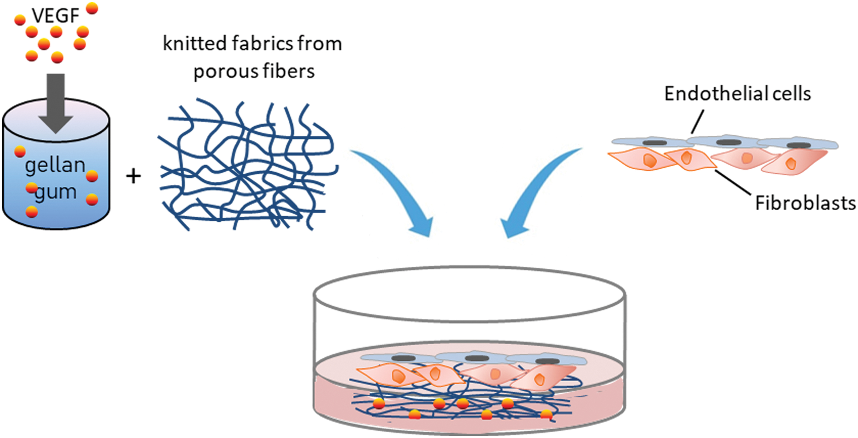
Schematic overview of co-culture experimentation for evaluation of porous fiber-based drug delivery system with incorporated VEGF. Knitted fabrics of melt-spun fibers were made from PLLA/PEO compounds, made porous by washing out the PEO compound and were filled with a hydrogel gellan gum as carrier for VEGF. PLLA/PEO, poly-
Experiment
Experimental design
Angiogenesis assay
The optimal concentration of VEGF as delivering drug from the porous fibers was evaluated using a collagen I angiogenesis assay. Therefore, HDMEC in monoculture were seeded semiconfluently on 24-well plates and subsequently cultivated in EBM-2 medium without supplements from the kit. A collagen I gel was prepared on ice (0.5 mL Collagen I [3 mg/mL]; 0.3 mL aqua dest.; 0.1 mL 10 × M199; 1 mL 10 × matrix buffer). Cell culture medium was removed and 350 μL of the collagen gel per well were transferred on top of the cells cultivated in a 24-well plate. After an incubation time of 30 min to allow coagulation of the gel, cells were treated with EBM-2 medium supplemented with different VEGF concentrations (0, 5, 50, 100 ng/mL) before cells were stained with Calcein AM (Firma) after 24 h, subsequently. VEGF concentration-dependent angiogenic induction of HDMEC was evaluated using a fluorescence microscope (Biozero Keyence, Neu-Isenburg, Germany).
Immunofluorescence staining
For immunofluorescent staining, cell culture samples were fixed in 3.7% paraformaldehyde for at least 5 min before washing with phosphate-buffered saline (PBS) and permeabilizing with 0.01% Triton-X/PBS. After washing again three times with PBS cell cultures were incubated with mouse anti-human CD31 (1:40; Dako, MO 08223) or mouse anti-human CD68 (1:200; Dako M0814) or mouse anti-human CD105 (1:25; Dako) diluted in a 1% bovine serum albumin/PBS solution for 60 min at room temperature. After washing three times with PBS, the cells were incubated with the secondary anti-mouse antibody Alexa 488 (Molecular Probes; MoBiTec, Göttingen, Germany) diluted 1:1000 in a 1% bovine serum albumin/PBS solution for 60 min at room temperature, protected from light. The cells were mounted with Fluoroshield (ImmunoBioScience Corp., Mukilteo, WA) and examined using a confocal laser scanning microscope (LeicaTCS-NT; Leica Microsystems, Wetzlar, Germany) and a fluorescence microscope (Nikon eclipse TS100; Nikon, Düsseldorf, Germany).
Image quantification
The immunofluorescently stained images (CD31) were analyzed using the image-processing software NIS Elements (Nikon). Microvessel-like structures were defined through CD31-positive staining, selected and extracted from the remaining images. The microvessel-like structures (%) were calculated from the total area of the images. For image quantification, three images of each experimental group were analyzed. All calculations were performed using MS Excel (Microsoft Office; Microsoft, München, Germany).
Enzyme-linked immunosorbent assay
About 200 μL of cell culture supernatants of the in vitro co-culture model consisting of HDF and HDMEC seeded on VEGF-containing and releasing porous fibers was collected after 24, 48, 96, and 168 h of co-cultivation. The concentration of VEGF was measured using DuoSet® E`LISA Development Systems according to the manufacturer's protocol (R&D Systems). A streptavidin-HRP colorimetric reaction was used to visualize protein concentrations and the optical density of each well was measured using a microplate reader (Tecan, Crailsheim, Germany) at a wavelength of 450 nm. Results are demonstrated as absolute values as indicated in the relevant figures.
Quantitative real-time reverse transcriptase–polymerase chain reaction
RNA was isolated using TRIZOL reagent (Sigma-Aldrich). Thus, one-well of the co-cultures was treated with 500 μL TRIZOL in one 1.5 mL tube and incubated for 5 min at room temperature. About 200 μL chloroform was added to each tube, followed by vortexing for at least 15 s. After an incubation time of 3 min, tubes were centrifuged for 15 min at 12,000 × g at 4°C. The aqueous phase containing the RNA was removed and transferred to a new tube before 500 μL isopropanol were added for RNA precipitation. After another centrifugation step (12,000 × g, 4°C, 10 min), supernatant was removed, the RNA pellet was washed in 1 mL ethanol and centrifuged again (7500 × g, 4°C, 5 min). The pellet was dried and resolved in 10 μL RNase free water before the RNA concentration was measured using a nanodrop spectrophotometer (NanoDrop, Wilmington, DE). To obtain a well-defined quantity of cDNA, 1 μg of extracted RNA was used for reverse transcription using an Omniscript RT kit according to the manufacturer's protocol (Qiagen, Hilden, Germany). For quantitative real-time polymerase chain reaction (PCR) the following primers were used for this study: Interleukin-6 (IL-6), tumor necrosis factor alpha (TNF-α), and E-selectin (all obtained from Qiagen). 60S ribosomal protein L13A (RPL13A) was used as endogenous control. Four nanograms of cDNA were used for one reaction. Quantitative real-time PCR was performed in triplicate with the following cycler program: 95°C for 10 min, 95°C for 15 s, and 60°C for 1 min, 40 cycles. To specify the length of the DNA fragments a dissociation stage was added to the program: 95°C for 15 s, 60°C for 1 min, 95°C for 15 s, and 60°C for 15 s. Relative gene expression was determined using the ΔΔCt method. Gene expression was compared by setting control cultures to 1 (reference value) as indicated in the relevant figures.
Cell viability analysis: MTS assay
Sterilized porous fibers from PLLA/PEO compounds were incubated in cell culture (EBM2 with supplements) medium for 3 days to generate an extraction medium of the proposed material. HDMEC and HDF were seeded at an appropriate density in each well of a 96-well plate (5000 cells/well) and cells were cultivated for 4 days in the generated extraction medium before cell viability was evaluated using an MTS assay. Therefore, aCellTiter 96® AQueous One Solution Cell Proliferation Assay Kit (Promega, Madison, WI) was used following the manufacturer's instructions. In brief, 30 μL of MTS solution were added to each well containing the sample in 150 μL of culture medium and incubated for 1 h at 37°C before the absorbance was measured at 490 nm with a plate reader (Infinite M200; Tecan). Blanks consisted of 150 μL of media with 30 μL of MTS solution incubated on plastic plates. All experiments were performed independently three times, each in triplicate.
Statistical analyses
All experiments were performed with at least three different donors. Data are presented as mean values ± standard deviation. Statistical significance was evaluated using paired Student's t-test. Statistical analyses were performed with MS Excel (Microsoft Office; Microsoft) and significance was assessed by *p < 0.03 or *p < 0.05, respectively.
Experimental results
Establishment of an in vitro cell culture model consisting of HDMEC and HDF to mimic cutaneous wound healing
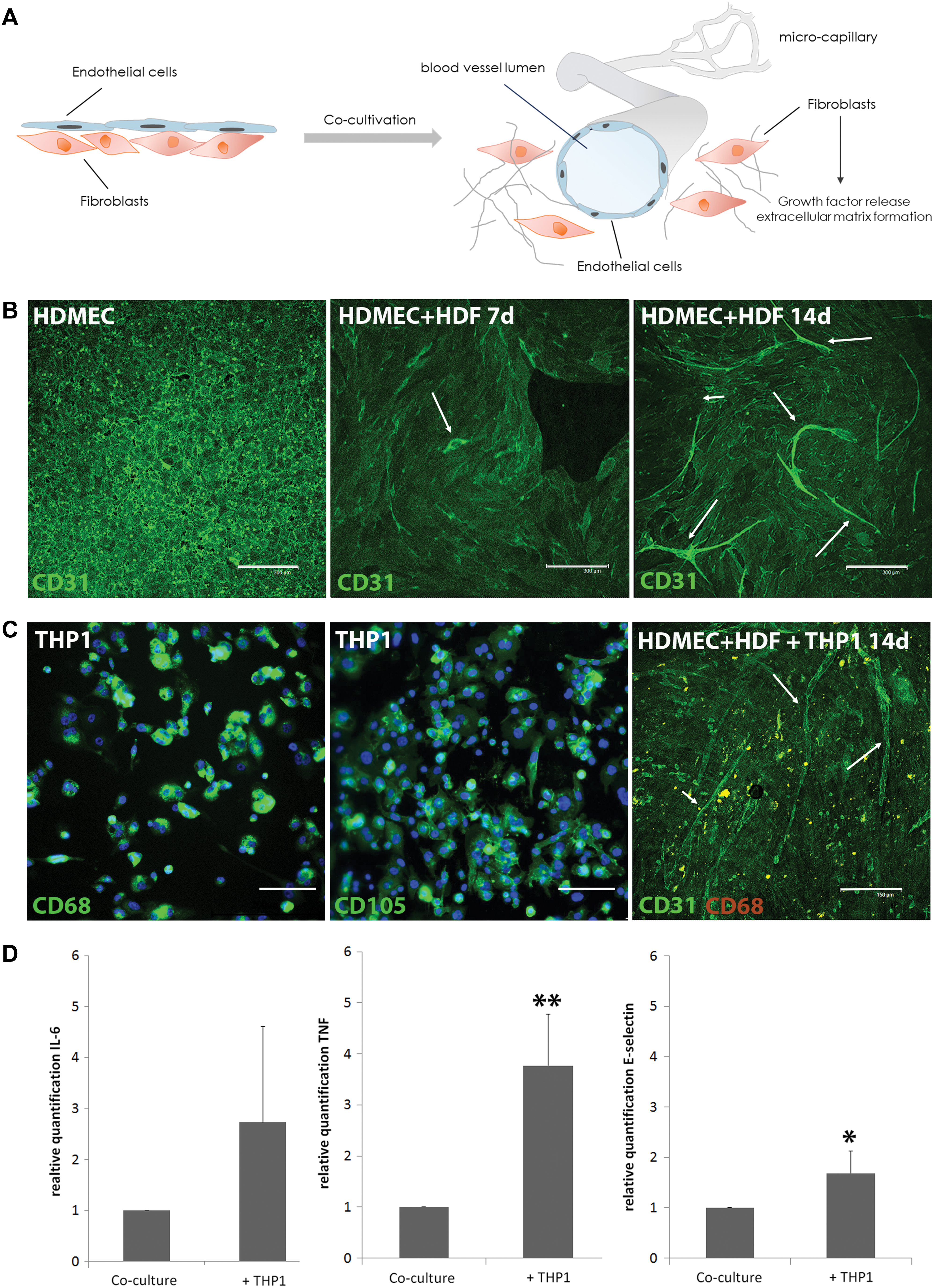
An in vitro wound healing model was developed by combining primary HDMEC with primary HDF in defined cell ratio and seeded in defined cell seeding order. Co-cultures were cultivated for 7 and 14 days, before capability of endothelial microvessel-like structure formation was analyzed as functional parameter for efficient wound healing (Fig. 2A). HDMEC in monoculture reveal a typical endothelial cell type-specific morphology, forming an endothelial cell monolayer with strong cell-to-cell contacts and a cobblestone-like organization documented using immunofluorescent staining of the endothelial marker CD31 (Fig. 2B, left). Co-culture cultivation of HDMEC with HDF for 7 days resulted in a slight angiogenic activation of HDMEC in the in vitro wound healing model, as documented by a more elongated, rearranged shape of the endothelial cells (Fig. 2B, middle). After 14 days of co-cultivation, endothelial cells strongly arranged into microvessel-like structures with large branches revealing a considerable angiogenic activation of HDMEC in co-culture with HDF in long-term culture (Fig. 2B, right). To analyze also pathophysiological wound healing processes, the co-culture model of HDMEC and HDF was further expanded by adding activated macrophages positively stained for CD68 and CD105 (THP-1, Fig. 2C) as wound healing-modulating factor. After 14 days of cultivation, CD31 staining of co-cultures treated with THP-1 revealed the presence of microvessel-like structures certainly exhibiting less branching points compared with controls without THP-1 stimulation (Fig. 2C, right). In triple cultures of THP-1, HDMEC, and HDF, activated THP-1 cells induced the process of inflammation within the in vitro wound model estimated through an upregulation of inflammation associated factors like IL-6, TNF-α, and E-selectin (Fig. 2D). Relative quantification of gene expression of proinflammatory factors TNF-α and E-selectin was significantly upregulated in THP-1-treated cultures compared with control co-cultures. Although IL-6 upregulation in THP-1-treated cultures cannot be assessed as statistically significant, a higher relative gene expression can also be observed for IL-6 in triple cultures consisting of THP-1, HDMEC, and HDF (Fig. 2D).
Porous fiber-based wound dressing: biocompatibility analyses
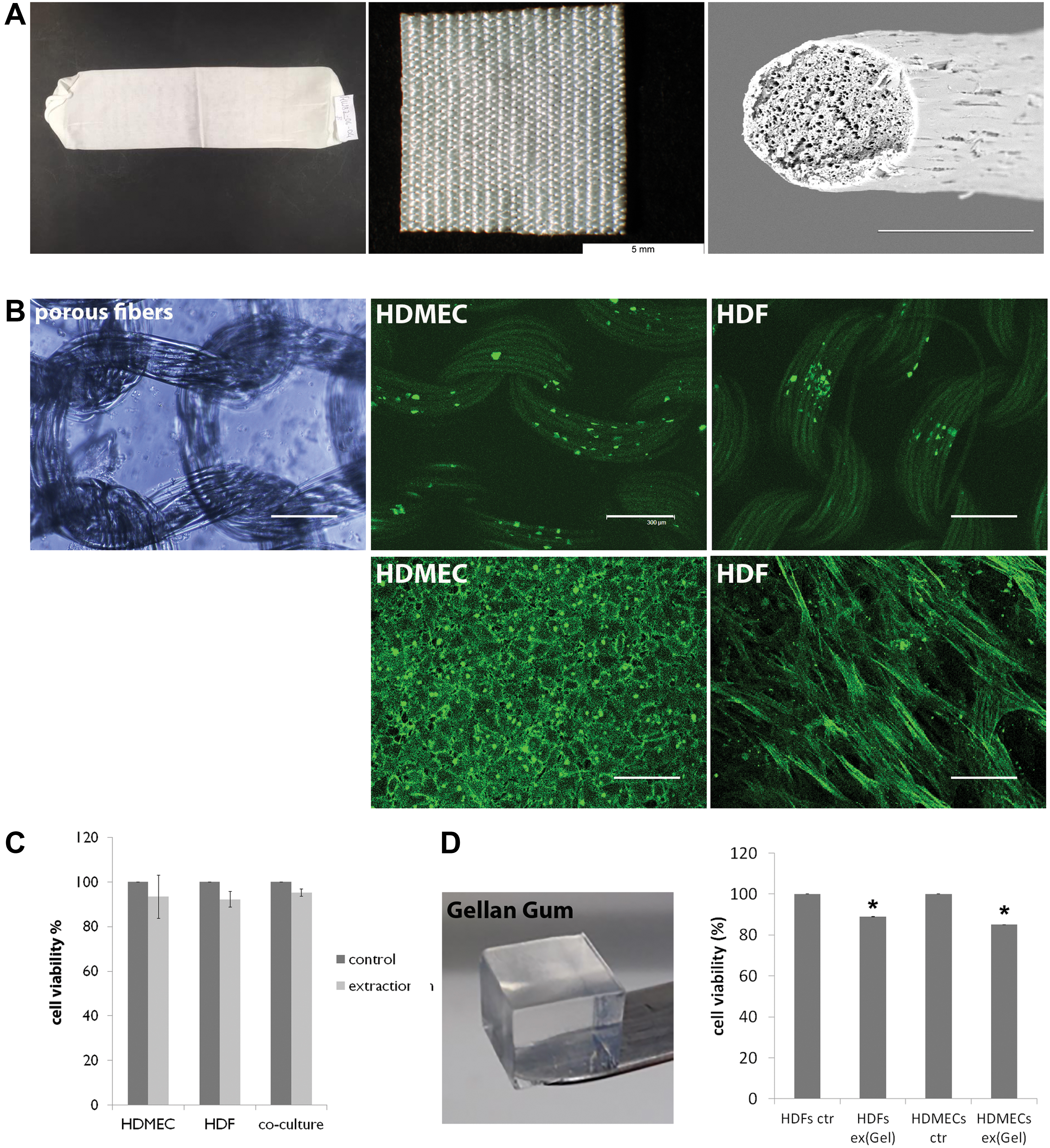
Melt spun PLLA/PEO blended polymer fibers were processed into knitted fabrics. Pores in the fibers were prepared by washing out PEO. An overview of a whole knitted fabric and a more focused view is given in Figure 3A (left and middle). Furthermore, porous structures were investigated using a scanning electron microscope, clearly demonstrating the high porosity of the cross-sectioned fibers (Fig. 3A, right). To evaluate the cell adhesion behavior as indirect indication for biocompatibility of the pure porous fibers, HDMEC and HDF in monoculture were seeded on top of the dressing material and were stained immunofluorescently with the appropriate cell-specific markers, CD31 and α-smooth muscle actin (Fig. 3B). In general, HDMEC and HDF can be detected on the surface of the knitted fabric (Fig. 3B, upper) and on the bottom of the cell culture plate (Fig. 3B, lower), exhibiting a normal cell type-specific morphology of both cell types after 24 h of cultivation. In addition, an extraction assay was performed to exclude any relevant cell disturbing or cytotoxic effects of the porous fibers (Fig. 3C) and of the hydrogel carrier for VEGF incorporation (gellan gum, Fig. 3D) on the used cell cultures of the in vitro wound healing model. Cultivation of HDMEC monoculture, HDF monoculture, and of co-culture consisting both cell types in extraction medium of porous fibers resulted in a slight decrease in cell viability with the highest metabolic activity decrease in HDMEC monoculture (Fig. 3C). In general, decrease of cell viability in material-extracted media cultivated cells cannot be assessed as statistically significant. Similar results can be obtained when appropriate cells were cultivated in extraction medium generated from the hydrogel (gellan gum). A slight decrease of metabolic activity can be observed when cells were cultivated in hydrogel extracted medium as well (Fig. 3D). Although this hydrogel mediated decrease in metabolic activity of HDMEC and HDF monocultures can be calculated as statistically significant, cell viability of both cell types cultivated in extraction medium was still observed at high levels of up to ∼90% (Fig. 3D).
Evaluation of porous fiber-based drug delivery system: VEGF release increases microvessel-like structure formation in vitro
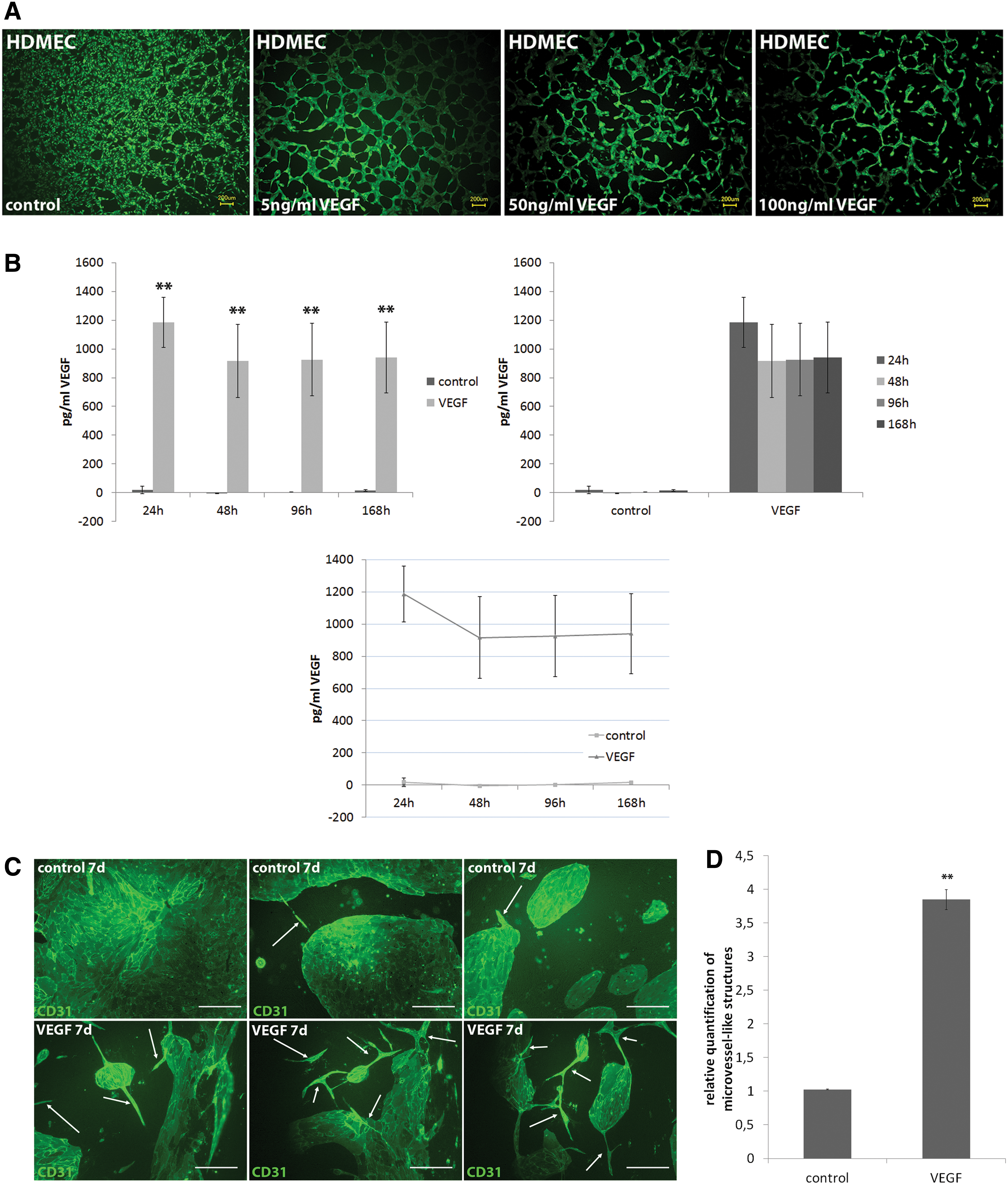
To estimate an appropriate VEGF concentration for further developing and establishing the porous fiber-based drug delivery system for wound healing applications, an angiogenesis assay based on a collagen I gel provided fundamental information with regard to VEGF concentration-dependent proangiogenic effects (Fig. 4A). Monocultures of HDMEC were covered with a three-dimensional collagen I gel and subsequently treated with different concentrations of VEGF (0–100 ng/mL). In contrast to the control (0 ng/mL VEGF), angiogenic induction of endothelial cells can be obtained when 5 ng/mL VEGF was applied for 24 h (Fig. 3A). Application of higher VEGF concentration resulted in abrogation of angiogenic sprouting of HDMEC in the gel (100 ng/mL). Concentration of VEGF as delivering drug incorporated in a gellan gum hydrogel in the porous fibers was adjusted to these results displaying the basis for further experiments. The In vitro wound healing model was cultivated on VEGF-releasing porous fiber-based wound dressing materials for 7 days of cultivation, before co-cultures were assessed for angiogenic response (Fig. 4C). Control co-cultures seeded on knitted fabrics from porous fibers without incorporated VEGF served as control. These control co-cultures reveal very slight angiogenic activation after 7 days of cultivation as documented by rearrangement of monolayered endothelial cell shape into a more elongated form (Fig. 4C, upper). In contrast, when co-cultures were cultivated on samples with incorporated VEGF in porous fibers, endothelial cells form huge microvessel-like structures after 7 days of co-cultivation and are highly organized into branches, thus documenting an ongoing process of angiogenesis induced in response to VEGF release from the fibers (Fig. 4C, lower). Quantification of microvessel-like structure formation confirmed the results displaying a significant higher percentage of vascularization when co-cultures were cultivated on VEGF-releasing wound dressings (Fig. 4D). The release of VEGF from porous fibers into cell culture supernatants was determined in the course of co-cultivation after 24, 48, 96, and 168 h and was compared with control co-cultures cultivated on samples of the pure fibers without incorporated VEGF (Fig. 4B). Concentration of VEGF in cell culture supernatants of co-cultures cultivated on VEGF containing fibers remained stable up to at least 7 days of co-culture experiments and was significantly higher compared with VEGF concentrations in co-cultures cultivated on fibers without VEGF incorporation where VEGF in supernatants can be hardly detected (Fig. 4B). Although the highest VEGF concentration can be documented in the supernatants of co-cultures on VEGF-releasing porous fibers after 24 h of cultivation, the concentration of VEGF after 48, 96, and 168 h can be constantly determined with concentrations up to ∼900 pg/mL.
Discussion
Wound healing is an important and complex process dependent on different cell types, growth factors, and chemical mediators interacting together in four highly controlled phases, involving hemostasis, inflammation, proliferation, and remodeling.11,14 Under physiological conditions tissue injury usually results in normal wound healing and complete regeneration, but may also fail and result in a chronic wound when underlying pathobiologic conditions interfere. Therefore, in vitro tissue-engineered models are essential to discover the pathogenesis of wound healing processes, identify new drug targets, and test newly developed wound dressing materials and therapeutics. Biological mimicry such as in vitro models based on co-culture techniques provides a beneficial strategy to investigate the behavior of cell types that are relevant for wound healing processes.15,16 During this study, the authors established an in vitro co-culture model for cutaneous wound healing consisting of primary HDMEC and primary HDF to study physiological and pathophysiological wound healing associated processes. In addition, this model was used to evaluate a porous fiber-based drug delivery dressing material for dermal wound healing. The major findings of this study are follows: (1) co-cultivation of HDMEC and HDF results in the formation of microvessel-like structures in long-term cultures and can be pathobiologically modified by adding macrophages; (2) the porous fiber-based drug delivery material is biocompatible with the in vitro wound healing model; (3) the porous fiber-based dressing material with incorporated VEGF enhances the process of angiogenesis in the in vitro co-culture model through a relatively stable VEGF concentration in the supernatant over 7 days of cultivation.
In normal human adult tissue, angiogenesis, the formation of new blood vessels, usually remains nonactive and is initiated after tissue injury during wound repair. 17 New blood vessel formation is an essential requirement for the wound healing process, because angiogenic capillary sprouts invade the wound and organize into a microvascular network to transport nutrients and oxygen to the wound area. 18 Because of the fact that microvascular endothelial cells are the basic component of blood vessels, their function is essential with regard to a functional angiogenesis. 9 Microvascular dysfunction is mainly responsible for the pathogenesis of chronic and nonhealing wounds and vascular lesions are the main cause of numerous problems in chronic wounds such as ischemic changes, infection, and ulcers. 19 During this study, the co-culture consisting of primary HDMEC and primary HDF reveal an induction of microvessel-like structure formation after 14 days of co-cultivation. HDMEC and HDF co-cultures have been established and investigated in several previous studies with regard to the cell-to-cell interaction.20–22 Most of these co-culture studies additionally used three-dimensional gels of extracellular matrix molecules. 20 Within the present co-culture model, fibroblasts are serving as matrix for the endothelial cells similar to the in vivo situation. By production of extracellular matrix in the dermis, fibroblasts are responsible for maintaining the structure of the ECM. 23 Furthermore, during the process of angiogenesis, fibroblasts of the connective tissue release chemical signals and growth factors that lead to activation of endothelial cells, finally leading to new blood vessel formation.14,24 In the context of co-culture experimentation, using fibroblasts seem to be important as regulators of angiogenesis, because HDMEC in monoculture form typical cobblestone-like monolayer instead of angiogenic sprouting. Further expansion of the wound healing model by adding macrophages as wound healing modulating factor results in an increased expression of proinflammatory factors like IL-6, TNF-α, and E-selectin, thus documenting an induced inflammatory response in the co-culture system through macrophages. During the process of wound healing, macrophages play a multifactorial role as they phagocytose necrotic tissue and pathogens and amplify the inflammatory response by secreting proinflammatory cytokines.14,25 It is well documented that the activated THP-1 cell line is able to induce an inflammatory response in vitro and the interaction of THP-1 cells and endothelial cells in various experimental settings has been investigated.13,26,27 In this study we were able to show that the simple combination of THP-1 with the in vitro wound healing model leads to an upregulation of the proinflammatory cytokines and provides a first indication that the co-culture-based in vitro model can be easily modified to induce an inflammatory-type response in vitro. Although our focus was not on immune response of macrophages and on phenotypic alteration of fibroblasts, fibrotic response that often occurs in prolonged tissue injury and chronic inflammation is still an unresolved medical problem and represents the purpose of further planned studies with a greater focus on the cellular mechanisms of chronic or inflamed wounds with the use of the in vitro wound healing model as major basis proposed here.
Moreover, the proposed in vitro wound healing model represents a convenient way to study cell–biomaterial interaction and to understand biomaterial-mediated mechanisms. This understanding allows modulation and adaption of the material to achieve desired biological functions. The development of drug delivery wound dressing materials displays a novel approach to improve the treatment of chronic wounds aiming at an enhanced regeneration and healing. For this purpose, a porous fiber-based wound dressing, incorporating VEGF in a hydrogel carrier was developed and was investigated for VEGF release and for proangiogenic effects in co-cultures of HDMEC and HDF. SEM imaging of the fiber cross sections document the successful creation of submicroscale pores in which the VEGF hydrogel can be stored. VEGF was completely released within the first 24 h and concentrations in cell culture supernatants remained stable during the course of co-cultivation up to at least 7 days of cultivation. HDMEC strongly respond to the released VEGF by organization into microvessel-like structures when seeded on the wound dressing material and compared with the control. Under physiological conditions, soluble factors, such as VEGF, are strictly regulated to induce the growth of new blood vessels in the early stages of injury. In chronic wounds of diabetic mice, the concentration of VEGF is significantly decreased when compared with normal healing wounds. 28 Although the delivery of VEGF to nonhealing wounds would have positive effects on the regeneration process, the controlled and sustained release of VEGF is still challenging for scientists because of the short half-life of VEGF. 29 Consequently, the development of novel delivery systems that can prevent growth factors from degradation by incorporation of the growth factors into a material would improve the controlled release and the possible therapeutic effect. 6 Accordingly, our study demonstrates that the incorporation of VEGF into the porous fibers results in a release of VEGF into cell culture supernatants that leads to a higher density of new capillaries in the wound healing model. During this study, VEGF was exemplarily chosen as growth factor for the incorporation into the porous fibers but can be easily replaced by another growth factor, cytokine, or any therapeutic drug to improve the process of regeneration in nonhealing wounds. Moreover, a dual delivery system would also be possible. Besides delivering of growth factors to enhance the regeneration process in nonhealing wound, antibiotic agents for local application might also be co-incorporated in the context of an infected wound. In addition, the release kinetics might by adjusted by modifications of the hydrogel carrier. This benefit in combination with the modifiable in vitro wound healing model makes the porous fiber-based drug delivery system a well-tuneable platform that might help to identify improving wound care products to enhance tissue regeneration. In conclusion, this study can contribute significantly to the understanding and improvement of drug delivering wound dressing materials in the context of treating chronic wounds.
Footnotes
Acknowledgments
The authors thank Verena Hoffmann for excellent technical support.
Disclosure Statement
No competing financial interests exist.
Funding Information
The study was funded by the Federal Ministry of Economics and Energy on the basis of a resolution of the German Bundestag (19523 BG).