




Abstract
Cell-based therapies require a large number of cells, as well as appropriate methods to deliver the cells to damaged tissue. Microcarriers provide an optimal platform for large-scale cell culture while also improving cell retention during cell delivery. However, this technology still presents significant challenges due to low-throughput fabrication methods and an inability of the microcarriers to recreate the properties of human tissue. This work proposes, for the first time, the use of methacryloyl platelet lysates (PLMA), a photocrosslinkable material derived from human platelet lysates, to produce porous microcarriers. Initially, high quantities of PLMA/alginate core–shell microcapsules are produced using coaxial electrospray. Subsequently, the microcapsules are collected, irradiated with ultraviolet light, washed, and freeze dried yielding PLMA microsponges. These microsponges are able to support the adhesion and proliferation of human adipose-derived stem cells, while also displaying potential in the assembly of autologous microtissues. Cell-laden microsponges were shown to self-organize into aggregates, suggesting possible applications in bottom-up tissue engineering applications.
Impact Statement
Microcarriers have increasingly been used as delivery platforms in cell therapy. Herein, the encapsulation of human-derived proteins in alginate microcapsules is proposed as a method to produce microcarriers from photopolymerizable materials. The capsules function as a template structure, which is then processed into spherical microparticles, which can be used in cell culture, cell delivery, and bottom-up assembly. As a proof of concept, this method was combined with lyophilization to process methacryloyl platelet lysates into injectable microsponges for cell delivery.
Introduction
Cell-based therapies have increasingly been proposed as a strategy to promote the regeneration of damaged tissue and organs.1–5 The delivery of cells is able to promote tissue recovery by replenishing lost cells and by enhancing the production and secretion of paracrine factors that stimulate endogenous proregenerative pathways.6,7 However, a notable challenge in implementing these therapies is the reduced retention rate of delivered cells in the affected tissue. 8
While incorporating cells within biocompatible scaffolds has been shown to increase cell retention, the implantation of these scaffolds generally requires invasive surgical procedures. 9 Minimally invasive alternatives are needed to reduce costs, as well as the discomfort caused due to extensive postoperative periods. A promising solution to this problem is injectable materials, such as fibrin gels, 10 self-assembling peptides, 11 and other polymers that display in situ gelling capability, 12 as well as cryogels, 13 microcapsules, 14 and microcarriers. 15
Microcarriers consist of small microparticle-based scaffolds, which have prominently been used as support structures in the culture of adherent cell lines in bioreactors, where they provide surfaces for cell attachment. These microcarrier-based cell culture systems can achieve high cell yields, allowing the mass production of cells for cell therapy and tissue engineering approaches.16–19 Additionally, the resulting cell-laden microcarriers constitute an optimal platform for cell delivery.
It has been reported that cell culture on microcarriers promotes the deposition of an extracellular matrix (ECM) that better resembles the cells' native ECM when compared with two-dimensional culture substrates, while also preserving cell phenotypes. 20 Furthermore, cell–microcarrier interactions can promote the self-assembly of large aggregates, providing a path toward the bottom-up assembly of tissues, which can be directed toward a wide variety of applications, from modeling diseases in vitro, developing regenerative patches to replace damaged tissues, or producing synthetic meat.21–24
Currently available microcarriers can differ in their composition, surface properties, mechanical properties, and porosity. Macroporous microcarriers present a sponge-like porous structure, which increases the available surface area for cell proliferation.18,25,26 These sponge-like microcarriers generate higher cell densities while maximizing the surface-area-to-volume ratio, thus achieving greater cell-to-microcarrier loading rates. In general, cells can also penetrate the inner structure of these microsponges provided that the dimensions of the pores are superior to 20 μm, sheltering the cells from mechanical stresses associated with stirring or injection.27,28
After transplantation, the success of biomaterial-based approaches depends on proper integration of the scaffolds into the existing tissue, which is limited by the inability of currently used materials to reproduce the properties of human ECM. Human-derived materials, obtained from sources, such as platelet lysates (hPL) 29 or perinatal tissues,30,31 are able to emulate the complexity of the ECM, which allows the development of platforms that better resemble human tissues. In addition, these protein-based materials contain a rich variety of bioactive molecules that promote regeneration.
The incorporation of hPL in biomaterial-based platforms has increasingly been pursued as a strategy for the sustained delivery of growth factors. hPL-loaded scaffolds have been shown to promote cell survival, 32 cell proliferation, 33 cell migration, 34 vascularization, 35 and osteogenesis, 33 while also improving the mechanical properties of hPL-loaded hydrogels. 36
Recently, our group has reported a method to chemically modify hPL with photocrosslinkable moieties. 37 This method has been used to synthesize methacryloyl platelet lysate (PLMA), a human-derived biomaterial that can be used to produce hydrogels by exposure to ultraviolet (UV) radiation. This novel material has already been applied in the development of a model of osteosarcoma,38,39 in the study of osteogenesis, 40 and in the development of porous scaffolds for xeno-free cell culture. 41
In this article, we propose a method to produce microcarriers for cell delivery by encapsulating a solution of a photocrosslinkable material in alginate microcapsules, providing a template for photopolymerization. This method was combined with ice templating to produce PLMA microsponges. It is envisioned that this method can be expanded to other photopolymerizable materials, as well as materials with alternative crosslinking mechanisms.
Materials and Methods
Methacrylic anhydride was used to introduce methacryloyl moieties in hPL, producing a photocrosslinkable protein-derived biomaterial, as previously described. 37 Briefly, hPL (StemCell Technologies, Canada) was thawed at 37°C and reacted with methacrylic anhydride (94%; Sigma-Aldrich, USA) in a 100:1 ratio to produce PLMA. The reaction mixture was kept at room temperature with constant stirring for 4 h. The PLMA was purified by dialysis with a 3.5K MWCO, 35 mm dry I.D. SnakeSkin™ dialysis membrane (Thermo Fisher Scientific, USA) against deionized water for 24 h. The solution was then sterilized through a Sartolab™ P20 0.2 μm filter (Sartorius, Germany), frozen with liquid nitrogen and freeze dried for 7 days in a freeze dryer (LyoQuest Plus Eco; Telstar, Spain) operating under vacuum at −60°C. Afterward, they were stored at 4°C until use.
The process used in the production of PLMA microsponges is summarized in Figures 1 and 2A. The PLMA is first entrapped in alginate microcapsules to produce a PLMA/alginate core–shell template structure. Microcapsules were produced using an electrospray system (Spraybase, Ireland, connected to a high-voltage power supply [LNC 30000-2; Heinzinger, Germany]) equipped with a coaxial needle (20G outer nozzle and 26G inner nozzle).
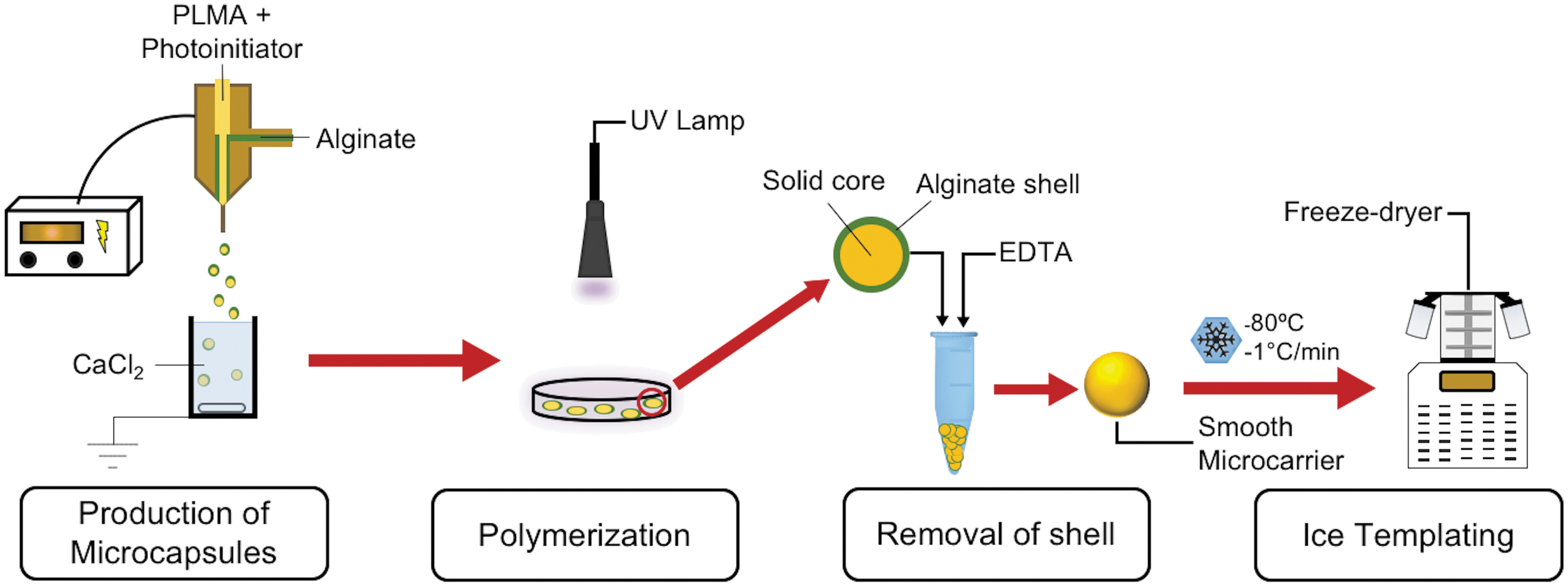
Schematic representation of the method reported in this article. A precursor solution is incorporated in alginate microcapsules to produce a template structure. The encapsulated precursor is then polymerized with an external light source. The alginate shell is then removed, providing smooth microcarriers. The preparation of dry porous microcarriers can then be performed by ice templating. EDTA, ethylenediamine tetraacetic acid; PLMA, methacryloyl platelet lysate; UV, ultraviolet.
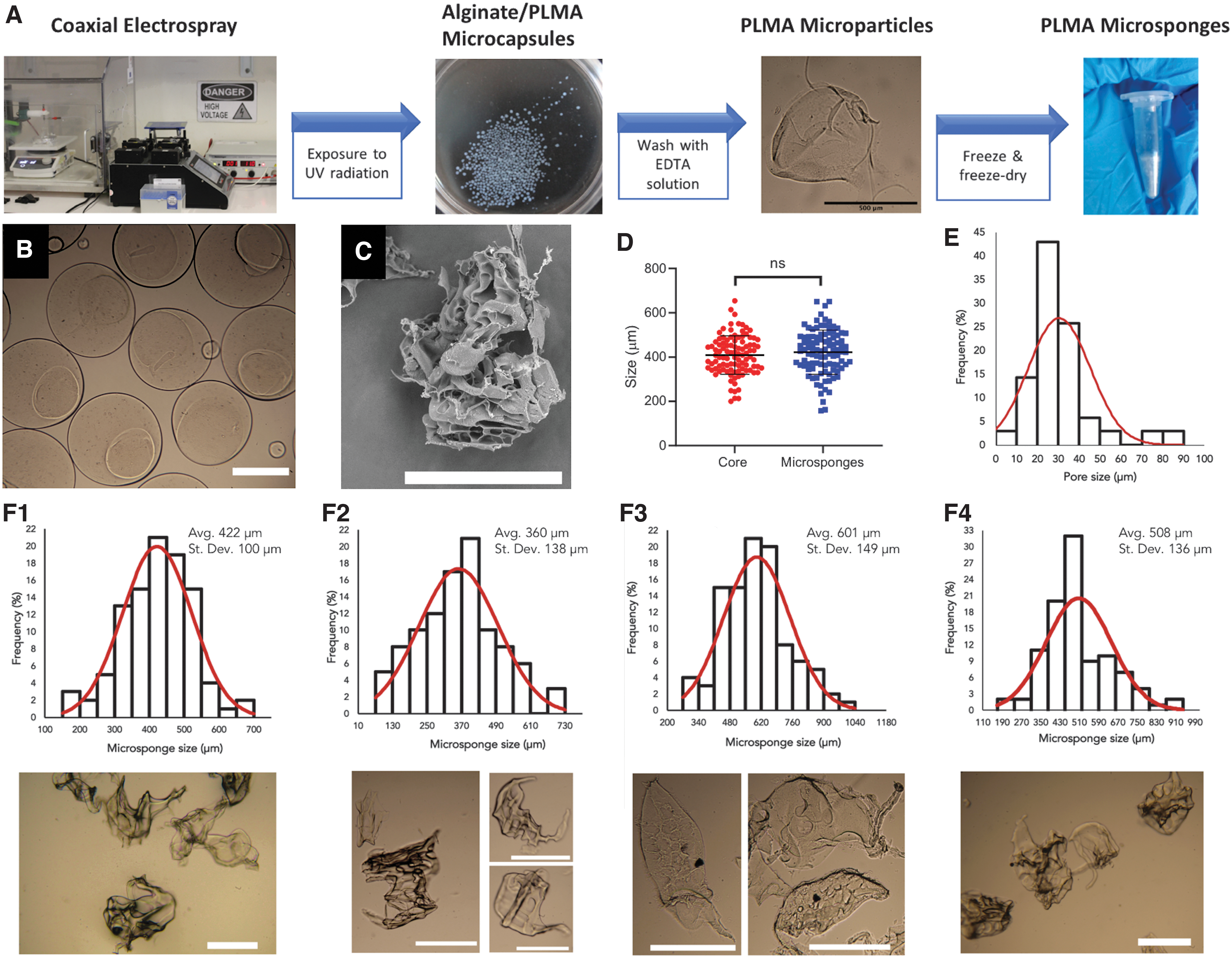
Preparation of PLMA microsponges.
A 15% (w/v) solution of PLMA was prepared in a 1% (w/v) solution of 2-hydroxy-4′-(2-hydroxyethoxy)-2-methylpropriophenone (Irgacure 2959; Sigma-Aldrich) in phosphate-buffered saline (PBS, pH 7.4; Sigma-Aldrich) and connected to the inner nozzle. A 1% (w/v) medium viscosity alginate solution (viscosity ≥2000 cP [2%, 25°C]; Sigma-Aldrich) in PBS was connected to the outer nozzle. Droplets were collected in a 0.1 M calcium chloride (CaCl2 anhydrous, ≥96.0% purity; Sigma-Aldrich) bath to crosslink the alginate shell. Flow rates were controlled using a programmable dual-drive syringe pump (Pump 33 DDS; Harvard Apparatus, USA).
The resulting core–shell capsules were subjected to irradiation (320–500 nm filter, 0.045 W/cm2) for 3 min using an OmniCure S2000 Spot UV Curing System (Excelitas Technologies Corp., USA). The microcapsules were collected and washed with a 0.2 M solution of ethylenediaminetetraacetic acid (EDTA, pH 7.0; Sigma-Aldrich) for 15 min. The resulting PLMA microparticles were then centrifuged at 1000 g for 5 min at room temperature, and washed with the EDTA solution a second time. The microparticles were then resuspended in distilled water and placed in Mr. Frosty® freezing containers (Nalgene; Nalge Nunc International, USA). Afterward, they were stored at −80°C overnight, and lyophilized overnight in a freeze dryer operating under vacuum at a temperature of −60°C, yielding PLMA microsponges.
To evaluate the injectability of PLMA microsponges, they were suspended in PBS, transferred to a syringe, passed through 21G, 25G, and 27G needles and collected. PLMA microsponges were examined through a Primo Star optical microscope (Zeiss, Germany) pre- and postinjection, and their dimensions were determined through analysis with ImageJ Software (NIH, USA). The dimensions of the microsponges were defined as an average of the length of their largest diameter and its lowest perpendicular diameter.
Additional examination of the structure of the microsponges was performed through scanning electron microscopy (SEM). After resuspension in PBS, PLMA microsponges were dehydrated in graded water/ethanol mixtures, dried in a critical point dryer, mounted on aluminum stubs, sputter coated with carbon, and visualized by SEM (Hitachi SU-70, Japan). Pore measurement was performed through analysis with ImageJ Software.
Experiment
Experimental design
PLMA microsponges
PLMA microsponges were prepared according to the previously outlined procedure. After production, the microsponges were stored at room temperature until use in cell assays.
Cell culture
Human adipose tissue-derived stem cells (hASCs) were isolated from human lipoaspirates by our group using a well-established procedure. 42 Liposuction tissue was obtained under a cooperation agreement between University of Aveiro and Hospital da Luz (Aveiro, Portugal) and approved by the Ethics Commission of Hospital da Luz. Informed consent was obtained from the donors. Cells were cultured using minimum essential medium (α-MEM) (Gibco®, Thermo Fisher Scientific), supplemented with 10% fetal bovine serum (Gibco, Thermo Fisher Scientific), and 1% antibiotic–antimycotic (containing penicillin, streptomycin, and amphotericin B; Gibco, Thermo Fisher Scientific). The flasks were placed in an incubator at 37°C in a humidified atmosphere with 5% carbon dioxide (CO2). Upon reaching ∼80% confluence, cells were enzymatically detached through incubation with trypsin (Gibco, Thermo Fisher Scientific) at 37°C for 5 min, followed by centrifugation at 300 g for 5 min.
Cell seeding on microsponges
An estimated 250 microsponges were preweighed and sterilized by 30 min of UV exposure. Afterward, they were suspended in α-MEM culture medium and transferred to μ-Slide 8-well coverslips (Ibidi, Germany). hASCs were then seeded in the microsponge suspension at a cell density of 150 hASCs/microsponge, and placed at 37°C in a humidified atmosphere with 5% CO2 for 7 days. Medium changes were performed every 2–3 days.
Live–dead assays
Live–dead fluorescence assays were performed to assess the viability of cells seeded on the microsponges, Samples were washed two to three times with PBS and incubated with calcein (1:500 in PBS; Thermo Fisher Scientific) and propidium iodide (1:1000 in PBS; Thermo Fisher Scientific) at 37°C for 15 min. After washing two to three times with PBS, the samples were resuspended in PBS and visualized by fluorescence microscopy (Axio Imager 2; Zeiss).
Quantification of cell proliferation
Cell proliferation was evaluated through DNA quantification using a Quant-iT PicoGreen dsDNA Kit (Thermo Fisher Scientific). At predetermined time points, the cell-laden microsponges were washed with PBS, resuspended in ultrapure water, and frozen at −80°C. To perform the DNA quantification assay, the samples were thawed at 37°C and placed in an ultrasound bath for 15 min to disrupt the cell membrane. DNA standards of concentration between 0 and 2 μg/mL were prepared. The samples and standards were then incubated with the PicoGreen reagent for 10 min in the dark, and their fluorescence measured (Ex 480 nm/Em 528 nm, Microplate Reader–Synergy HTX with luminescence, fluorescence, and absorbance; BioTek, USA). Three independent experiments were performed for each condition and time point.
Cell morphology analysis
Analysis of cell morphology was performed using 4′,6-diamino-2-phenylindole (DAPI)/phalloidin staining. Cell-laden PLMA microsponges were fixed using a 4% formaldehyde solution (Sigma-Aldrich) in PBS for at least 2 h. Samples were incubated at room temperature in a solution of phalloidin (Flash Phalloidin Red 594, 1:40 in PBS; BioLegend, USA) for 45 min. The samples were then washed with PBS and counterstained with DAPI (1:1000 in PBS, Thermo Fisher Scientific) for 5 min. After washing with PBS, the microsponges were observed using a LSM 900 confocal fluorescence microscope (Zeiss).
Injection of cell-laden microsponges
To evaluate the possibility of using the microsponges as a platform for cell delivery using injectable devices, tubular polydimethylsiloxane (PDMS, Sylgard184; Dow Corning) molds were prepared by mixing silicone elastomer and curing agent in a 10:1 ratio (w/w). A 21G needle was used as a template to produce a cylindrical cavity. PLMA microsponges were seeded with hASCs and cultured at 37°C in a humidified atmosphere with 5% CO2 for at least 4 h to allow cells to attach to the microsponges. Cell-laden microsponges were then injected into the tubular mold, and placed in an incubator at 37°C in a humidified atmosphere with 5% CO2 for 2 days.
Statistical analysis
Statistical significance was evaluated through unpaired t-tests using GraphPad Prism (Version 8.4.2) Software at the significance level of 0.05.
Experimental Results
Fabrication of PLMA/alginate core–shell microcapsules
Initial experiments showed that low-viscosity sodium alginate was unable to entrap the PLMA in spherical microcapsules. When switching to medium viscosity alginate, it was found that concentrations of 2% (w/v) and higher were very viscous, resulting in elongated microcapsules and frequent clogging. Thus a 1% (w/v) medium viscosity sodium alginate solution was used to produce the shell of the microcapsules. By employing a flow rate of 15 mL/h for alginate and 1 mL/h for PLMA, while applying a voltage of 11 kV, it was possible to generate microcapsules with a total diameter of 819 ± 76 μm, a core diameter of 402 ± 80 μm, and shell thickness of 209 ± 43 μm (Fig. 2B). The microcapsules generated with these parameters displayed a spherical shape, with an overall aspect ratio of 1.06 ± 0.06 and a core aspect ratio of 1.42 ± 0.28.
Preparation of PLMA microsponges
Liquid core–shell microcapsules were collected and irradiated to promote photopolymerization of the core. The microcapsules were prepared in short intervals and irradiated immediately after production, because the photoinitiator is continuously released from the microcapsules through diffusion, preventing the polymerization of PLMA after extended periods of time. After crosslinking, the alginate shell was removed by washing the microcapsules with an EDTA solution, producing PLMA microparticles. To produce a sponge-like porous network, the microparticles were frozen in a freezing container to maintain a constant freezing rate of −1°C/min. The frozen microparticles were then freeze dried. In addition to the generation of a porous network, this drying procedure also facilitates the transport and storage of the microsponges.
The microcapsules described in the previous section were used to generate microsponges with an average diameter of 422 ± 100 μm (Fig. 2D). The dimensions of the microsponges were not significantly different from the diameter of the cores of the initial microcapsules, confirming the suitability of the core–shell microcapsules as a template structure. A slight increase in their dimensions was nevertheless observed, due to the formation of a porous structure after freeze drying. The PLMA microsponges displayed an average pore diameter of 30 ± 15 μm (Fig. 2E).
The dimensions of the microparticles produced using this technique can be modulated by adjusting production parameters, allowing them to be tailored toward different applications. By increasing the applied voltage to 14 kV, the dimensions of the microsponges were reduced to 360 ± 138 μm. On the other hand, by doubling the PLMA flow rate to 2 mL/h while keeping the alginate flow rate at 15 mL/h, it was possible to increase the size of the microsponges to 601 ± 149 μm. These results show that both the strength of the applied electric field and the flow rate of the PLMA solution can be used to control the average diameter of the final microsponges, with greater voltages leading to smaller microsponges. Additionally, higher PLMA flow rates result in larger cores, and thus, larger microsponges, which is consistent with previously published reports.23,43,44
The effect of increasing both flow rates while keeping the ratio between them constant was also investigated. In this case, the microsponges presented an average diameter of 508 ± 136 μm. As such, doubling both flow rates also increases the dimensions of the microsponges, however, the increase is not as significant as the one observed when only the PLMA flow rate is doubled. This could be caused by a greater compression of the PLMA core by the alginate shell, reducing its dimensions, indicating that it may be possible to achieve greater control over the final average sizes of the microsponges by carefully adjusting both flow rates.
Cell seeding and proliferation
To evaluate the possibility of using these microstructures as a cell delivery platform, hASCs were seeded on the surface of the microsponges. Live–dead assays showed that hASCs adhered to the surface of the microsponges at early time points and that a majority of cells remained viable for up to 7 days in culture. Cell proliferation was confirmed by an increase in DNA content over time (from 293 ± 53 ng/mL at day 2 to 732 ± 185 ng/mL at day 7) (Fig. 3B). Moreover, analysis of cell morphology performed by DAPI/Phalloidin (Fig. 3C), showed that hASCs displayed a spreading behavior on the surface of the microsponges by day 2 of culture.
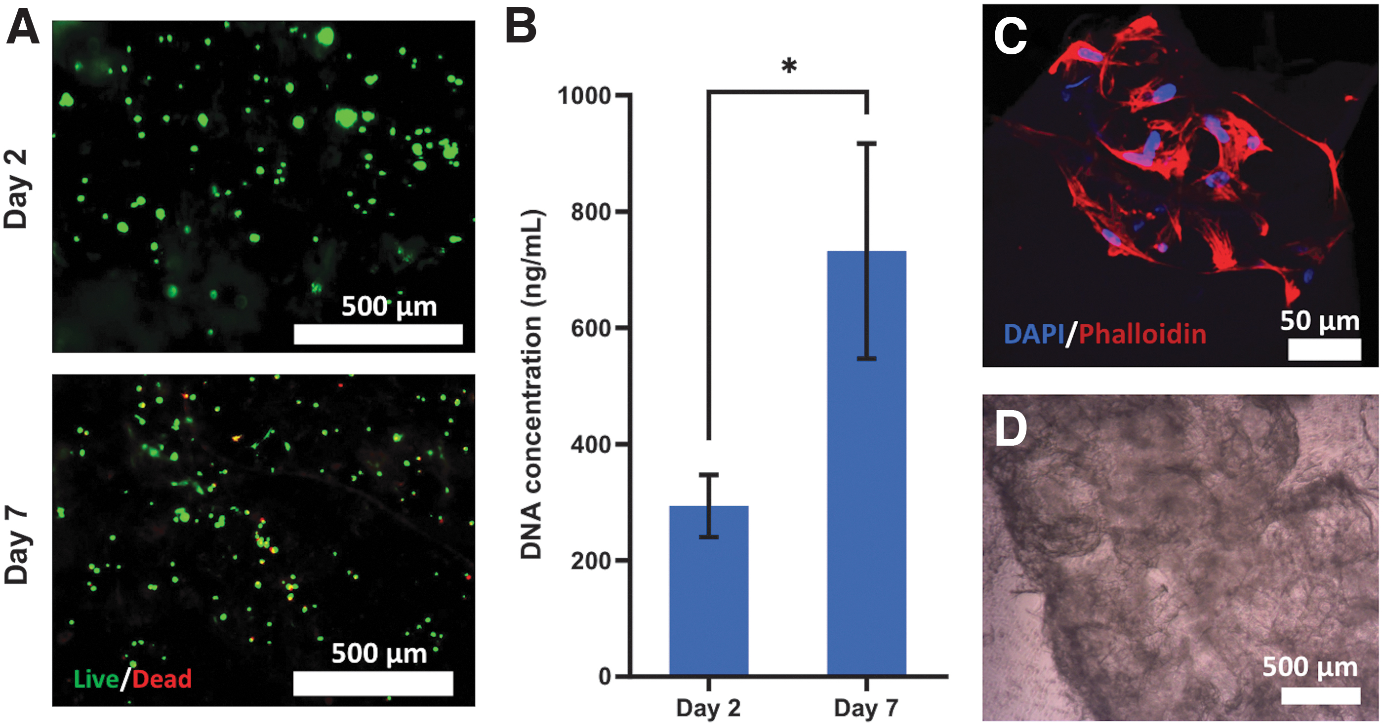
hASC viability, attachment, and proliferation on PLMA microsponges.
Interestingly, the self-assembly of cell–microsponge aggregates was observed in cell culture wells even in the absence of external stimuli, as displayed on Figure 3D.
Injectability of the microsponges
To be used as an injectable scaffold for cell delivery, microsponges should be able to pass through the needle of a syringe without compromising their structural integrity. The injectability of the microsponges was assessed by passing a suspension of microsponges through 21G, 25G, and 27G needles. Regardless of their dimensions, the microsponges successfully pass through the needles without causing a blockage. To assess whether the integrity of the microsponges was maintained, the dimensions of the microsponges were measured after injection (Fig. 4). The analysis of the microsponges and their size distribution postinjection shows that the size and shape of the microsponges was maintained after passing through 21G and 25G needles, indicating that their integrity was preserved.
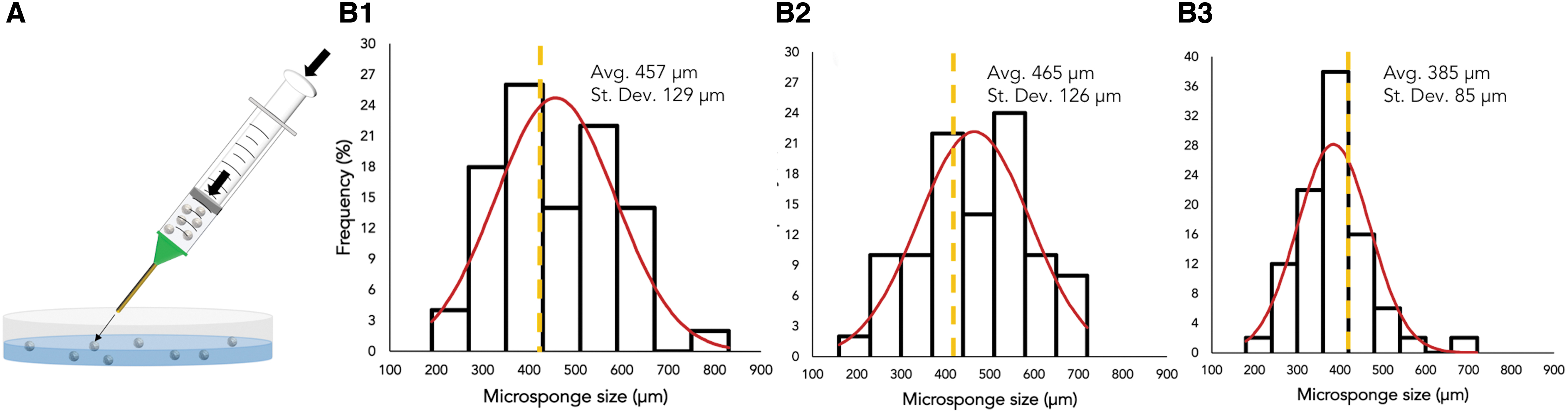
In fact, the average diameter of the microsponges increased to 457 ± 129 μm and 465 ± 126 μm after injection through 21G and 25G, respectively. This increase was caused by the aggregation of some microsponges after injection. A slight decrease in average microsponge dimensions was observed after passing through a 27G needle, which indicates that the microsponges may have been damaged. As such, the structural integrity of larger microsponges cannot be guaranteed after passage through a 27G needle.
To assess the possibility of using the PLMA microsponges both as injectable cell carriers and building blocks for bottom-up tissue engineering, cell-laden microsponges were injected through a 21G needle into PDMS molds. After 2 days, cell culture medium was injected into the mold to remove the resulting constructs, demonstrating that the microsponges were able to assemble into fiber-shaped constructs, thus assuming the shape of the mold (Fig. 5C, D). This demonstrates that the cell-laden microsponges are able to be molded into a predefined shape, denoting the ability to adapt to the shape of fractures and defects in vivo. Cell morphology analysis also indicates that the cells are not evenly distributed throughout the microsponges. Instead, they produce localized cell clusters on their surface.
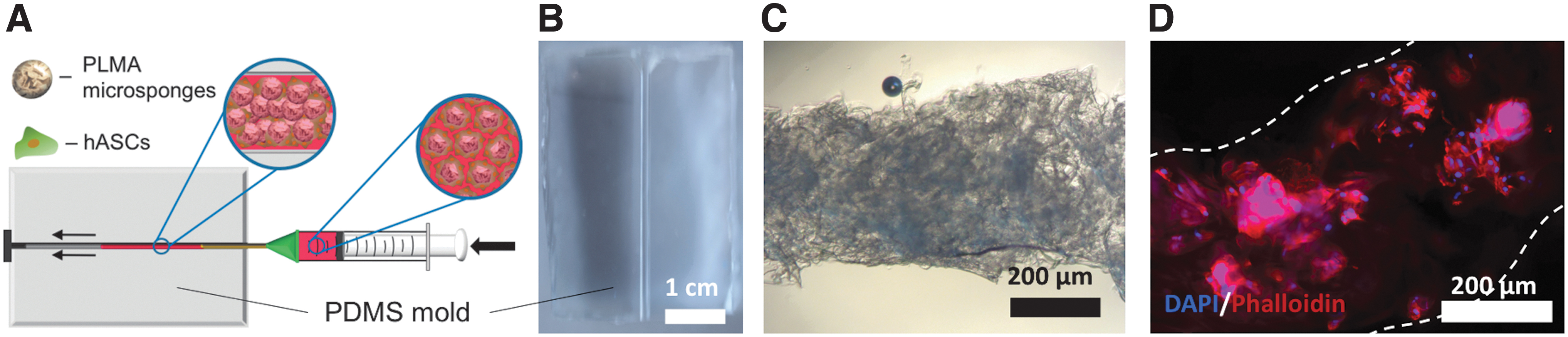
Injection of cell-laden microsponges in vitro.
Discussion
Alginate microcapsules have previously been reported as a suitable platform for the culture and storage of cells and spheroids.44,45 In this work, it is proposed that alginate microcapsules can also be used to secure a photocrosslinkable precursor solution, providing support during polymerization and allowing the production of microcarriers with controlled dimensions.
Although microfluidic devices are already used to produce monodisperse microcarriers of controlled size from photocrosslinkable materials, such as GelMA,46–48 the implementation of this technology in clinical applications has been hindered due to low production rates. 49 Microfluidic devices are typically limited to flow rates in the 0.5–10 μL/min range, preventing mass production of microparticles.48,50,51 By using coaxial electrospray, it is possible to employ higher flow rates, thus increasing production rates up to 1000-fold. Moreover, this technology can be scaled up through multiplexed electrospray setups,52–54 enabling the high-throughput generation of microcapsules and microsponges for clinical application.
The use of animal-derived materials in the development of cell delivery platforms raises animal welfare and health concerns, as they can transmit zoonotic diseases or trigger an immune reaction. Furthermore, the regenerative potential of materials isolated from animal sources is limited by their inability to accurately recreate the properties of human tissues.55–58
hPL constitutes a safe, easily accessible resource that can be harnessed to develop novel therapeutic platforms. hPL contains a rich cocktail of bioactive proteins, including cytokines, chemokines, and a wide variety of growth factors,59,60 which has prompted its use in cell culture as a xeno-free alternative to animal-derived serums61,62 and its incorporation into implantable biomaterial scaffolds.63–65
In this study, the production of microcarriers derived exclusively from hPL is reported for the first time. Electrohydrodynamic atomization was used to produce PLMA microparticles, which were frozen at a controlled freezing rate and freeze dried to introduce macroporosity. The resulting microsponges display an average pore size of 30 μm, which is comparable to the pore dimensions of commercially available macroporous microcarriers used in three-dimensional cell culture, commonly situated in the 10–50 μm range.66,67 As such, it is envisioned that the pores on the surface of the microsponges will allow cells to migrate to the interior of the microsponges, vastly increasing the available surface area for cell culture as well as the quantity of cells that can be produced and delivered.
Future studies could focus on the application of alternative parameters during ice templating, producing microsponges with different porosities, pore dimensions, and pore distribution, to assess the influence of these parameters on cell migration and proliferation. 68 Changing the freezing rate, the degree of modification of PLMA or the concentration of PLMA are all possible strategies that can be explored to adjust pore dimensions.27,69
The method described in this study is able to generate microcarriers with dimensions ranging from 100 to 1000 μm, depending on the production parameters. To guarantee the supply of oxygen and nutrients to cells grown on the PLMA microsponges, smaller diameters were preferred. In vascularized tissues, all cells are located within 200 μm of the nearest capillary, which is considered the maximum distance that ensures proper diffusion of oxygen.70,71 As such, macroporous microcarriers should not exceed a diameter of ∼400 μm, as it may result in cell death within their inner porous network. As such, when selecting the default parameters for microsponge production, a low PLMA flow rate was chosen (1 mL/h), to produce microsponges with reduced dimensions.
While an applied voltage of 14 kV produced microsponges with an average diameter below 400 μm, the use of higher voltages also introduces greater heterogeneity in the dimensions of the microsponges, which could lead to differences in cell behavior between microsponges. As such, a voltage of 11 kV was selected as the default parameter so as to not compromise the reproducibility of cell behavior.
PLMA microsponges showed promise as injectable cell carriers, as they were able to pass through needles as small as 25G without causing a blockage, significantly altering their shape and dimensions or damaging their internal porous structure, an encouraging prospect regarding their future application in a clinical setting. Further research will be necessary to assess the mechanical properties of the microsponges, as well as their stability, both in vitro and in vivo.
When seeded with cells, the microsponges were shown to promote cell adhesion, proliferation, and spreading. hASCs were selected for cell assays, as they can be isolated directly from each patient through a simple and minimally invasive procedure, similar to hPL. 42 The delivery of hASCs has been pursued as a therapeutic strategy, as these cells can produce and secrete proregenerative factors that promote angiogenesis.72,73
As such, PLMA microsponges seeded with hASCs could constitute a fully autologous and humanized cell delivery platform for personalized regenerative medicine. Moreover, cell-laden PLMA microsponges self-organized into larger aggregates, a promising prospect for the bottom-up assembly of tissue constructs. 74
Conclusions
In summary, a scalable method to prepare highly porous microcarriers from photocrosslinkable biomaterials has been described in this article. Liquid core–shell microcapsules are first produced using coaxial electrospray and processed into spherical microcarriers, which can be lyophilized to generate an internal porous network. This method was successfully used to produce human protein-derived microsponges supporting hASC attachment and proliferation. We can envisage the production of fully personalized carriers using the blood of the patients, thus creating a fully autologous platform for cell delivery. Furthermore, cell-laden microsponges displayed the ability to self-assemble into larger constructs. As such, it is proposed that the PLMA microsponges described in this study can be employed as an injectable platform for cell delivery and bottom-up assembly.
Footnotes
Acknowledgment
The authors are grateful to Inês A. Deus for critical point drying of the samples for SEM.
Authors' Contributions
B.M.F.L.: investigation, methodology, visualization, and writing—original draft; M.C.G.: investigation, visualization, and writing—review and editing; C.A.C.: conceptualization, funding acquisition, methodology, project administration, resources, supervision, and writing—review and editing; J.F.M.: conceptualization, funding acquisition, methodology, project administration, resources, supervision, and writing—review and editing. All authors have read and agreed to the final version of the article.
Disclosure Statement
No competing financial interests exist.
Funding Information
The authors would like to acknowledge the financial support of the Portuguese Foundation for Science and Technology (FCT) for project Beat (PTDC/BTM-MAT/30869/2017) and the individual contract 2020.01647.CEECIND of C.A.C. This work was developed within the scope of the project CICECO-Aveiro Institute of Materials, FCT Ref. UIDB/50011/2020 and UIDP/50011/2020, financed by national funds through the FCT/MCTES. The authors would also like to acknowledge financial support by the European Research Council (ERC) for project ATLAS (ERC-2014-ADG-669858). The work was also partially supported by the European Union (EU) Horizon 2020 for the project InterLynk, Grant agreement: H2020-NMBP-TR-IND-2020, Project ID: 953169.