




Abstract
Femur head necrosis, also known as osteonecrosis of the femoral head (ONFH), is a widespread disabling pathology mostly affecting young and middle-aged population and one of the major causes of total hip arthroplasty in the elderly. Currently, there are limited number of different clinical or medication options for the treatment or the reversal of progressive ONFH, but their clinical outcomes are neither satisfactory nor consistent. In pursuit of more reliable therapeutic strategies for ONFH, including recently emerged tissue engineering and biomaterials approaches, in vivo animal models are extremely important for therapeutic efficacy evaluation and mechanistic exploration. Based on the better understanding of pathogenesis of ONFH, animal modeling method has evolved into three major routes, including steroid-, alcohol-, and injury/trauma-induced osteonecrosis, respectively. There is no consensus yet on a standardized ONFH animal model for tissue engineering and biomaterial research; therefore, appropriate animal modeling method should be carefully selected depending on research purposes and scientific hypotheses. In this work, mainstream types of ONFH animal model and their modeling techniques are summarized, showing both merits and demerits for each. In addition, current studies and experimental techniques of evaluating therapeutic efficacy on the treatment of ONFH using animal models are also summarized, along with discussions on future directions related to tissue engineering and biomaterial research.
Impact statement
Exploration of tissue engineering and biomaterial-based therapeutic strategy for the treatment of femur head necrosis is important since there are limited options available with satisfactory clinical outcomes. To promote the translation of these technologies from benchwork to bedside, animal model should be carefully selected to provide reliable results and clinical outcome prediction. Therefore, osteonecrosis of the femoral head animal modeling methods as well as associated tissue engineering and biomaterial research are overviewed and discussed in this work, as an attempt to provide guidance for model selection and optimization in tissue engineering and biomaterial translational studies.
Introduction
Femur head necrosis or osteonecrosis of the femoral head (ONFH) is a pathological process in which destruction of blood supply to the femoral head results in bone osteonecrosis. 1 It mostly occurred in young and middle-aged population, accounting for about 30% of all orthopedic diseases. It is estimated that about 20 million people around the world currently suffer from ONFH and there are 8.12 million cases in China, causing a heavy burden on society and families. 2 More than 80% of untreated ONFH patients experience femoral head collapse within 1 to 3 years, and eventually have to undergo artificial hip replacement. 3 These facts make ONFH one of the most challenging and formidable diseases in orthopedics.
ONFH can be clinically divided into traumatic and nontraumatic type based on its causes. 4 Traumatic ONFH is commonly caused by an acute mechanical disruption of blood supply to the femoral head. The initial lesions occur in weight-bearing zone of femoral head, and trabecular bone under femoral head is subjected to abnormal mechanical stimulation, resulting in stress fractures. Regarding limited bone repair capability of patients, femoral head eventually collapses. For nontraumatic ONFH, on one hand, minor trauma is an inducible factor and often combined with other factors such as intraosseous blood pressure and fat embolism to cause femoral head necrosis. Besides, nontraumatic ONFH patients are commonly associated with the use of corticosteroid or excessive drinking.
The clinical difficulties in ONFH treatment are mainly related to insufficient blood supply to lesion site. 5 Early treatments of ONFH, including drug therapy, physical therapy, core decompression, osteotomy, bone grafting, and so on, are suggested to slow down its progression to avoid artificial hip replacement. However, the role of drug therapy and physical therapy in prognosis has not been well established and the clinical outcomes vary significantly in medical practices. These methods are difficult to change pathological tissue/cell metabolism in femoral head, and fundamentally solve blood supply problem in lesion site. Due to the progressive nature of ONFH and the lack of relevant research, it is difficult to guarantee long-term effect of hip-preserving strategy since there is no universal standard for the best surgical procedure. 6
Recently, the use of tissue engineering and biomaterials to reconstruct or improve femoral head blood supply has attracted a lot of attention. Tissue engineering and biomaterial methods are mainly based on cells, growth factors, scaffold materials, and local drug delivery systems,7–10 which are effective methods for reconstructing bone tissue and vascular network, although their clinical translation is still challenging. Therefore, a reliable animal model is of extreme importance for ONFH studies by tissue engineering and biomaterial approaches. Suitable and reliable animal models can reflect the changes of microenvironment in vivo under pathological conditions and reflect the progress of human diseases to some extent. 11
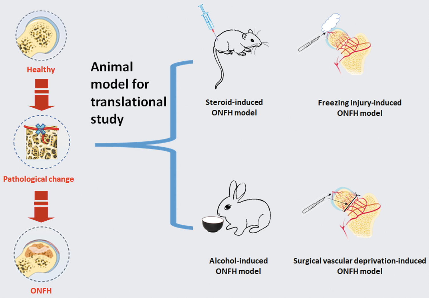
So far, researchers have established various types of ONFH animal models to evaluate outcomes of tissue engineering and biomaterial-based treatments for ONFH. And yet, there is still a lack of standard animal modeling method due to variations in research purposes and experiment conditions. As an attempt to provide better understanding of ONFH animal models and their application, this article summarizes several methods for establishing ONFH experimental animal models and their related researches, and discusses the advantages and disadvantages of these models, providing guidance for model selection and optimization in tissue engineering and biomaterial translational studies (Fig. 1).

Methods of modeling, evaluation, and treatment of femoral head necrosis.
Current In Vivo Animal Models of ONFH
Steroid-induced ONFH models
Steroid is a metabolomic hormone synthesized and secreted by adrenal cortex, highlighted with anti-inflammatory, antiallergic, antitoxin, immunosuppressive, and antishock potentials. However, steroids are proved to be involved in bone matrix protein decomposition, overexcretion and absorption of calcium and phosphorus, osteoblast activity inhibition, and protein mucopolysaccharide synthesis reduction, resulting in femoral head necrosis, osteoporosis, and other bone and joint complications after decades of clinical practices. 12 Therefore, steroid-induced ONFH is adopted as the most commonly used animal modeling strategy in research (Table 1, Fig. 2).13,14
Establishment of ONFH animal models using different approaches.
Construction of ONFH Animal Model and Relevant References
MPS, methylprednisolone; MPSL, methylprednisolone acetate; ONFH, osteonecrosis of the femoral head; SD, Sprague-Dawley; N.A., not available.
Usually, ONFH animal model can be generated through intramuscular injection of steroid.15–18 For example, Zhao et al. 19 injected steroid methylprednisolone acetate (MPSL) into the right gluteus medius muscle of rabbits (20 mg/kg), and a 75% incidence of ONFH occurrence after 2 weeks was reported by histopathological examination of the femur and humerus.
Different from sole steroid injection, a combination of steroid and endotoxin for ONFH modeling is developed for better clinical condition simulation. 20 Clinically, steroid therapy is often used in the treatment of severe acute respiratory syndrome, asthma, rheumatoid arthritis, and systemic lupus erythematosus. 21 In this process, endotoxin released by the bacteria is found to act as an exogenous heat source, stimulating severe immune responses, 22 causing inflammation, and microcirculatory disturbance, even endotoxin shock and disseminated intravascular coagulation.23,24
Inspired by this observation, the use of steroids combined with endotoxin to construct steroid-induced ONFH models is increasingly adopted, which not only shortens the modeling time and improves the modeling success rate but also simulates the actual physiological response. 25 For example, Li and Wang 26 established a rat model of steroid-induced ONFH by intraperitoneal injection of endotoxin 10 mg/kg for 2 days, followed by intraperitoneal injection of methylprednisolone 10 mg/kg for 3 days. After model established, osteonecrosis occurrence rate in model group was twice as high as that in control group interfered with bone morphogenetic protein (BMP) signal pathway inhibitors, while osteonecrosis was not observed in the blank group.
Motivated by combined use of endotoxin and steroids, allogeneic serum was also used to induce severe immune response to consequently generate steroid-induced ONFH model. Steroids with allogeneic serum can induce severe immune response and inhibit bone cells from producing collagen and elastic fibers, leading to steroid-induced ONFH. 27 For example, Wang et al. used equine serum in combination with MPSL to establish animal models with a success rate of 84.62% and a mortality rate of 13.33%. 28
The steroid-based modeling method was also used in large animals such as pigs, but the outcomes are complicated. In a work, Drescher et al. reported that a high-dose methylprednisolone infusion reduced femoral head blood flow after 24 h, 29 but no evidence of ONFH was found in these pigs.30,31
In summary, steroid-based modeling methods can mimic the etiology of ONFH and reveal simplicity and low costs in small animal models. Compared to the only use of steroid, modeling method of combined endotoxin or allogeneic serum is more consistent with the clinical pathological features of ONFH, along with shortened modeling time and more obvious phenotype of osteonecrosis, and therefore is accepted by more scholars. However, reliability of this modeling method in large animals is still questionable and there are so far no standards in the steroid type, dosage, medication methods, and so on.
Alcohol-induced ONFH models
Chronic alcoholism is closely related to ONFH, mainly related to abnormal fat metabolism and osteoporosis after excessive drinking.32–34 The method of alcohol-included ONFH models can simulate the progress of clinical pathology. The main methods are local injection of alcohol and alcohol liquid feed.35–37 Local injection of alcohol is most commonly used in large animals such as sheep and dogs, such as Zhu et al., who make an arc incision in the area below the rotor and then drill the medullary canal in the center of the femoral head under the guidance of X-rays. 38 ONFH model was constructed with bone wax sealing hole after alcohol was injected into bone marrow canal by catheter.
The alcohol liquid feed method imitates human ONFH by filling the animal with alcohol. Excessive drinking can reduce bone cell metabolism and cause osteoporosis, paving bone trabecula thinning and reduction of local stress concentration area. 39 For example, Sprague-Dawley rats were fed a liquid diet containing 8% ethanol.40,41 After 6 weeks, 8 of 10 rats had obvious ONFH sign with decrease in bone density of femoral head.
Compared to other modeling methods, alcohol diet method is easy to operate and reveals high success rate. Despite that, there is a risk of femoral neck fracture and alcohol leakage, 42 resulting in inaccurate doses. Moreover, animal models induced by local injection of alcohol cannot simulate the pathological development of human ONFH. 42 Although alcohol poisoning is closely related to ONFH, the time/dose of alcohol intake leading to alcohol-induced ONFH has not been determined.
Femoral head necrosis induced by freezing injury
The animal model of femoral head necrosis using liquid nitrogen freezing method generally chooses animal rabbits, because of its large size and easy operation. 43 Liao et al. transected the round ligament of the femoral head of New Zealand rabbits in the experiment, and immediately use liquid nitrogen to contact the surface of the femoral head causing damage to the vascular endothelial cells, and then affecting the normal function of blood vessels. 44 This procedure can generate blood reperfusion injury in the frozen femoral head, which further develops avascular necrosis. 45 In another study, Li et al. established the ONFH small-tailed Han sheep model by drilling holes in the weight-bearing area of the femoral head at the head neck junction, and then inserting a cryoprobe into the tunnel to induce vascular hypothermic injury in the femoral head. 46
Compared with hormone modeling, liquid nitrogen freezing method exhibits advantages of short cycle, high repeatability, no involvement of other organs, no chemical residues, and limited mortality rate. However, the disadvantages include that it cannot correspond to the clinical pathogenesis and mechanism, and it is so far mostly used for treatment and prognosis research. Therefore, in case of ONFH pathogenesis investigation, this method is probably not appropriate.
Femoral head necrosis induced by surgical vascular deprivation
Traumatic ONFH is usually caused by acute mechanical interruption of blood supply to the femoral head, 47 along with femoral neck fracture and hip dislocation as common etiology. The experimental animals are mainly rats and immature pigs.48,49 Traumatic necrosis of the femoral head is usually caused by vascular ligation. For example, Liu et al. transected the ligament artery of rat femoral head after surgical separation of the femoral head, and then ligated the femoral neck with nonabsorbable suture. 48 This method destroyed the blood vessels around the femoral neck and lead to avascular necrosis of the femoral head.
According to the literature, vascular ligation modeling method requires simple operation procedure, but has high modeling success rate, high femoral head surface collapse rate, and accurate necrosis scope. The disadvantage of this method is that it cannot simulate the pathogenesis of most human ONFH. Nevertheless, it is still a reliable modeling method for simulating avascular necrosis of the femoral head, especially recommended for the pursuit of restoring the mechanical properties of the femoral head or increasing the blood supply of the femoral head.
Current Animal Studies on the Treatment of ONFH
Since osteonecrosis areas are generally difficult to heal spontaneously, most ONFH patients inevitably develop femoral head collapse or degenerative arthritis. Therefore, it is necessary to develop effective ONFH treatment strategy in animal models for medical practice. Currently, nonoperative and operative treatment strategies have been proposed, and some have been clinically adopted. Moreover, the uses of tissue engineering and biomaterials to improve bone blood supply have become a new research hotspot (Table 2).
Tissue Engineering and Biomaterial Approaches for the Treatment of ONFH
BM-MSC, bone marrow mononuclear cell; BMP-2, bone morphogenetic protein-2; CMC/ALG/BMSC/EPC, carboxymethyl chitosan/alginate/bone marrow mesenchymal stem cell/endothelial progenitor cell; MSC, mesenchymal stem cell; PCL/nHA, polycaprolactone/nano-hydroxyapatite; VEGF, vascular endothelial growth factor; N.A., not available.
Nonsurgical treatments
Nonsurgical treatment of ONFH usually includes medication and physical therapy. Drug therapy has been reported for the early-stage treatment of ONFH, but with limited positive outcomes. Physical therapy methods include high-energy shock wave therapy and hyperbaric oxygen therapy, which can reportedly induce the formation of new blood vessels and promote bone cell proliferation and differentiation, as well as subsequent bone formation, but their role in the prognosis has not been well established and clinical outcomes vary significantly in medical practices.50–52
Surgical treatments
Surgical treatments usually refer to core decompression, bone grafting, and osteotomy. Core decompression surgery removes necrotic bone, reducing the pressure on the femoral head, stimulating bone cell growth, and improving the microcirculation of blood vessels.53–55 However, the decompression hole with large diameter may destroy the mechanical support structure of the femoral head and even cause iatrogenic collapse. Bone grafting surgery refers to the removal of dead bone by fenestration of the femoral head or neck, followed by transplantation of blood-rich bone into the voids. 56 In clinical practice of bone grafting surgery, however, the bone flaps struggled with the problem of shifting or falling off after surgery.57,58
Moreover, due to the large trauma area of the femoral head and changes in the normal anatomical structure, hip replacement becomes less possible for patients in the later stage if osteotomy fails. 59 Therefore, osteotomy has been rarely used in clinical practice in recent years.
It is also important to clarify that the aforementioned surgical treatments cannot fundamentally change the pathological tissue/cell metabolism in femoral head and cannot avoid surgical trauma, bringing more risks for ONFH patients in poor health conditions. When the ONFH progresses to irreversible late stage, hip replacement is still the last resort for patients. Regarding the costs of surgery and limited lifespan of hip implants, it is highly demanding to develop new treatments to delay or even reverse the progress of ONFH for patients.
Tissue engineering and biomaterial approaches for the treatment of ONFH
Approaches using cells and cell derivates
Mesenchymal stem cells have been shown to promote osteogenesis and vascularization of bone, and thus have been studied in combination with other surgical approaches for better clinical outcomes in ONFH treatment (Table 2).60,61 For example, steroid-induced ONFH in adult female Japanese white rabbit was treated by core decompression surgery combined with bone marrow mesenchymal stem cell (BMSC).62,63
After 12 weeks, the bone mineral density (BMD), bone volume, and the number and diameter of new vessels in the core decompression plus mesenchymal stem cell group were significantly higher than those in the control groups. Xu et al. reported that the combination of BMSCs and endothelial progenitor cells promoted steroid-induced ONFH in New Zealand white rabbit by promoting osteogenesis and angiogenesis and reducing adipogenesis. 10 Exosomes are extracellular vesicles secreted by cells and are known to transmit intercellular signals, promote self-repair of damaged cells, and participate in regulation of cell growth, migration, and vascular development.64,65 In a recent study, exosomes of hypoxic preconditioned BMSCs had positive effects on angiogenesis and avoidance of osteonecrosis in the steroid-induced rat animal model. 66
Although cell therapy has revealed promise for ONFH treatment in animal models, its therapeutic potential for clinical use remains questionable. 67 For example, cell therapy is often associated with immune risks and it is not clear which source of stem cells is more appropriate for ONFH treatment. Also, the microenvironment in vivo varies in individuals and largely affects the survival, differentiation, and consequent therapeutic effect of cells. 68 In addition, it is not clear how many cells are needed for each patient to achieve optimal results for different degrees of ONFH.
The use of growth factor
The use of growth factor in ONFH treatment can effectively stimulate endothelial cells, endothelial progenitor cells, osteoblasts, BMSCs, and other cell migration and proliferation and promote the formation and maturation of blood vessels. There are many growth factors involved in the regulation of angiogenesis, including vascular endothelial growth factor (VEGF), BMP, insulin-like growth factor, fibroblast growth factor, and platelet-rich plasma (PRP). For example, Tong et al. treated ONFH in a mouse model with injection of PRP into caudal vein and histological results showed that PRP reduced humoral and cellular immune responses and reduced the degree of ONFH. 69 However, angiogenic factors have a short half-life and are difficult to be locally delivered to necrosis loci with accurate concentration and distribution control.
Growth factors combined with gene therapy have also been explored. The gene fragment carrying growth factor was transfected into seed cells, which enchanced the ability of promoting blood vessel and bone formation. 8 For example, Guzman et al. characterized MSCs that were genetically modified to overexpress platelet-derived growth factor-BB (PDGF-BB-MSCs) in vitro and then implanted into ONFH rabbit model. 70 Gene-modified PDGF-BB-MSCs as an adjunct to core decompression can enhance bone regeneration and angiogenesis in early ONFH therapy and reduce the progression of osteonecrosis. Despite the promising outcomes of PDGF-BB-MSCs, such gene-modified MSC strategy needs to be confirmed to be safe for the perspective of regulatory science and more studies are demanded before its translation to clinical application.
Drug delivery
Drug delivery system can precisely control drug release and target the lesion site by various methods such as modifying drug and the use of biomaterials as carriers. In an ideal drug delivery system, local drug concentration can maintain a proper level for a sustained time, which is very attractive and important for targeted treatment of ONFH. For ONFH treatment, so far, many drug delivery systems have been developed, including chitosan, 10 gelatin,71,72 alginate, 10 and hydroxyapatite microspheres, 73 virus or liposome carrier, 1 and microosmotic pumps (Table 2). 74 For example, gelatin microspheres loaded with growth factor VEGF could continuously release the growth factors, activate Wnt signaling pathways, and increase osteogenic and angiogenic factors, thus facilitating ONFH repair. 71
In another study, Dailiana et al. used a micro-osmotic pump to continuously administer VEGF over a period of 14 days. In a canine ONFH model induced by freezing injury and treated with the micro-osmotic pump method, a significant increase in bone volume/total volume was observed, along with an increase in trabecular bone thickness, and a reversal of osteonecrosis. 74 Histological results showed that FGS with 30% porosity could enhance bone regeneration and had excellent biomechanical properties in bone marrow canal.
Scaffold for ONFH treatment
With better understanding of the therapeutic effects of biomaterials, scaffold is increasingly adopted to reconstruct or repair necrotic bone tissue.46,75 The pore structure of scaffold not only creates an environment for bone cells to differentiate and proliferate but also forms a strong interface between the scaffold and bone tissue, providing reliable mechanical support for femoral head.72,73,76 For example, Maruyama et al. 77 prepared a functional gradient scaffold (FGS) made of polycaprolactone and β-tricalcium phosphate by 3D printing to implant in a steroid-induced ONFH in a New Zealand white rabbit model.
Maruyama et al. 78 further combined this scaffold with bone marrow-derived monocytes and demonstrated that functionally gradient scaffolds could improve the growth of necrotic rabbit femoral heads and injection of bone marrow-derived monocytes can reduce the area of osteonecrosis. Hydroxyapatite with good biocompatibility and osteoconductivity was incorporated into porous scaffolds to promote bone tissue growth onto the pore wall and into the pores.7,79–81 Kang et al. 82 treated steroid-induced ONFH in rabbits with strontium-doped calcium polyphosphate (SCPP) scaffold and autologous bone marrow mononuclear cells (BM-MNCs). The results showed that the combination of SCPP scaffold and BM-MNCs could enhance VEGF expression and promote osteogenesis, which may improve angiogenesis and allow the incorporation and remodeling of new trabecular bone without mechanical weakening.
In addition, piezoelectric ceramics, such as hydroxyapatite and barium titanate composites, have been shown to promote bone repair and reconstruction. 83 These developments have highlighted the direction of making the scaffold bioadaptable to bone regeneration in femoral head.
Bone cement
Bone cement is simple and effective for treating irregular bone defects, relieving postoperative pain, and improving bone structural stability. The use of acrylic bone cement such as polymethylmethacrylate (PMMA) in core decompression surgery has demonstrated to improve microcirculation, prevent osteonecrosis collapse, and increase the success rate of surgery, while the thermal effects of PMMA cross-linking may result in aseptic loosening. 84
In addition to inert and nonbiodegradable acrylic bone cements, calcium phosphate cements (CPCs), which have Ca/P similar to human body and degradability in vivo to stimulate the growth of new bone, have also been studied. For example, Chang et al. combined propylene fumarate and calcium phosphate bone cement, mixed with different doses of ginsenosides, and their results showed the potential of Rg1-binding bone cement in stimulating neovascularization and providing sufficient mechanical strength for the femoral head. 85 CPCs can also be used to deliver functional ions, drugs, cells, and growth factors for the repair of the femoral head, but cases are rare.
Magnesium screw
Biodegradable magnesium is a newly emerging biodegradable material for manufacturing reconstructive bone screws, which first perform the initial fixation function and then degrade over time without the need for costly secondary resections. Magnesium screws were successfully used to stabilize the graft and restore blood supply to the femur. 86 Katiella et al. implanted magnesium alloy combined with BMSC therapy in liquid nitrogen freezing-induced ONFH in New Zealand white rabbits and demonstrated the potential of magnesium alloy in promoting new bone formation to repair ONFH. 87 The potential of biodegradable magnesium screws for vascularized bone graft fixation in patients with avascular necrosis of the femoral head was also investigated in humans. 58
Compared with conventional methods without flap fixation, magnesium screws could improve the stability of flap in situ, and larger bone is formed in the fusion area of the screw and flap, which is beneficial to the recovery of the operation. The challenges of magnesium screws are how to match its degradation process to the regeneration process of bone in complicated physiological and mechanical environment and to construct a tissue regenerative environment with its degradation products (Mg2+ ions, H2, Mg oxides, etc.).
Experimental Techniques of Evaluating Therapeutic Efficacy in Animal Models
General observation and visual inspection
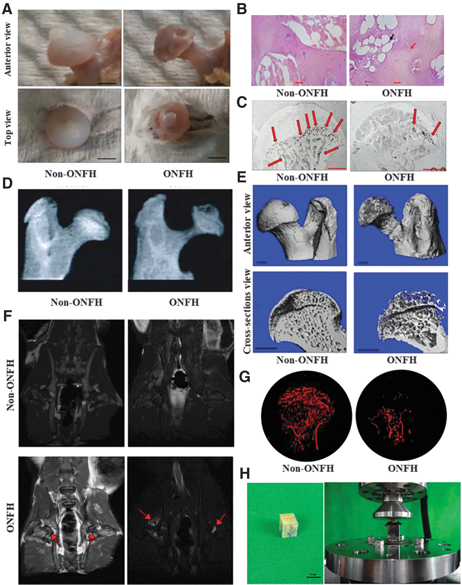
After the experimental animals are sacrificed, their femoral heads are taken out completely and usually the color, contour, cartilage surface, and articular surface of the femoral heads are inspected with naked eyes. Compared to healthy or non-ONFH group, femoral heads from ONFH group exhibit irregular femoral head contours, signs of collapse, and rough and pale articular cartilage (Fig. 3A). After avascular necrosis of the femoral head model is constructed, animals are often observed to be lethargic, their daily activities are reduced, hair loss of luster, and their food intake and body weight decreased compared with those before modeling. In addition, model animals often show different degrees of lameness, and the strength of the affected limbs is weakened. 48
Histopathological examination
In experimental models, the most commonly used method to identify ONFH is histopathology. Compared with the non-ONFH group, HE staining of bone tissue in the necrotic area of the model showed that the bone trabecula was sparse or fragmented and the proportion of bone cells empty lacunae increased, bone marrow hematopoietic cells decreased, adipocyte proliferation or hypertrophy, microvascular occlusion or missing. (Fig. 3B).33,77 To observe the microvascular status in the necrotic area, ink perfusion experiment can be used to observe the number or density of blood vessels to assess the blood supply to the femoral head (Fig. 3C). Compared to other imaging methods like angiography, this method is simple and cost-efficient to implement. 88
Imaging evaluation
In the late stage of ONFH, some large animals like emu show typical bone density decline and cystic degeneration of femoral head on plain X-ray films (Fig. 3D). Micro-computed tomography (micro-CT) can reveal the bone structure more clearly than X-rays and in three dimensions (Fig. 3E).89–91 In addition, BMD and bone volume fraction at the central coronal plane of the femoral head can be measured by peripheral dual-energy X-ray absorptiometry before and at the late stage of ONFH, respectively, to evaluate the changes of BMD in the femoral head region. 92
In the early stage of ONFH, when bone does not seem abnormal on X-ray imaging, magnetic resonance imaging is a highly sensitive method for diagnosing ONFH and can be used to evaluate the disease model of ONFH (Fig. 3F). The low intensity of the t1-weighted image signal, high intensity of the t2-weighted image signal, and high fat-suppression image signal intensity in the head and neck junction area are used as criteria for the diagnosis of ONFH. 93 In addition, digital subtraction angiography can visualize the distribution, shape, and location of blood vessels, assisting in better understanding the progress of the disease (Fig. 3G). 94
Biomechanical study
Biomechanical evaluation of the femoral head specimen is a simple and reliable method to assess the bone quality, revealing the changes in bone strength of the femoral head and the changes in microstructure of the femoral head during collapse of ONFH. 55 Once the bone specimen was subjected to axial compression displacement control at a certain speed in the material testing machine (Fig. 3H), the ultimate strength, yield strength, and elastic modulus of the femoral head could be calculated to evaluate the biomechanical properties of the femoral head. 95
Finite element analysis is a widely used computational method for mechanical engineering and has been recently adopted in ONFH-related research. Based on the 3D reconstruction of necrotic femoral heads from CT or micro-CT images, finite element analysis can be used to model and simulate how the interested regions of necrotic femoral heads respond to external deformation, and then generate mechanical parameters such as the stress, strain, and elastic modulus to evaluate the biomechanical properties of the femoral heads. 96
Challenges and Prospects
Among the different construction methods of ONFH animal models, steroid- and alcohol-induced methods can simulate pathogenesis of ONFH, but there are no standard recommendations for the dosages of alcohol and hormones, and success rate of these two methods is still not satisfactory. Larger animal models are more similar to humans in terms of bone morphology, fracture healing, and so on, but modeling methods and mechanistic study of large animal models are comparatively lacking. Animal models of traumatic femoral head necrosis cannot perfectly match the clinical pathogenesis and mechanism of ONFH, accompanied with damage to the surrounding tissue, so it is more recommended for treatment outcome and prognosis investigation.
When constructing ONFH animal models, there is a risk that researchers are prone to consider difficulty of modeling before the underlying pathogenic mechanism of the model. Clinical treatments of ONFH have revealed that patient's condition varies and many individual factors need to be considered in the treatment, such as the patient's age, morbidity degree, and osteoporosis. Unfortunately, current modeling methods hardly reflect these individual factors. In addition, the body weight and walking gait of the animals are different from that of human beings, which pose a challenge to reliability of ONFH model.
For researches on tissue engineering or biomaterial-based treatment, there is a lack of advanced methods for postimplantation evaluation, such as single cell sequencing, immunoregulation, and so on. The current biomaterials or tissue engineering approach are mostly used as void filler after core decompression surgery or bone fixation without bioactive or smart functionality. Specifically, lack of bioadaptive materials suitable for the whole process of tissue repair, and how to realize the design and use of tissue engineering materials such as piezoelectric response, spatiotemporal adaptation, and environment-responsive functionality remain major challenges in this area.
To evaluate the efficacy and mechanisms of different therapies for ONFH, construction of experimental animal models should resemble clinical scenarios to produce satisfactory experimental data. Therefore, when choosing a proper animal model, there are many factors to consider. First of all, the physiological structure and histological characteristics of experimental animals should be selected to represent characteristics of human diseases. Second, different stages of disease progression in humans also need to be mimicked by animal models. Third, abided by the research ethics for animal study, animal models should be reasonably and carefully designed according to the characteristics and research purposes. Fourth, from a biomechanical point of view, the size and structure of femoral head and weight-bearing situation of the limbs should be considered.
Footnotes
Authors' Contributions
J.M., Y.S., and H.Z. wrote the original draft. H.Z., L.Y., and P.Z. proposed the topic and content of the draft, ensured the integrity of the study from conception to completion. L.Y., P.Z., H.Z., X.L., Y.B., and C.L. contributed to acquisition of funding and proofreading. X.J. contributed to technical editing.
Acknowledgments
The authors thank Hongshui Wang and Mingjun Li for their assistance during article preparation.
Disclosure Statement
No competing financial interests exist.
Funding Information
This work was supported by grants from the National Key Research and Development Program of China (No. 2020YFC1107401 to Lei Yang), National Natural Science Foundation of China (82025025 to Lei Yang; 81772405 and 81572100 to Ping Zhang; 81601863 to Xinle Li, 31801585 to Yanjie Bai), Full-time Talents Program of Hebei Province of China (2020HBQZYC012 to Lei Yang), Natural Science Foundation of Hebei Province of China (No. E2021202003 to Huan Zhou), Natural Science Foundation of Jiangsu Province (Grant No. BK20180837 to Yanjie Bai), Postdoctoral Science Foundation (Grant No. 2018T110546 to Yanjie Bai), and Changzhou Sci & Tech Program (No. CQ20200023 to Chunyong Liang).