
Abstract
Due to the prevalence of cardiovascular disease in the United States, small-caliber vascular grafts for coronary bypass surgery continue to be in high demand. Human umbilical arteries, an underutilized resource, were decellularized using zwitterionic (3-[(3-cholamidopropyl)dimethylammonio]-1-propanesulfonate [CHAPS]) and ionic (sodium dodecyl sulfate [SDS]) detergents and evaluated as potential vascular grafts. Vessels were tested for decellularization efficacy, mechanical integrity, and recellularization potential. Hematoxylin and eosin staining and DNA quantification revealed moderate to successful removal of cells in both conditions. While CHAPS-decellularized vessels displayed collagen structure most similar to intact tissue, both CHAPS- and SDS-decellularized vessels demonstrated burst pressures lower than that of intact tissue. Alcian Blue staining and sulfated glycosaminoglycan (sGAG) quantification indicated the preservation of sGAG content after both decellularization pathways. Both conditions were also capable of recellularization with human umbilical vein endothelial cells, and the use of a basic fibroblast growth factor treatment did not have a significant effect on the density of adhered cells after 5 days. Whole CHAPS-decellularized vessels were successfully recellularized. Additionally, an evaluation of the effects of freeze–thaw cycles was performed. In summary, human umbilical arteries present a promising alternative for small-caliber vascular grafts due to their high availability and ability to be decellularized and recellularized for safe and successful implantation.
Impact Statement
Coronary heart disease accounts for one of nine deaths in the United States each year. Bypass surgery has been shown to decrease the risk of heart attack; however, many patients do not have a suitable saphenous vein, which is required to redirect blood flow around their blocked arteries. In this study, we evaluate decellularized umbilical artery as a potential small-diameter vascular graft based on its mechanical properties and its recellularization potential.
Introduction
Heart disease is the leading cause of mortality, accounting for 20.6% of deaths in the United States in 2020. 1 Specifically, coronary artery disease can be treated by utilizing the patient's saphenous vein for coronary artery bypass; however, 20–30% of saphenous veins are unsuitable. 2 While synthetic grafts made from expanded polytetrafluoroethylene, polyethylene terephthalate, and polyurethanes are successful as large-diameter vascular grafts, they are unsuccessful for small diameters (<6 mm).3,4 The most significant modes of failure are thrombus formation and poor patency due to incompatible mechanical properties. 5
Extracellular matrix (ECM) grafts made from decellularized tissues present a physiologically similar, low-cost alternative to synthetic vascular grafts. Decellularization removes protein, lipid, and nucleotide remnants and reduces immunogenicity of foreign ECM by eliminating cellular antigenic components. 6 ECM grafts consist of biological components such as fibronectin, collagen, elastin, laminin, and glycosaminoglycans, which provide binding sites for cell adhesion and stimulate cell proliferation. Since decellularization can affect density, orientation, and size of key structural components such as collagen, decellularization methods aim to preserve the original ECM structure and mechanical properties. 5 While umbilical veins have been studied as vascular grafts, umbilical arteries, which possess a more suitable diameter and mechanical integrity for small-caliber vascular grafts, have been scarcely studied and have not been recellularized as whole vessels for in vivo implantation.7–9
Studies utilizing 3-[(3-cholamidopropyl)dimethylammonio]-1-propanesulfonate (CHAPS) and sodium dodecyl sulfate (SDS) with umbilical arteries present promising pathways to cell removal.6–11 While SDS effectively removes cellular material, it is a harsher anionic detergent that denatures proteins, degrades collagen, and reduces mechanical integrity. 12 CHAPS, a zwitterionic detergent, retains more collagen and mechanical strength but is less effective at removing cellular material. Additionally, freezing and thawing tissue is well established as a decellularization method, but it is unknown if multiple freeze–thaw (F-T) cycles affect the mechanical properties and structure of decellularized umbilical artery. 13
The acellularity of synthetic and ECM grafts has been linked to failure by thrombus formation. This is due to exposed collagen in ECM grafts and inflammatory polymeric materials such as polyurethane in synthetic grafts, which stimulate clotting factors. Endothelialization acts as a barrier between graft material and flowing blood; however, trans-anastomotic endothelialization is difficult in humans compared with small animal models. 14 Studies involving the in vivo implantation of in vitro re-endothelialized decellularized porcine carotid artery grafts have yielded success in preventing thrombosis and maintaining patency. 15 To promote endothelialization, basic fibroblast growth factor (bFGF), a common supplement, was utilized to increase endothelial cell proliferation. 16
The most significant issues present in small-diameter vascular grafts are the mismatch in mechanical properties and thrombosis. Resulting complications, including neointimal hyperplasia, aneurysm, and thrombosis, can lead to a loss of patency and graft failure. The aim of this study was to assess which umbilical artery decellularization method would circumvent these challenges by maintaining physiologically similar mechanical properties and supporting endothelial cell adhesion.
Materials and Methods
Umbilical artery isolation and decellularization
Fresh, discarded human umbilical cords between 10 and 30 cm were obtained from Weill Cornell Medical College. They were immediately rinsed in phosphate-buffered saline (PBS; Corning Life Sciences), and 5 cm segments of umbilical arteries were isolated by sharp dissection. Artery segments were selected based on the absence of blood clots. Tissues not immediately decellularized were stored in PBS supplemented with 1% penicillin–streptomycin (Corning) at 4°C for up to 2 weeks.
Arteries were decellularized through one of the two pathways (Fig. 1):
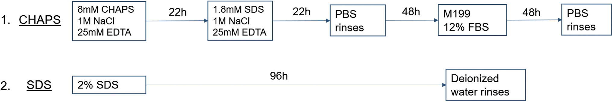
CHAPS-based (1) and SDS (2) decellularization pathways visualized. CHAPS, 3-[(3-cholamidopropyl)dimethylammonio]-1-propanesulfonate; EDTA, ethylenediaminetetraacetic acid; FBS, fetal bovine serum; PBS, phosphate-buffered saline; SDS, sodium dodecyl sulfate. Color images are available online.
1. CHAPS-based pathway: Twenty-two hours in CHAPS buffer (8 mM CHAPS [Sigma–Aldrich, St. Louis, MO], 1 M NaCl [Sigma–Aldrich], and 25 mM ethylenediaminetetraacetic acid [EDTA] [Sigma–Aldrich] in PBS), followed by 22 h in SDS buffer containing 1.8 mM SDS (Sigma–Aldrich), 1 M NaCl, and 25 mM EDTA in PBS, 48 h of PBS washes, and 48 h in M199 basal media (Corning) with 12% fetal bovine serum (FBS) (BioTC, Wayne, NJ). 8 Arteries were rinsed in PBS between solution changes and at the culmination of the decellularization process. (Protocol derived from Gui et al. 8 )
2. SDS pathway: Ninety-six hours in 2% SDS solution (in deionized water) refreshed every 24 h. Arteries were rinsed with deionized water between solution changes and at the culmination of the decellularization process.
All steps were performed at 37°C under agitation in an orbital shaker with 50 mL solution per 5 cm segment. Decellularization methods were compared using histological staining, burst pressure, and recellularization.
Histological staining
One centimeter segments of whole intact (non-decellularized) and decellularized arteries (cut from the 5 cm segment after decellularization) were fixed in 4% formaldehyde (Sigma–Aldrich) for 48 h, stored in ethanol, and sent to VitroVivo Biotech (Rockville, MD) for paraffin embedding and hematoxylin and eosin (H&E) staining. Sections were stained with picrosirius red (PSR) (American Mastertech, Lodi, CA) or Alcian Blue (Newcomer Supply, Middleton, WI). Modified Harris Hematoxylin (Epredia, Kalamazoo, MI) was used as a nuclear counterstain. Histology samples were imaged via brightfield microscopy at 2.5 × , 10 × , 20 × , and 40 × magnification (Zeiss ApoTome1).
DNA quantification
To evaluate the DNA content of intact and decellularized umbilical arteries, segments were oven dried at 60°C for 24 h and then finely ground using a Kimble Pellet Pestle. Samples were weighed and digested with a lysis buffer containing Proteinase K (Thermo Fisher Scientific) for 16 h at 55°C. DNA was isolated using magnetic beads, diluted in elution buffer, and measured photometrically at 260 to 280 nm.
Sulfated glycosaminoglycan quantification
Umbilical artery segments were dried and finely ground as previously described. Samples were weighed and digested in a 125 μg/mL papain buffer (Sigma–Aldrich) for 16 h. Sulfated glycosaminoglycan (sGAG) content was determined by addition of a 31 μM solution of dimethylmethylene blue (Sigma–Aldrich) in 55 mM formic acid, pH 3, followed by measurement of absorbance at 520 nm. sGAG content was calculated by interpolation to a standard curve. The curve was generated using 18, 35, and 70 μg/mL heparan sulfate.
Burst pressure testing
Intact and decellularized vessels were secured to barb adapters at both ends using superglue. On one end, a barb-NPT adapter was attached to a pressure sensor (Automation Products Group, Inc., Logan, UT). On the other end, a barb-female luer lock adapter was attached to a 5 mL syringe, which injected PBS into the vessel at constant pressure. A slow-motion camera was used to record the highest pressure before failure.
2D seeding: recellularization of umbilical artery sheets
Cell culture
Human umbilical vein endothelial cells (HUVECs) were provided by Dr. Sina Rabbany from Hofstra University. HUVECs—transfected with E4ORF1 of the AdE4 gene complex for survival—were expanded in a Heracell 150i incubator (Thermo Scientific) with 5% CO2 and 20% O2.
17
HUVECs were fed with M199 basal media (Corning) supplemented with 20% heat-inactivated fetal bovine serum, endothelial growth supplement (R&D, Minneapolis, MN), heparin (Sigma–Aldrich), HEPES buffer (Corning),
2D static seeding: recellularization of arterial sheets
Vessels were exposed to one F-T cycle before cell seeding. Following decellularization, the vessels were sterilized in 70% ethanol at room temperature overnight and were rinsed in PBS. Micro-scissors were used to split one wall of the vessel so it lay as a flat sheet, and patches ∼3 × 5 mm in size were placed into a 96-well plate with the lumen-side facing up. Select samples were incubated in 20 ng/mL bFGF (CAT. No. 100–18B; PeproTech) for 24 h at 37°C before seeding.
Sheets were coated in 20 μg/mL fibronectin (Sigma–Aldrich) for 1 h and rinsed in PBS. A 30 μL suspension of HUVECs was seeded at a density of 2 × 10⁶ cells/mL and allowed to adhere for 1 h before being fed with cell culture media. After 5 days, recellularized sheets were rinsed in PBS, fixed in 4% formaldehyde, and stained with 4′,6-diamidino-2-phenylindole (DAPI) nuclear stain.
To quantify density of adhered cells, three confluent regions on each sheet (n = 9) were imaged using a fluorescence microscope (Zeiss Axio Vert.A1) at 5 × , and blinded counters manually recorded the number of nuclei in each image. The results were averaged to obtain cell density per mm2. To assess seeding success, an 8 × 8 array of 5 × image tiles was collected to visualize the whole sheet. Based upon this image, observations were made regarding seeding consistency and overall cell coverage. Sheets with <10% cell coverage were excluded due to poor seeding technique.
Cell roundness was quantified by circularity of cell nuclei using ImageJ. Four 10 × magnification images for DAPI nuclear counterstain of each condition were converted to 8-bit images and automatically thresholded using Renyi entropy. All cells in each image were characterized by analyzing particles ranging in size from 20 to infinity. Circularity for each cell was calculated based on the following equation: circularity = 4π × area/perimeter. 2 A circularity value of 1 indicates a perfect circle and 0 indicates an elongated polygon.
3D static seeding: recellularization of whole artery
Whole vessels were sterilized as previously described. Barb-luer lock adapters were fixed to both ends of the vessel and capped with male luer lock plugs to contain solutions. Vessels were injected with fibronectin (20 μg/mL) in cell culture media and incubated for 1 h. A suspension of 3 × 10⁶ HUVECs/mL was injected into the vessel until capacity. The vessel was secured in a custom-fabricated stainless-steel frame to encourage even seeding throughout the lumen (Fig. 2a, b). The frame was placed in a conical and rotated 90° every 15 min for 3 h. Cells were gravity-fed with 1 mL of cell culture media every 24 h for 5 days before fixation in 4% formaldehyde. Each vessel was cut into several sheets and evaluated using the DAPI staining and imaging methods previously described.
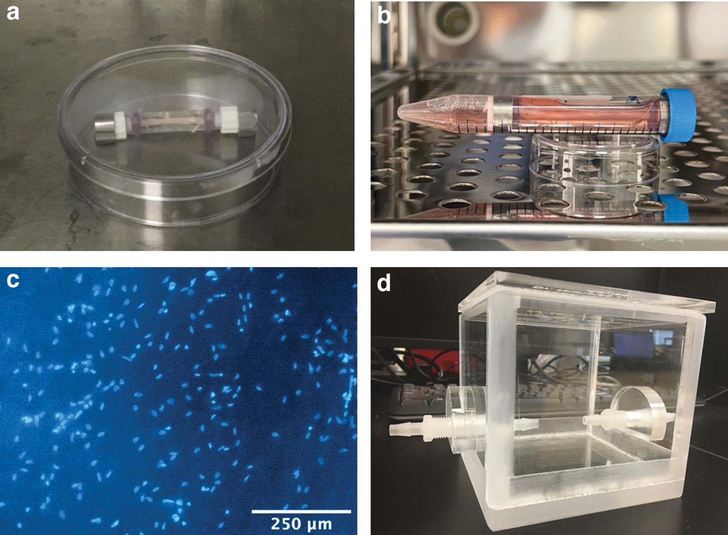
Representative images of a cannulated, whole CHAPS-decellularized vessel with cell suspension during the seeding process
F-T testing
F-T procedure
Cords were immediately frozen at −80°C upon receipt and stored for over 6 months. Arterial segments from the same cords were frozen and thawed according to the custom procedure presented in Figure 3. All steps were performed in dry tubes.

F-T procedure for three F-T cycles. F-T, freeze–thaw. Color images are available online.
After each cycle, one artery segment from the same cord was placed in PBS supplemented with penicillin–streptomycin at 4°C for storage before decellularization. This resulted in samples which had undergone one, two, or three F-T cycles. Samples were compared using tensile testing, DNA testing, and recellularization.
Tensile testing
Vessels were cut into dog bone shapes with the aid of a custom 3D-printed stencil (Fig. 4a, b). 18

Dog bone shape specimen stencil as modeled in AutoCAD (Autodesk, San Rafael, CA) with measurements in millimeters
Samples were stored in PBS and lightly dried on a task wipe before testing.
Gauge length, and thickness and width of the narrowest section of the tissue were measured using a digital caliper. The vessel was pulled at a rate of 0.08 mm/s until failure (Fig. 4c). The resulting data were used to calculate true strain and true stress, which allowed for the calculation of the maximum tensile stress and Young's Modulus of each sample (Fig. 4d).
2D static seeding: recellularization of arterial sheets
HUVECs and CHAPS-decellularized sheets (without bFGF) were prepared as previously described. A 100 μL suspension of 52,800 cells was added to achieve a density of 165,000 cells/cm2 per well. The following day, sheets were moved to new wells. Sheet-seeded HUVECs were cultured for 5 days, and media was refreshed daily. Cells were fixed, stained, and counted as previously described.
Statistical analysis
After using the Anderson–Darling test to determine if the data were normally distributed, a two-tailed Student's paired t-test was used to determine statistical significance at a p-value of <0.05 using Microsoft Excel (Microsoft, Redmond, WA). All data are expressed as mean ± standard deviation. All sample sizes (n = 3) describe biological replicates unless stated otherwise.
Experiment
Assessment of decellularization
Umbilical arteries were decellularized using CHAPS and SDS detergents. CHAPS-decellularized vessels were opaque with a yellow hue due to 48 h incubation in M199 (Fig. 5a). The tissue was less stiff and more collapsible upon transverse compression than the intact or SDS-decellularized vessels. SDS-decellularized vessels were white in color, stiff, and often tortuous. Artery diameters were ∼1 mm and slightly decreased after decellularization. Removal of cells was confirmed histologically and by DNA quantification (Fig. 5b).
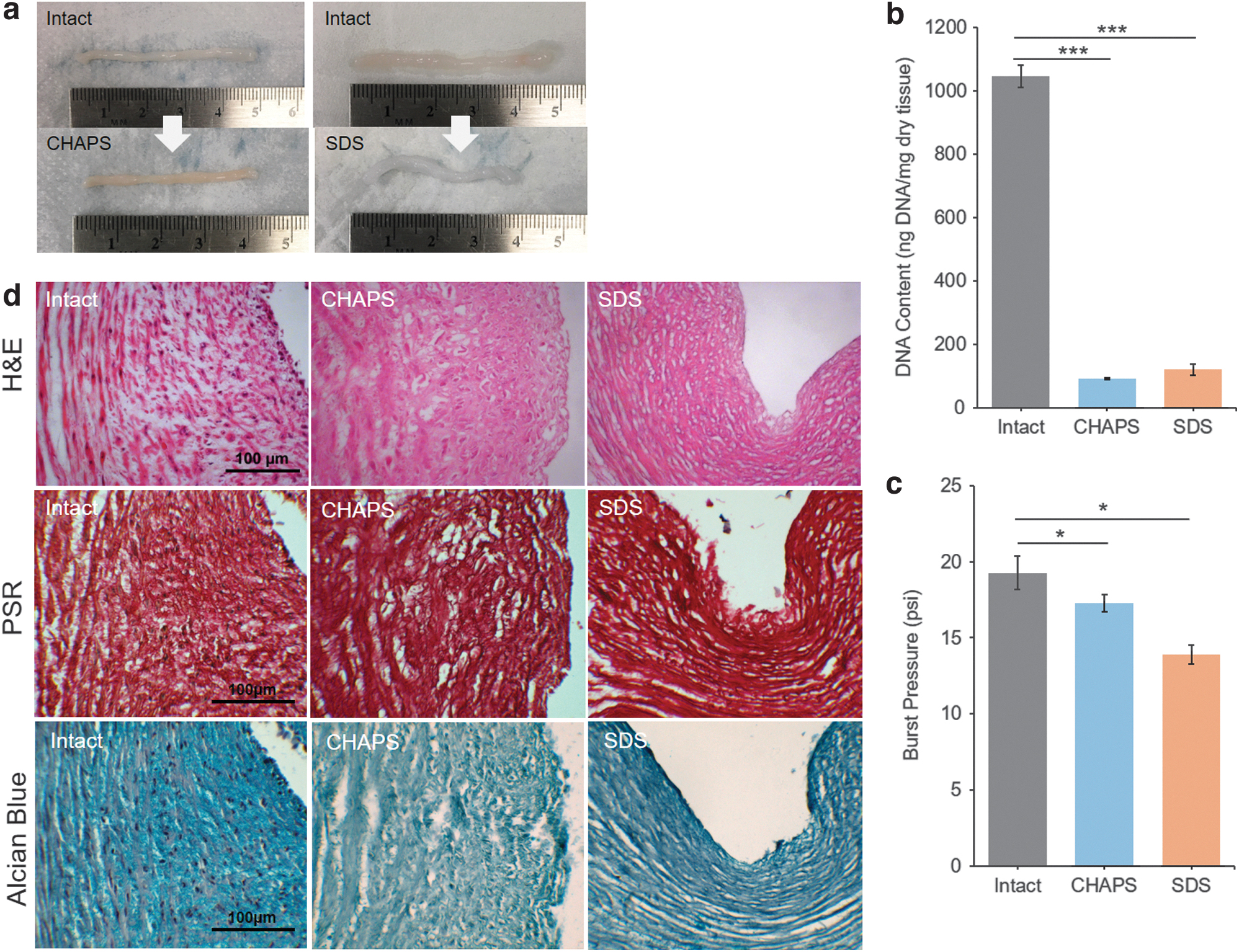
Effects of decellularization on umbilical arterial tissue's physical characteristics. Representative images of intact, CHAPS-decellularized (left), and SDS-decellularized (right) vessels
Well-formed nuclei could be identified throughout each layer of the intact arterial wall. These were absent in the CHAPS- and SDS-decellularized vessels; however, diffuse nuclear material appeared as hematoxylin-stained strands throughout the SDS condition. DNA content (ng DNA/mg dry tissue) was 1046.9 ± 34.5, 92.7 ± 3.5, and 121.7 ± 17.5 for intact, CHAPS-decellularized, and SDS-decellularized vessels, respectively. A statistically significant removal of 91.1% and 88.3% of DNA was observed for CHAPS- and SDS-decellularized vessels, respectively (p < 0.001).
Total sGAG content (μg sGAG/mg dry tissue) was 0.91 ± 0.13, 1.15 ± 0.25, and 0.70 ± 0.30 for intact, CHAPS-decellularized, and SDS-decellularized vessels, respectively. Statistically similar sGAG content was observed between intact and decellularized samples (p > 0.22). There was no significant difference in sGAG content between CHAPS- and SDS-decellularized vessels (p = 0.16).
Burst pressure was assessed to be 19.27 ± 1.12, 17.30 ± 0.56, and 13.90 ± 0.62 psi for intact, CHAPS-decellularized, and SDS-decellularized vessels, respectively (Fig. 5c). Burst pressures of both decellularized conditions were lower than burst pressure of intact vessels, indicating a loss mechanical properties. SDS-decellularized vessels had 27% lower average burst pressure than that of intact vessels (p = 0.010), whereas CHAPS-decellularized vessels showed a 10% decrease in burst pressure (p = 0.027).
Collagen (type I and III) structure was qualitatively evaluated through PSR histological staining (Fig. 5d). Intact tissue displayed a collagen network, which decreased in porosity from the tunica adventitia to the intima. While both CHAPS- and SDS-decellularized tissues appeared porous in structure throughout the tunica media, the collagen structure of the SDS condition was more organized and compacted. No damage to the intimal layers that would cause concern for recellularization was observed. Alcian Blue staining indicated that GAGs were still present after decellularization. The organization of GAGs in each condition appeared to mirror that of collagen.
Recellularization of arterial sheets with HUVECs
To compare recellularization potential of decellularization conditions, the luminal surface of arterial sheets was seeded with HUVECs. Cells showed successful adhesion to both CHAPS- and SDS-decellularized tissues (Fig. 6a, b) with slightly rounder nuclear morphology on SDS-decellularized sheets (Fig. 6c, d). A circularity of 0.31 ± 0.13 and 0.39 ± 0.15 was measured for cells cultured on CHAPS- and SDS-decellularized sheets, respectively. Five days was sufficient to grow a confluent endothelial layer.
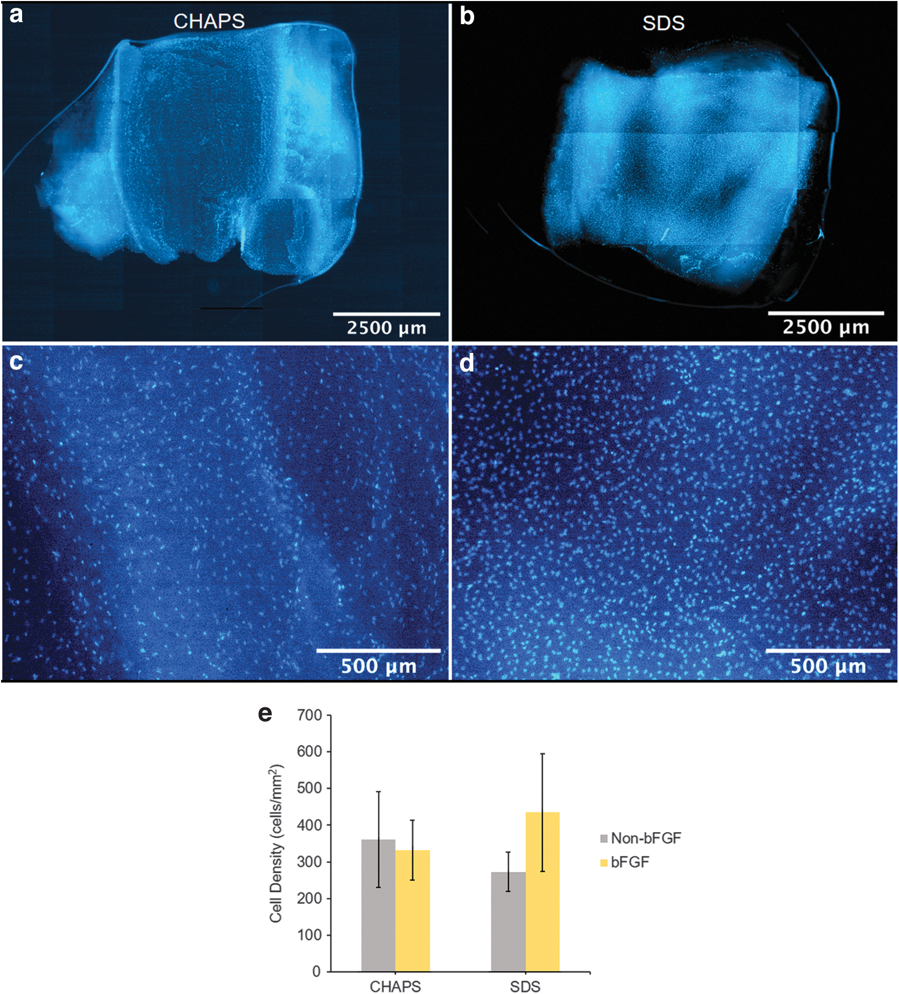
Recellularization of umbilical artery sheets. Representative 5 × and 8 × 8 array of 5 × tile images of DAPI-stained recellularized CHAPS- and SDS-decellularized sheets with bFGF treatment
Manual quantification of DAPI images (three regions on each sheet) revealed similar adhered cell density on all conditions. Without bFGF treatment, CHAPS-decellularized sheets had 25% higher cell density than SDS-decellularized sheets (p = 0.22) (Fig. 6e). Cell density was higher after bFGF treatment in the SDS condition (p = 0.24), but there was no difference in the CHAPS condition (p = 0.69). Comparing both after bFGF treatment, the SDS condition had 31% higher cell density than the CHAPS condition (p = 0.38). Cell density on CHAPS-decellularized sheets with and without bFGF treatment was 331.63 ± 81.50 and 360.08 ± 130.65 cells/mm2, respectively, whereas cell density on SDS-decellularized sheets with and without the same treatment was 434.59 ± 161.04 and 272.43 ± 53.10 cells/mm2, respectively.
Whole vessel recellularization
Whole CHAPS-decellularized vessels were recellularized using a custom stainless-steel frame and barb-plug setup (Fig. 2a, b). After 5 days, DAPI staining revealed successful adhesion of cells to the lumen of the vessel (Fig. 2c). SDS-decellularized vessels did not have successful recellularization due to their tortuosity, which created uneven seeding conditions.
A chamber was constructed from acrylic sheets and polypropylene barb adapters for future shear stress conditioning of the endothelial layer in recellularized whole vessels (Fig. 2d).
The effect of F-T cycles on tissue decellularization, mechanical strength, and recellularization potential
One to three F-T cycles before decellularization led to no significant change in recellularization potential, as the density of adhered cells was 374.48 ± 90.85, 365.48 ± 26.76, and 384.98 ± 112.50 cells/mm2 for tissue frozen and thawed one, two, or three times, respectively (Fig. 7a).

Comparison of one to three F-T cycles on decellularization. Density of adhered cells after recellularization experiment (n = 3 per F-T condition)
No significant textural differences were observed among samples of different F-T cycle number. Longitudinal tensile testing revealed no significant differences in ultimate tensile strength across the three F-T conditions (Fig. 7b). While the three F-T conditions had the lowest Young's modulus (Fig. 7c), this was not statistically significant (p = 0.18).
Discussion
Development of a mechanically compatible and anti-thrombogenic small-caliber vascular graft has long been of interest. While synthetic and xenogeneic alternatives are being extensively studied, decellularized human umbilical arteries still have potential as small-caliber grafts. 8 There is no agreement on the best decellularization technique for blood vessels; however, SDS and CHAPS detergents are the most commonly used agents for decellularization of umbilical arteries.19,20 In this study, we compared our own 2% SDS-only decellularization pathway, previously utilized in porcine heart and lung decellularization, to a protocol established by Gui et al using CHAPS and SDS buffers.8,10
H&E histology revealed that Gui et al's CHAPS-focused pathway was more effective than our SDS method. While SDS is known to be a harsh detergent, cell nuclei fragments remained post-decellularization. Interestingly, Gui et al similarly reported a “diffuse smear” of nuclear material in H&E stains of umbilical arteries decellularized in CHAPS and SDS buffers without successive incubation in FBS. 18 As serum nucleases in FBS can cleanly remove residual DNA, this step may be required in our SDS method. Although Tuan-Mu et al found success decellularizing for 12–48 h with 0.1–1% SDS, decellularized segments of tissue were only 2 mm long as opposed to 5 cm used here. 20
Of note, all presented histology data contain fresh non-decellularized tissue, once frozen–thawed CHAPS-decellularized tissue, and fresh SDS-decellularized tissue. These F-T cycles do not indicate a suggested pretreatment; these were the circumstances under which limited tissue supply could be utilized. While a few factors differed from Gui et al's CHAPS technique, including an additional extra F-T and the occasional incomplete dissolution of EDTA and NaCl, histology revealed similar ECM structure post-decellularization. This suggests that CHAPS and SDS may be most crucial in determining the final microstructure of decellularized tissue.
For biochemical analysis, total DNA and sGAG content were quantified. The incomplete removal of DNA in the SDS condition and more effective DNA removal by CHAPS decellularization shown in H&E histology were confirmed by DNA quantification. sGAG levels of SDS- and CHAPS-decellularized vessels were similar to that of non-decellularized vessels. The slight increase in sGAG content in CHAPS-decellularized vessels was likely due to the loss of cell mass after decellularization. sGAGs are vital for collagen structure formation in ECM.
Upon decellularization with SDS, sGAGs are potentially removed, allowing collagen fibers to move more freely, whereas in CHAPS-decellularized vessels, sGAGs are preserved, maintaining even distribution of collagen fibers. 21 Visualization of GAG distribution by Alcian Blue staining mirrored that of the collagen in PSR stains. The differences in stain color across conditions were attributed to the use of Modified Harris Hematoxylin as a nuclear counterstain. Overall, the DNA and sGAG content before and after decellularization was comparable to other studies.8,21
Maintenance of graft mechanical integrity is critical for successful integration into the native vasculature. Collagen and elastin fibers are responsible for imparting strength and elasticity. 22 PSR stains revealed that intact and CHAPS-decellularized tissues had similar density and distribution of collagen, whereas SDS-decellularized tissue had a more porous, net-like architecture. While both decellularized tissues had significantly lower burst pressures compared with intact tissue (p = 0.026), CHAPS-decellularized tissue had a significantly higher burst pressure than SDS-decellularized tissue (p < 0.01).
This difference may be due to the collagen network porosity observed in PSR stains of SDS-decellularized vessels. Similarly, Tuan-Mu et al demonstrated a decrease in the mechanical properties of umbilical arteries after decellularization with SDS. 20 Not only has SDS been known to degrade collagen but also compact the collagen and elastin structure of heart valves, resulting in a loss of detailed collagen structure.23,24 This may explain the tendency of the vessel to become tortuous. Overall, burst pressure results of the intact and CHAPS conditions were similar to values previously reported. 8
While studies report that endothelial cells can populate acellular umbilical arteries, explanted vessels only demonstrated 60% cell coverage. 25 Thus, recellularization of grafts is necessary before implantation. Recellularization was successful on both CHAPS- and SDS-decellularized sheets, indicating maintenance of ECM proteins and luminal topology. Cell nuclei appeared to have a rounder morphology on SDS-decellularized sheets when compared with CHAPS-decellularized sheets, suggesting more cell spreading. This may be due to the greater stiffness of the SDS-decellularized sheets. The higher fibronectin concentration used here before cell seeding may have aided in the cellular repopulation compared with other umbilical artery studies.8,26 Fibronectin has been shown to persist for up to 8 weeks when coated on decellularized aortic conduit implants, resulting in improved endothelialization in vivo.
Previously, Conklin et al showed an increase in cell proliferation due to bFGF in studies spanning only 1 to 3 days. 27 As we utilized a higher seeding density, cell growth may have plateaued in both treated and nontreated conditions before the 5-day time point. Alternatively, BrdU assays may be more effective at determining whether bFGF promoted endothelial cell proliferation during the first 3 days. Rajabi et al demonstrated that 9% of infused bFGF remained after 4 h of passive absorption on SDS-decellularized heart tissue. 28 Despite its short half-life, bFGF absorbed onto collagen sponges and biohybrid hydrogels has shown sustained release lasting between 7 and 14 days. 29
A small increase in adhered cell density with addition of bFGF (p = 0.24) in the SDS condition may be due to sequestration of bFGF within the porous ECM microstructure. ECM porosity is desired as it can sequester growth factor for increased and controlled release.29,30 Additionally, bFGF can improve the strength of cell adhesions, which help cells resist lifting due to fluid flow. 27 Thus, the potential of bFGF to improve preconditioning outcomes may yet be observed with the application of shear stress.
It is well established that freezing and thawing can be used as a method for decellularization and long-term tissue storage.22,31,32 However, freeze–thawing is known to be a harmful process for tissues, causing protein degradation and cell lysing. Typical F-T cycles intended for decellularization involve snap-freezing, often in cryoprotectants or other liquid buffers, but dry freeze–thawing may be less harmful to tissue as there is less intracellular ice crystal formation.33–35 Previous studies have reported freeze–thawing as having no effect on the mechanical properties of femoral arteries, whereas umbilical arteries showed a loss in compliance but no change in stiffness after one F-T cycle and greater residual DNA after decellularization compared with fresh arteries.20,31
Furthermore, others have reported that rapidly freezing umbilical arteries using a vitrification solution creates stiffer decellularized vessels compared with non-decellularized vessels. 21 We observed that arteries exposed to one to three dry F-T cycles before CHAPS decellularization suffered no significant detrimental impact on major mechanical and biochemical properties of interest. Therefore, our data may support the feasibility of incorporating multiple dry F-T cycles into future decellularization and long-term storage protocols.
A whole CHAPS-decellularized vessel was successfully recellularized using a custom setup and a higher cell seeding density than 2D sheets. According to Gui et al, umbilical arteries can expand from a diameter of 1.5 to 4.5–5.5 mm when exposed to physiological pressure. 8 Therefore, the seeding density may need to be further increased to account for the higher surface area produced by vessel dilation upon injection with media. To the best of our knowledge, whole decellularized umbilical arteries have been repopulated with Wharton's jelly mesenchymal stromal cells, but not yet HUVECs. 36
Those findings indicated that static seeding, followed by dynamic bioreactor seeding, can increase cell distribution uniformity when compared with dynamic seeding solely by agitation. Similarly, we created a chamber to expose seeded HUVECs to flow-induced shear stress to precondition them for physiological pressures. This method of preconditioning has been extensively studied and shown to increase the strength of cell adhesion and decrease the likelihood of thrombosis, suggesting positive in vivo outcomes.26,37,38
In brief, detergent-decellularized human umbilical arteries have significant potential as non-immunogenic, mechanically compatible, allogeneic, recellularization-capable, small-caliber vascular grafts. These findings offer another promising detergent-based pathway for decellularization of umbilical arteries as well as methods for the recellularization of other decellularized small-diameter vessels. In future studies, umbilical arteries will be heparin-functionalized to improve recellularization and the endothelial layer preconditioned with fluid shear stress. Recellularized and preconditioned vessels can then be implanted into rat models to test for immunogenicity, thrombogenicity, and long-term patency.
Footnotes
Acknowledgments
All discarded human tissues were generously provided through an approved Weill Cornell Medical Center Institutional Review Board protocol. Cell populations were gifted by Dr. Sina Y. Rabbany from Hofstra University.
Authors' Contributions
V.W.: Conceptualization, methodology, formal analysis, investigation, validation, writing—original draft, writing—review and editing, and visualization. S.G.: Investigation and validation. M.S.: Conceptualization and methodology. N.M.: Conceptualization, methodology, validation, resources, writing—review and editing, supervision, project administration, and funding acquisition.
Disclosure Statement
No competing financial interests exist.
Funding Information
No funding was received for this article.