
Abstract
Acquired anterior glottic webs (AGW) can lead to abnormally elevated phonatory pitch, dysphonia, and airway obstruction requiring urgent intervention. In this study, we construct a novel AGW rabbit model using heat injury by a laryngoscopic way. A primary study was conducted to identify the injury depth in rabbits' vocal folds (VFs) by graded heat energy, and the heat energy for the incurrence of epithelial layer, lamina propria, and muscular layer (ML) injury was 25, 30 and 35 W, respectively. Then, four different models were designed based on the depth and degree of the injury to determine the optimal procedure for AGW formation. Morphological features, vibratory capacity, and histopathologic features of the AGW were correspondingly evaluated. The procedure for conferring the heat injury to the depth of ML and the extent of anterior commissure and middle part of bilateral VFs showed the highest success rate of AGW formation (95%, 19/20). For its low cost, effectiveness, and stability for AGW formation, the heat injury rabbit model with a laryngoscopic approach may provide a new platform for testing novel anti-adhesion materials and bioengineered therapies.
Impact Statement
Tissue engineering based on biomaterials has been a very hot research field and may be introduced to prevent the acquired anterior glottic web (AGW) formation. However, lacking a widely recognized animal model for AGW has limited the trial of anti-adhesion materials in the larynx. In this study, we have developed a novel rabbit model for AGW formation by conferring a heat injury under a laryngoscope; this model is cheap, effective, and stable for the anti-adhesion materials and bioengineered therapies. Thus, this research would arouse crucial interest and be widely employed.
Introduction
Ascribed as a bridge of fibrous scarring tissue covered superiorly and inferiorly by the epithelium in between the free edges of true vocal folds (VFs), glottic web can be congenital or acquired. 1 Regardless of age, it could be secondary to radiotherapy, reflux, external trauma or intubation, iatrogenic trauma, and rarely inflammatory process. 2
Acquired anterior glottic webs (AGW) represent the most prevalent of glottic web, which usually happen in laryngeal endoscopic surgery for lesions involving both sides of the VFs and the anterior commissure. 3 Based on its severity and size, symptoms could range from abnormally elevated phonatory pitch, dysphonia, and airway obstruction requiring urgent intervention. 4 Various methods, including endoscopic fibro-mucosal flaps, butterfly mucosal flap technique, and mitomycin C,1,5,6 have been devised to address the unwanted re-adhesion in the anterior glottis. However, despite a myriad of techniques have been employed, AGW still remains as a difficult-to-treat clinical entity to the laryngologist. As a result, the advanced strategies of the AGW management are still urgent to be developed.
In recent years, a variety of biomedical materials such as Interceed™ and Seprafilm™ have been developed to prevent the postoperative adhesion within the thoracic cavity, abdominal cavity, and nasal cavity. 7 Various biomaterials such as collagen, bovine pericardium, and dextran were attempted to prevent VF scarring with preliminary results.8,9 Therefore, tissue engineering based on biomaterials may be introduced for the prevention of AGW formation. To evaluate various treatments and effectiveness of various biomaterials for AGW, elaborate studies using stable animal models would be required before clinical trials. To ensure high-quality research results, a method for constructing an AGW model in a graded manner as a platform for testing novel anti-adhesion material and regenerative medicine needs to be established.
In this study, we have introduced and validated a novel rabbit model for AGW formation through heat injury under a laryngoscope (Fig. 1). To construct an effective and stable AGW model, this study has comprised three parts. First, a graded heat energy was screened to determine the injury of epithelial layer (EL), lamina propria (LP), and muscular layer (ML). Second, four different models were designed based on the depth and injury degree to explore the optimal procedure of AGW formation. Finally, morphological features, vibratory capacity, and histopathologic features of the AGW were correspondingly evaluated by laryngoscopy, high-speed photography, and histology.
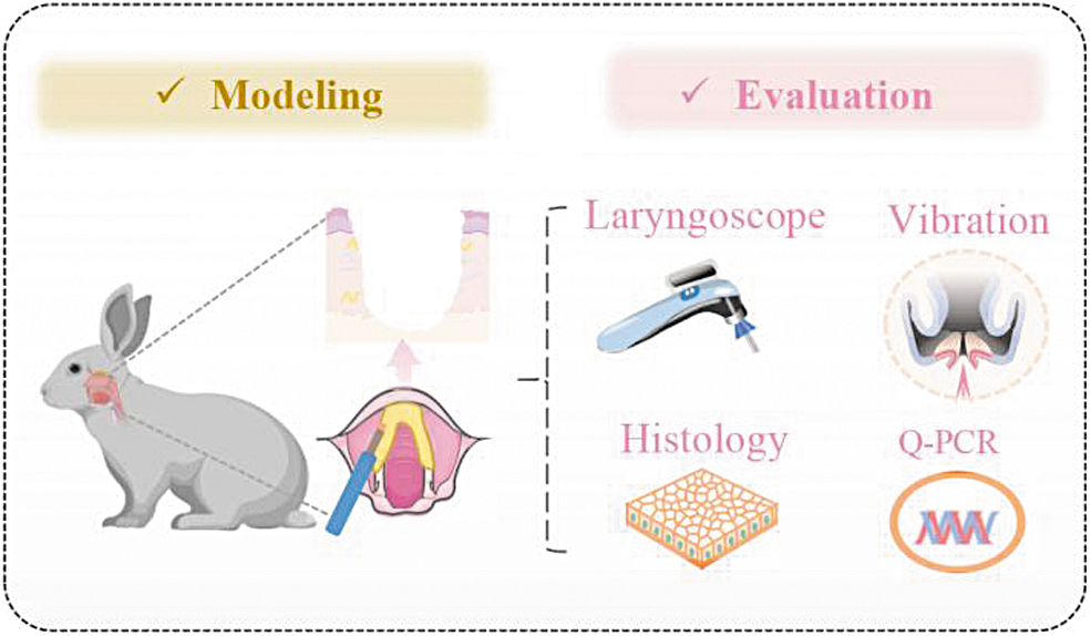
Schematic diagram of the experiment. Q-PCR, quantitative real-time PCR.
Materials and Experiments
Surgical procedure and laryngoscopic evaluation
The experimental protocols were approved by the Sichuan University Animal Care and Use Committee (No. 20220304045). Intravenous injection of pentobarbital sodium was chosen to anesthetize the New Zealand white rabbits with the weight of 2.4–2.6 kg. The heat injury was imposed by high-frequency electrocautery (GD350-P; Shanghai Hutong) (Supplementary Movie S1), and the subcutaneous injection of carprofen was used to ease the pain of operation. Besides, penicillin was used 30 min before surgery and 24 h after surgery to prevent infection. The weight, sputum production, wheezing, and dyspnea of all animals were monitored daily. At 0, 2, 4, and 8 weeks after injury, laryngoscopy was carried out by visual laryngoscope (VLS2-01; Zhejiang UE Medical Corp., China) (Supplementary Fig. S1).
High-speed imaging of excised larynx
An excised laryngeal setup and a high-speed camera (2000 fps, HKM-A2001-MCHM2000; Beijing Microview Co., Ltd., China) were used to assess the VF mucosal wave ex vivo. The larynxes were harvested immediately after the laryngoscopy at 8 weeks, and, to visualize better, the whole supraglottic structure was removed. Subsequently, the bilateral arytenoid cartilages were sutured together. Then, 25 mm Hg of air was applied via a tube linked to the trachea to lead VF vibrations. The area viability (pixels) was expressed as:
Here, Amax and Amin represented the maximal and minimal glottal gap area (pixels) during vibration, respectively. The mucosal wave oscillations were traced by the pixels of amplitudes that a point was farthest and closest from the midline, respectively. The images were analyzed by HSCI V1.0.0 (2022SR0319624; West China Hospital, China).
Histological evaluation
After harvest, the larynxes were sequentially fixed in 4% paraformaldehyde, dehydrated, embedded, and sequentially sectioned. The slices were subjected to hematoxylin and eosin (HE), Masson staining, and Sirius red staining (Servicebio, China).
For Masson staining, the collagen volume fraction (CVF%) referred to the proportion of blue staining area of collagen in the total tissue area. Sirius red staining could show the collagen I (Col I) and collagen III (Col III) in the normal VFs, scarring VFs and AGW, and further visualized under polarized light mode (Olympus VS200, Japan).
Anti-α-smooth muscle actin (SMA) (ab32575; Abcam) was used as the primary antibody for immunofluorescence and the goat serum as the negative control. The IntDen was calculated by the ImageJ software as:
where IntDen represented the gray value of integrated density, A represented the number of pixels, and M represented the mean gray value of positive α-SMA staining.
The ImageJ software (NIH, Bethesda, MD) was further applied to analyze.
Quantitative real-time PCR
Total RNA in VFs was extracted by TRIzol reagent according to the manufacturer's protocol followed by reverse transcription (RR047; Takara, Japan). The cDNA of GAPDH, COL1A1 (encoding Col I), COL3A1 (encoding Col III), and ACTA2 (encoding α-SMA) was mixed with the SYBR Green master mix (RR820; Takara, Japan), and GAPDH served as a housekeeping gene (Supplementary Table S1). The relative level of gene expression was expressed as the 2−△△Ct method.
Statistical analysis
The difference between the groups was analyzed by Student's T test or one-way analysis of variance using the GraphPad Prism software. All data are expressed as the mean ± standard deviation. p < 0.05 was regarded as statistically significant (*p < 0.05, **p < 0.01, ***p < 0.001, ****p < 0.0001).
Results
Exploration of the depth of VF injury by graded heat energy in rabbits
The formation of AGW is closely associated with the depth and degree of injury. Should the heat injury be limited to the EL, it is extremely difficult to form AGW. Thus, a preliminary study was conducted to identify the injury of EL, LP, and ML by the graded heat energy. Following the heat injury, the larynxes were harvested for HE staining for the determination of the depth of VF injury (Fig. 2). The ultimate energies of heat injury were as follows:

HE staining of the injured VFs at 0 week with by graded heat energy. EL, epithelial layer; HE, hematoxylin and eosin; LP, lamina propria; ML, muscular layer; VFs, vocal folds.
The normal (energy = 0 W, contact duration = 3 s);
The injured EL (energy = 25 W, contact duration = 3 s);
The injured LP (energy = 30 W, contact duration = 3 s);
The injured ML group (energy = 35 W, contact duration = 3 s) (Supplementary Movie S1).
Exploration of the procedures of AGW formation in rabbits
To optimize the method for AGW formation, four different procedures were designed according to the depth and extent of the injuries (Fig. 3):
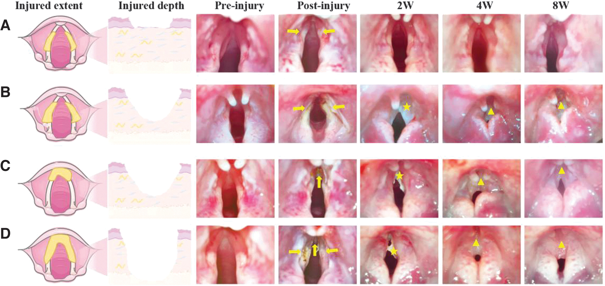
Laryngoscopic observation of the VFs before and after the heat injury at 0, 2, 4, and 8 weeks (arrows, injured regions of VFs; asterisks, granulation tissues; triangles, AGW).
Group A (n = 20): the depth = the EL (energy = 25 W, contact duration = 3 s), and the extent = middle part of bilateral VFs;
Group B (n = 20): the depth = the LP (energy = 30 W, contact duration = 3 s), and the extent = middle part of bilateral VFs;
Group C (n = 20): the depth = the LP (energy = 30 W, contact duration = 3 s), and the extent = anterior commissure;
Group D (n = 20): the depth = the ML (energy = 35 W, contact duration = 3 s), and the extent = the anterior commissure and middle part of bilateral VFs.
Two weeks after the heat injury, bilateral VFs in group A have been completely epithelized under laryngoscopic examination (Fig. 3A), whereas some yellowish-white granulation tissues were noted in the other three groups. By weeks 4 and 8, the granulation tissues were resolved and covered with normal mucosa. Notably, compared with the normal VFs with an inverted “V”-shaped anterior commissure, the AGW showed an inverted “U”-shaped scar band with a broad angle margin by 8 weeks, indicating that the modeling was successful (Supplementary Movie S2 and Fig. 3B–D).
At 4 weeks, successful AGW formation was verified in 51 of 80 rabbits (63.8%). The rate of AGW formation in groups A–D was 5% (1/20), 70% (14/20), 85% (17/20), and 95% (19/20), respectively (Supplementary Table S2). As a whole, the procedure of conferring the heat injury to the depth of ML and the extent of the anterior commissure and middle part of bilateral VFs attained the highest rate for AGW formation (95%, 19/20). At 8 weeks, VFs in appearance under laryngoscope of the above four different models were stable.
Notably, although no adhesion was observed in some VFs, their appearance under a laryngoscope showed yellow streaks, which was different from pre-heat injury (Fig. 3A). Three outcomes were noted after the VF injury, including fully restoration to normal, scar formation, and adhesion formation. For the next step, we have further evaluated the difference in the vibratory capacity and histopathologic features between normal VFs, scarred VFs, and AGW.
Evaluation of the vibration capacity of the AGW in rabbits
Using a high-speed digital camera system, the vibratory motion of the normal VFs, scarred VFs, and AGW were estimated on 8 weeks after the injury (Fig. 4A). Compared with normal VFs, which presented regular vibration and complete glottal gap closure, the scarred VFs showed stiffened mucosal wave and glottal insufficiency. The vibration may even vanish in the AGW (Fig. 4B and Supplementary Movie S3). To further compare the variability and amplitude of the normal VFs, scarred VFs, and AGW, the glottal gap area and mucosal wave oscillations were calculated (Fig. 4C). Contrast to the normal VFs, the maximal glottal gap area and the area variability of the scarred VFs and AGW were significantly reduced (p < 0.05, Fig. 4D). The amplitude from four cycles showed that mucosal wave oscillations of the scarred VFs had deteriorated significantly. Meanwhile, the mucosal wave oscillations of AGW were flat (Fig. 4E).

High-speed photography of the normal VFs, scarred VFs, and AGW.
These results revealed that the vibratory capacity of VFs in both AGW and the scar has seriously deteriorated, compared with that of the normal VFs. While the vibratory capacity VFs in the AGW was the worst, which indicated successful modeling and could offer evidence for testing novel anti-adhesion materials and bioengineered therapies in terms of functions. Next, we further evaluated the differences in histological features among the normal VFs, scarred VFs, and AGW.
Evaluation of the histological features of AGW in rabbits
On histological evaluation, a fibrotic band and excessive collagen depositions were observed by HE staining at the anterior commissure and middle part of bilateral VFs at 8 weeks after the injury (Fig. 5A). As shown by Masson, collagen fibrils in the scarred VFs and AGW were arranged densely and exhibited as thicker bundles compared with the normal VFs. Moreover, the CVF% of AGW showed significant increase at the anterior commissure and middle part of bilateral VFs, followed by those of the scarred VFs (p < 0.05, Fig. 5B).
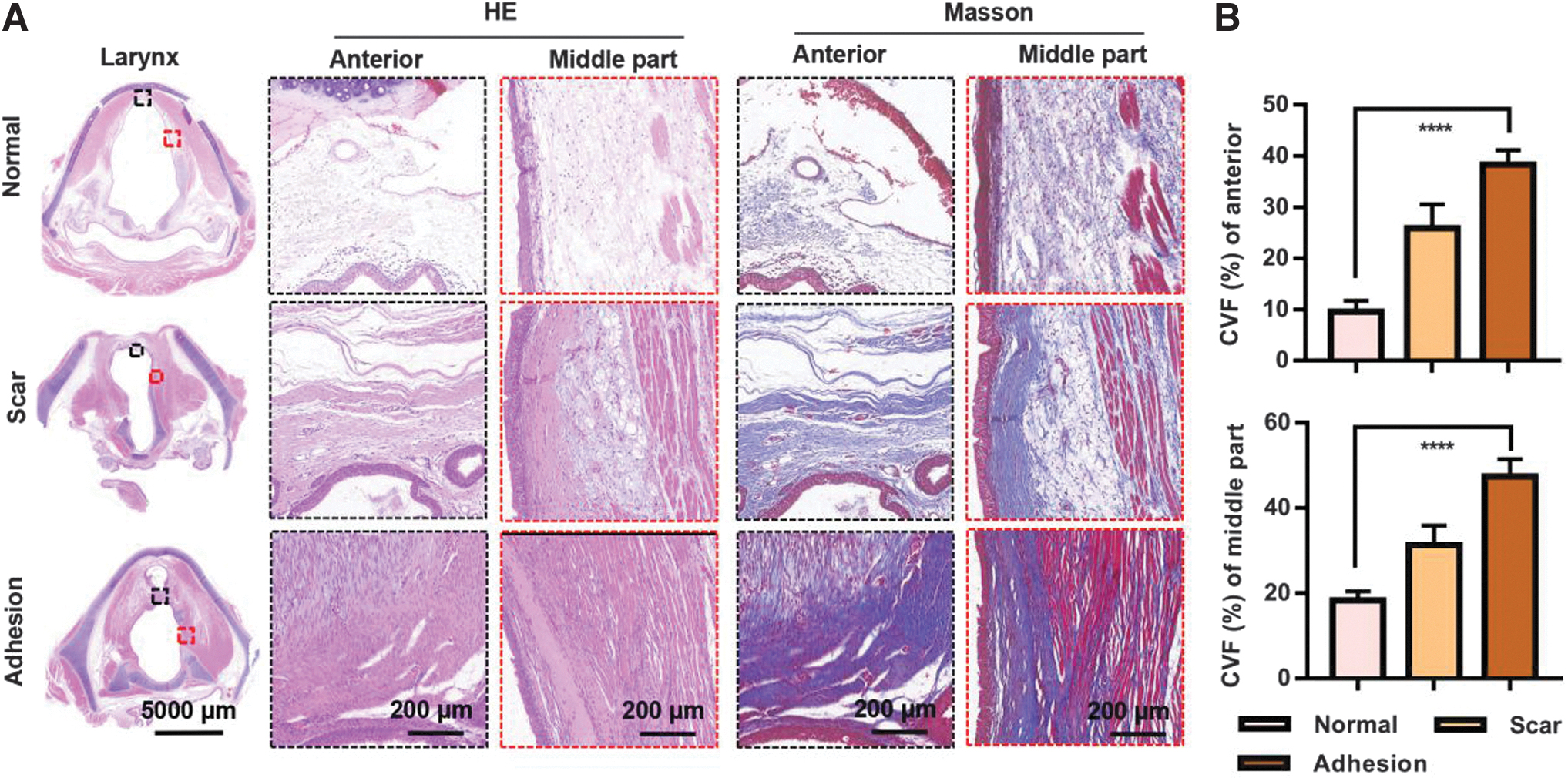
HE and Masson staining of the normal VFs, scarred VFs, and AGW at 8 weeks
Sirius red staining could be capable to explore distribution of different collagens. Col I presented yellow-red birefringence and Col III showed yellow-green birefringence under polarized light.10–12 Compared with the normal VFs, which presented aligned collagen fibers with sparse and gracile yellow-green and yellow-red birefringence, while collagen bundles in the AGW showed more strongly red fibers, followed by those of the scarred VFs (Fig. 6A). Above results were in accord with the statistical results (Fig. 6C–E). Positive staining of the α-SMA was observed in hypertrophic scars, which represented fibrosis. 13 As shown by immunofluorescence, the AGW showed a greater amount of positive α-SMA staining compared with the normal and scarred VFs (Fig. 6B, F).
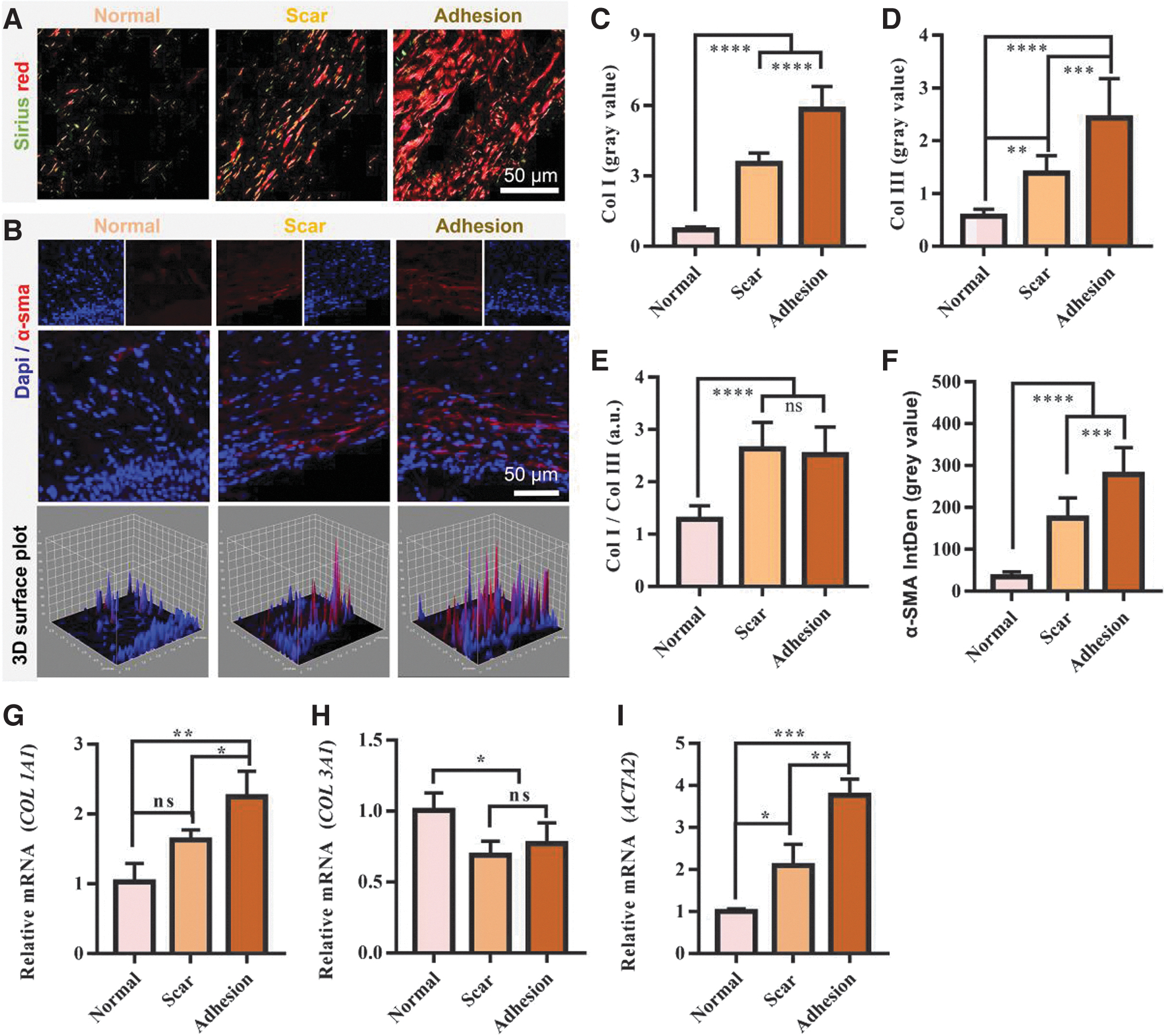
Analysis of the fibrosis of normal VFs, scarred VFs, and AGW.
The corresponding relative mRNAs of COL1A1, COL3A1, and ACTA2 by quantitative real-time PCR were analyzed at 8 weeks after the injury. Results showed that the COL1A1, COL3A1, and ACTA2 expression in the VFs of AGW increased significantly, followed by those of the scarred VFs (p < 0.05, Fig. 6G–I). Briefly, the results suggested that the fibrosis of AGW were more active compared with that of the normal VFs and scarred VFs, which further indicated successful modeling and could offer evidence for testing novel anti-adhesion materials and bioengineered therapies, histopathologically.
Evaluation of the growth and survival of different staging AGW in rabbits
The Cohen's classification has defined four types of AGW according to the degree of glottic narrowing, 14 which was used to evaluate the four different heat injury modeling in this study (Fig. 7A). Here, the rates of web-grade I to web-grade IV were 21.6% (11/51), 23.5% (12/51), 19.6% (10/51), and 35.3% (18/51), respectively. Among them, the rate of AGW formation in groups A–D was 5% (1/20), 70% (14/20), 85% (17/20), and 95% (19/20), respectively (Supplementary Table S2).
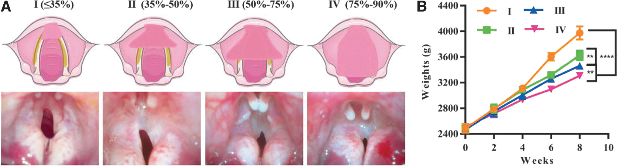
Evaluation of the growth and survival of differently staged AGW in rabbits according to Cohen's classification.
Depending on the stage of the web formation, the AGW may result in dysphonia, dysphagia, and airway restriction.6,15,16 Web-grade I and web-grade II probably are asymptomatic or only with mildly impedient phonation; it could be left untreated or delivered to voice therapy. 17 More severely, web-grade III and web-grade IV may appear respiratory distress, laryngeal obstruction, or recurrent respiratory infections requiring urgent treatment (Supplementary Movie S4). 4 When airway obstruction occurs, the patient or animals may die of asphyxia if the airway should not be opened in time. Thus, we evaluated the growth and survival of different staging AGW in rabbits for testing the effectiveness of novel anti-adhesion materials and bioengineered therapies in improving symptoms and reducing mortality.
In this study, rabbits of web-grade IV gained the least weight, followed by web-grade III, web-grade II, and web-grade I (p < 0.05, Fig. 7B). Besides, the swallowing function influenced food intake and growth (Supplementary Movie S5). In this study, 16 of the 51 rabbits with AGW (31.4%) died mainly due to the sputum blockage or glottic area reduction with the formation of the web. The mortality rates for web-grade I and web-grade IV were 0 (0/11) and 61.1% (11/18), respectively (Supplementary Table S3). Among them, the mortality rates of procedures A to D were 0% (0/1), 28.6% (4/14), 35.3% (6/17), and 31.5% (6/19), respectively.
Above results indicated that the severity of the symptoms has varied according to the different staging of AGW. Thus, another objective of novel anti-adhesion materials and bioengineered therapies in the future is to reduce the mortality and improve swallowing function of advanced AGW.
Discussion
Lack of a widely recognized animal model for AGW has limited the trial of anti-adhesion materials in the larynx. In this study, we have developed a novel rabbit model for the AGW formation by conferring a heat injury under a laryngoscope. The heat energy for the incurrence of injured EL, LP, and ML was 25, 30 and 35 W, respectively. The procedures of AGW formation were evaluated through multidimensional approaches, including laryngoscopy, high-speed photography, and histopathology. The formation of AGW was verified in 51 of the 80 rabbits (63.75%). The procedure for conferring the heat injury to the depth of ML and the extent of anterior commissure and middle part of bilateral VFs showed the highest success rate of AGW formation (95%, 19/20). In particular, we have staged the AGW according to Cohen's classification and assessed the corresponding growth and survival, which may provide more clinic evaluations of therapeutic strategy, especially the anti-adhesion materials and bioengineered therapies.
Compared with previously described models, the model of AGW formation in this study has the following strengths. First, the laryngoscopic approach is easily performed with sound visualization by a laryngologist who is familiar with laryngeal anatomy and expertise in endoscopic operation. Consequently, the procedure is less invasive and more efficient compared with conventional laryngofissure methods. 18 Also, antibiotics may be unnecessary for the prevention of postoperative wound complications.
Second, we have constructed the model for AGW formation by graded heat energy. Previously described methods to confer a VF injury to the animals included cup-forceps biopsy, 19 VF stripping, 20 and CO2 laser. 16 In theory, cup-forceps biopsy, as a widely used method for conferring VF injury in animals, could control the extend of the mucosal defect matching the size of cup. 21 Actually, it tended to surpass the cup size. 22 The VF stripping, usually ripping the entire length of the membranous VF, resulted in uncontrollable injury of the thickness and extent of VFs. 15 CO2 laser, widely used in heat injury modeling,10,16,23,24 was more consistent with the clinical practice. However, it was of great challenge to operate in small animals such as rabbits. Taken together, the main problem with the above methods was the difficulty in standardization of the depth and dimension of the wound. Again, details of the modeling process have remained unclear, and the evaluation methods were not comprehensive in these studies.
As a result, the applicability of the previous studies was limited due to the nonrepeatability and inconsistency of the modeling process. 25 In this study, we screened the heat energy of injured EL, LP, and ML, which was 25, 30 and 35 W, respectively. Moreover, we have compared four procedures for the induction of AGW, which respectively mimic actual clinical scenes including the operation of polyps, Renke's edema, VF leukoplakia, and laryngeal carcinoma involving the anterior commissure. The procedure for conferring the heat injury to the depth of ML and the extent of anterior commissure and middle part of bilateral VFs showed the highest success rate of AGW formation (95%, 19/20). Above step-ladder experiments have helped to construct a stable model for AGW formation. Therefore, we recommend to use this procedure for conferring the heat injury to the depth of ML and extent of anterior commissure and middle part of bilateral VFs for future testing on novel anti-adhesion materials and bioengineered therapies.
Third, we have evaluated the differences in histological features and vibration functions, and further staged the AGW by the Cohen's classification and evaluated the clinical characteristics, which may provide a platform for testing novel anti-adhesion material and regenerative medicine.
Last but not the least, compared with larger animals such as dogs, pigs, and monkeys, experiments with rabbits are cheap, with the cost for the purchase and experiment of large animals being 20 to 30 times higher. Herein, this is the first study of a rabbit model for AGW formation through heat injury under a laryngoscope.
There are some limitations in this study. First, up to date, in clinical practice, the most common factor that causes VFs heat injury is CO2 laser; however, it is of great challenge to operate in small animals such as rabbits. Besides, we constructed a stable heat injury AGW formation model through graded energy, which is effective for further evaluation of anti-adhesion materials and bioengineered therapies. Second, the mortality rates for web-grade III and web-grade IV were 40.0% (4/10) and 61.1% (11/18), respectively; this high mortality might be unacceptable for animal studies. Actually in clinic, the urgent intervention was required under web-grade III and web-grade IV, which may show various severe symptoms. The future objective of anti-adhesion material and regenerative medicine for web-grade III and web-grade IV is to alleviate respiratory distress and reduce the mortality rate, precisely.
In conclusion, a rabbit model for the AGW formation induced by heat injury with a laryngoscopic approach has been successfully developed. The method is cheap, effective, and stable for the preclinical evaluations of therapeutic strategies, including the anti-adhesion materials and bioengineered therapies.
Footnotes
Acknowledgments
We gratefully acknowledge the technical assistance of Research Core Facility of West China Hospital (Lin-Qiao Tang) and Animal Experiment Center of West China Hospital (Xiao-Ling Yang).
Authors' Contributions
H.Y. and H.-Q.X. designed the research. J.-J.H. and C.-Y.Z. conducted the experiments and wrote the article. R.W., X.-X.L., and M.-J.C. assisted in the experimental process and data collection. M.X. and Y.-L.J. conducted the data analysis. Jesse L.L., H.-Q.X., and H.Y. edited and reviewed the article. This article has been approved by all coauthors.
Disclosure Statement
No competing financial interests exist.
Funding Information
This work has been jointly sponsored by a Post-Doctor Research Fund from West China Hospital, Sichuan University (No. 2021HXBH005), Key Research and Development Program of Sichuan Provincial Department of Science and Technology (No. 2021YFS0216), and the “1.3.5” Project for Disciplines of Excellence of West China Hospital, Sichuan University (No. ZYJC18002).
References
Supplementary Material
Please find the following supplemental material available below.
For Open Access articles published under a Creative Commons License, all supplemental material carries the same license as the article it is associated with.
For non-Open Access articles published, all supplemental material carries a non-exclusive license, and permission requests for re-use of supplemental material or any part of supplemental material shall be sent directly to the copyright owner as specified in the copyright notice associated with the article.