
Abstract
Current tissue engineering (TE) methods utilize chondrocytes primarily from costal or articular sources. Despite the robust mechanical properties of neocartilages sourced from these cells, the lack of elasticity and invasiveness of cell collection from these sources negatively impact clinical translation. These limitations invited the exploration of naturally elastic auricular cartilage as an alternative cell source. This study aimed to determine if auricular chondrocytes (AuCs) can be used for TE scaffold-free neocartilage constructs and assess their biomechanical properties. Neocartilages were successfully generated from a small quantity of primary neonatal AuCs of three minipig donors (n = 3). Neocartilage constructs had instantaneous moduli of 200.5 kPa ± 43.34 and 471.9 ± 92.8 kPa at 10% and 20% strain, respectively. TE constructs’ relaxation moduli (Er) were 36.99 ± 6.47 kPa Er and 110.3 ± 16.99 kPa at 10% and 20% strain, respectively. The Young’s modulus was 2.0 MPa ± 0.63, and the ultimate tensile strength was 0.619 ± 0.177 MPa. AuC-derived neocartilages contained 0.144 ± 0.011 µg collagen, 0.185 µg ± 0.002 glycosaminoglycans per µg dry weight, and 1.7e-3 µg elastin per µg dry weight. In conclusion, this study shows that AuCs can be used as a reliable and easily accessible cell source for TE of biomimetic and mechanically robust elastic neocartilage implants.
Impact Statement
This study outlines the method of scaffold-free neocartilage tissue engineering from auricular chondrocytes (AuCs). The inherent ability of AuCs to produce elastic fibers offers advantages in regenerating essential craniofacial elements such as pinnae, nose, temporomandibular disc, and epiglottis. Unlike costal cartilage, which ossifies with age and requires an invasive procedure to obtain cells, auricular cartilage remains elastic and is more accessible. In addition, scaffold-free neocartilage manufacturing eliminates detrimental effects associated with exogenous scaffold degradation. The method described here allows the manufacture of a large quantity of scaffold-free elastic neocartilages from a small amount of primary AuCs that can be obtained with a minimally invasive procedure. The elastic neocartilages would suit the auricular, nasal, temporomandibular disc, and laryngeal cartilage regeneration and reconstruction.
Introduction
The craniofacial cartilage loss due to microsomia, trauma, thermal, chemical, cryogenic burns, or tumor resection poses significant morbidity, loss of function, and esthetic and psychological issues to patients 1 Tissue engineering (TE) methods have advanced drastically in recent decades and offer solutions that can resolve limitations associated with traditional autologous grafting techniques. TE combines cells, scaffolds, and biomechanical stimuli to construct a new tissue.2–4 Using exogenous scaffolds presents the risk of scaffold degradation and toxicity, immunogenicity, and scaffold-driven alteration of the cell phenotype. 5 The scaffold-free TE method eliminated scaffold-related pitfalls and was successful in cartilage TE. 6 This methodology allows chondrocytes to self-assemble and recapitulate the properties of native tissue where the extracellular matrix (ECM) is produced by the cells instead of being provided exogenously.6–8 Previous studies utilized both articular chondrocytes and costal cartilage as a cell source for TE of craniofacial cartilages, such as temporomandibular joint disc (TMJD).9,10 Although costal chondrocytes yield implants with robust ECM and high mechanical integrity, 11 their use for autologous applications is not as attractive because of the significant second-site morbidity needed to harvest cells. Furthermore, costal cartilage is not an elastic type of cartilage; thus, costal chondrocytes are not programmed to generate elastic fibers. 12 In addition, costal cartilages have an age-dependent tendency to undergo ossification, 13 rendering them even more limited for use as a cell source, especially in adults. Cumulatively, these limitations call for exploring an alternative novel cell source to achieve the same goal. Given their elastic cartilage nature, auricular chondrocytes (AuCs) are an attractive cell source for TE of maxillofacial cartilages. 14 Craniofacial surgeons can apply TE products manufactured from AuCs to regenerate or reconstruct TMJD, auricular tissue, 15 and epiglottis. 16
The temporomandibular joint (TMJ), or jaw joint, is a synovial joint essential for everyday functions such as talking and chewing. Temporomandibular joint disorder (TMD) is usually characterized by multiple symptoms such as headaches, neck, back, and TMJ pain. 17 This disorder affects 5–12% of the world’s population, with a high prevalence among younger persons and women. 18 TMJD derangement has been described as the primary source of TMD and degeneration of the entire joint. 19 TMJD conditions include disc displacement, thinning, and perforation.20,21 Current TMD treatments vary from symptomatic therapy for patients in the early stages of TMD to invasive surgery in advanced disease. 22 The progress in cartilage TE technology offers promising tissue replacement options for patients with TMD. 11 However, current cartilage TE methodologies remain limited in their ability to imitate the native TMJD structure fully. Specifically, elastic fibers are an essential constituent of the TMJD, and in the human disc, their highest concentration is in the posterior and lateral attachments. 23 In 1975, one report presented a similar distribution of elastic fibers in minipigs. 24 Thus, AuCs could serve as a potentially beneficial cell source for TMJD reconstruction, and minipigs can be used as a translational model.
Auricular tissue reconstruction remains one of the most significant challenges for clinicians, as no current techniques or materials have been able to imitate the elastic properties of native auricular cartilage. 15 Although costal cartilage grafts have shown promising results, their reproducibility, need for repeat surgical readjustments, and donor site morbidity remain significant limitations. 15 Auricular cartilage is defined as an elastic type of cartilage because of the presence of elastic fibers, 25 whereas costal cartilage is not. Thus, the elastic properties required for auricular repair have yet to be achieved with the rib cartilage source. 15 Hence, an additional advantage of AuCs is their native ability to generate elastic fibers. TE methods have yet to explore AuCs as a cell source for tissue-engineered neocartilage. The utilization of these cells not only eliminates the donor site morbidity complications but also allows for an elastic cartilage source to be used in auricular reconstruction.
Laryngeal cancers remain to be one of the most common head and neck malignancies. 26 These cancers are often linked to glottic cancers, which can be surgically treated with subtotal laryngectomies with epiglottal reconstruction; 16 however, patient age, severity of the cancer, and other comorbidities have limited this treatment’s ability to be used in most patients. 27 TE technologies have tried to reconstruct the larynx completely utilizing stem cells. However, the difficulty in controlling stem cell differentiation and their heterogeneity remain to be one of the biggest challenges for larynx regeneration. 28 In addition, these cells cannot mimic the normal elastic makeup of the larynx. 29 Therefore, cartilage TE methods utilizing an elastic cell source such as auricular cartilage can positively impact the combined efforts of laryngeal reconstructions.
The first objective of this study was to examine the population kinetics of AuCs expanded in monolayer. The second objective of this work was to generate cartilage constructs derived from AuCs using the established scaffold-free self-assembling methodology. 11 Objective three was to characterize and report the biochemical, biomechanical, and histological properties of AuC-derived cartilage constructs. We hypothesized that dozens of mechanically robust and biologically comparable cartilage constructs can be manufactured from a small amount of primary AuCs.
Method
Study design (Fig. 1)
AuCs were isolated from neonatal porcine ear notches. Tissue was minced and enzymatically digested into a single-cell suspension. Cells were counted and then frozen or plated immediately for expansion. AuCs underwent several weeks of monolayer expansion followed by 10 days of suspension aggregate culture to induce redifferentiation of cells. Following aggregate culture, cells were seeded in high density into custom-made agarose wells where cells self-assembled into cartilage tissue. Tissue was grown for an additional 28 days following seeding. Upon completion of the cartilage tissue-engineering protocol, the tissue was subjected to mechanical, biochemical, and histological analysis.
Experiment
Tissue collection/cell isolation
Triangular ear notch samples (0.2 × 0.6 × 0.7 cm on average) were harvested from three 2-week-old Yucatan Minipigs following the standard procedure of pig labeling for identification purposes where ear notches are typically discarded. Sterile instruments were used to collect the tissue and to separate the auricular cartilage from the skin of the pina. The cartilage was cut into 0.5 mm pieces and transferred into a 6-well plate to digest in 2–3 mL of 2 mg/mL collagenase solution (Worthington Biochemical Corp.; 290 active units/mg DW) supplemented with 3% fetal bovine serum (FBS) (Atlanta Biologicals) for 18 h at 37°C and 5% CO2 with orbital rotation at 60 RPM. Upon digestion, the cells were rinsed, and a cell count was obtained via hemocytometer using Trypan blue vital staining. Chondrocytes that were not immediately used were stored in 90% FBS and 10% dimethyl sulfoxide (Corning© DMSO, Mediatech Inc.) and cryopreserved in liquid nitrogen.
Chondrocyte expansion in monolayer
AuCs were seeded into cell culture flasks with expansion media (Table 1) until cells reached 80% confluency. Cultures were maintained in a humidified atmosphere at 37°C and 5% CO2. Chondrocytes were passaged until an adequate number of cells was reached for the aggregate redifferentiation phase. Media was replaced every 3–4 days or at every passage.
Chondrogenic Expansion Media
For passaging, 0.05% trypsin (Gibco 0.05% Trypsin-EDTA 1X, Cat#25300-054) was added to the monolayers and left for 10 min at 37°C. Cells were collected, spun down at 400 RPM, and were further digested in 2 mg/mL collagenase solution supplemented with 30 µL/mL (3% v/v) FBS for 1 h at 37°C to remove any residual ECM. Once the collagenase digestion was complete, cells were counted and assessed for viability with Trypan blue vital staining.
Population doubling and expansion kinetics
Population doubling times were determined based on a previously described protocol. 30 At each passage, cells were seeded at a constant density of 4 × 10⁴ cells per cm2 and allowed to grow to 80–90% confluency. Cells were counted and assessed for viability with Trypan blue vital staining. The duration between each passage was recorded for a total of 25 passages.
Aggregate redifferentiation
Following sufficient expansion in the monolayer, AuCs were suspended in chondrogenic media (Table 1) and seeded into 90 mm petri plates coated with 20 mg/mL agarose at a density of 1 million cells/mL in a total volume of 20 mL. In the first 24 h, cells were cultured at 37°C in 5% CO2 with orbital rotation at 60 RPM for 24 h. After 24 h, plates were left in the same conditions but static for 10 more days. The media was replenished every 3–4 days during this 10-day period. On the 11th day, aggregates were digested into single-cell suspension with 15 mL/plate of 0.05% trypsin for 45 min at 37°C, followed by digestion with 2 mg/mL collagenase solution supplemented with 30 μL/mL (3% v/v) FBS for 90 min. Cells were then spun down at 400 RPM and counted. Cell viability was assessed via Trypan blue vital stain.
Scaffold-free cartilage TE utilizing self-assembly
Cells were seeded at a volume of 8 million cells in a seeding volume of 300 µL of chondrogenic media into custom-made 7 × 12 mm agarose molds. After 4 h, constructs were topped off with an additional 2 mL of chondrogenic media. Seeded constructs were fed fresh chondrogenic media every other day. Cells were unconfined from their molds 4–5 days after seeding and transferred into 6-well plate with 7 mL of chondrogenic media. Unconfined constructs were left to grow for 4 weeks and fed fresh chondrogenic media every 3–4 days. Constructs were subjected to biomechanical, biochemical, and histological analyses on day 28 of self-assembly.
Biochemical testing
The wet and dry weights of all constructs were obtained before and after 24-h lyophilization, respectively. Then, these lyophilized segments were digested in a papain solution (125 µg/mL papain + 5 mM N-acetyl-L-cysteine + 5 mM EDTA [anhydrous] + 100 mM phosphate buffer) at 1 µL of solution per 1 µg of sample dry weight at 60°C for 18 h. Collagen content was quantified with the OH-proline assay (Sigma Aldrich) following the manufacturer’s instructions. The collagen content was then calculated based on a bovine collagen standard curve. Sample values were compared with both native TMJ values11,31 and costal chondrocyte TE construct values 11 retrospectively.
The concentration of sulfated glycosaminoglycans in each sample was determined via a glycosaminoglycan (GAG) Blyscan Assay (Bicolor Life-Science Assays) based on 1,9-dimethylmethyl blue binding, per manufacturer’s instructions. Sample values were compared with both native TMJ values and costal chondrocyte TE construct values. 11
The DNA content was assessed via a Pico Green assay (Invitrogen Quanti-iT PicoGreen dsDNA Assay kit) based on pico green fluorescent dye binding to double-stranded DNA, following the manufacturer’s instructions. From this data, we were able to infer the number of cells per sample.
The concentration of elastin was determined via a Fastin Elastin Assay (Biocolor Life-Science Assays) based on synthetic porphyrin binding of water-soluble derivative α-elastin per the manufacturer’s instruction. Sample values were compared between TE and native auricular tissue from neonatal minipigs.
Mechanical testing
Mechanical testing was based on the protocol previously described. 32 For the tensile testing, constructs were cut into dumbbell-shaped samples while maintaining a length and width of 4:1. Imaging software (ImageJ, U.S. National Institutes of Health, Bethesda, Maryland) was utilized to determine the thickness and width of each dumbbell-shaped sample from a high-resolution photograph. A uniaxial tension test was performed using Instron 5565 (Norwood, MA). Each segment was strained at a rate of 1%-gauge length per second to generate stress/strain curves from load-displacement data. Load, elongation, width, and thickness were analyzed with software (Matlab, MathWorks, Natick, MA) via a custom-coded program that plots load normalized to cross-sectional area and strain. These plots were used to define the specimens’ tensile stiffness (Young’s modulus) and ultimate tensile strength (UTS).
For compressive testing, 3 mm in diameter circular samples were obtained with a punch-biopsy knife. Samples were placed in phosphate-buffered saline at room temperature for a uniaxial, unconfined stress-relaxation test via the Instron 5565 (Norwood, MA). This test strained the samples to 10% and 20% sequentially and allowed a subsequent relaxation for 10–20 min, respectively. From this test, instantaneous (Ei) and relaxation moduli (Er) were calculated using a custom-made Matlab program. 32
Histology
Tissues-engineered constructs were formalin-fixed and embedded in paraffin following standard procedure. 33 Hematoxylin and eosin, Safranin-O with Fast Green counterstain, and Picosirius Red stains were applied on 5-µm sections according to previously published standard protocols. 33 For the comparative purposes, elastin staining (Virchow Van Gieson) was applied onto sections of TE constructs sourced from auricular and archived costal chondrocytes as well as archived sections of adult and neonatal auricular cartilage and minipig TMJD at five anatomical areas (lateral, medial, central, posterior, and anterior). 33 The images were obtained using CellSense/Olympus software and an Olympus microscope (OlympusOptical Co., LTD, model: BX40F4) equipped with a color camera (Olympus DP72). All histological preparations were subsequently evaluated by a board-certified histopathologist (N.V.).
Results
Cell collection
Triangular ear notch samples measured approximately 0.2 × 0.6 × 0.7 cm. After the removal of skin ear notches weight was 7.84 ± 1.844 mg. On average, 0.5 × 10⁶ primary cells were recovered from one triangular ear notch with a total area of 0.081 cm2. Upon digestion of the ECM with collagenase, cell viability was 90–100%.
Macroscopic morphology and dimensions of auricular neocartilage constructs
The gross morphology of the cartilage constructs was smooth and flat (Fig. 2A). The average dimensions were 6.25 ± 2.15 mm (width), 12.20 ± 2.28 (length) mm, and 0.777 ± 0.135 mm (thickness).
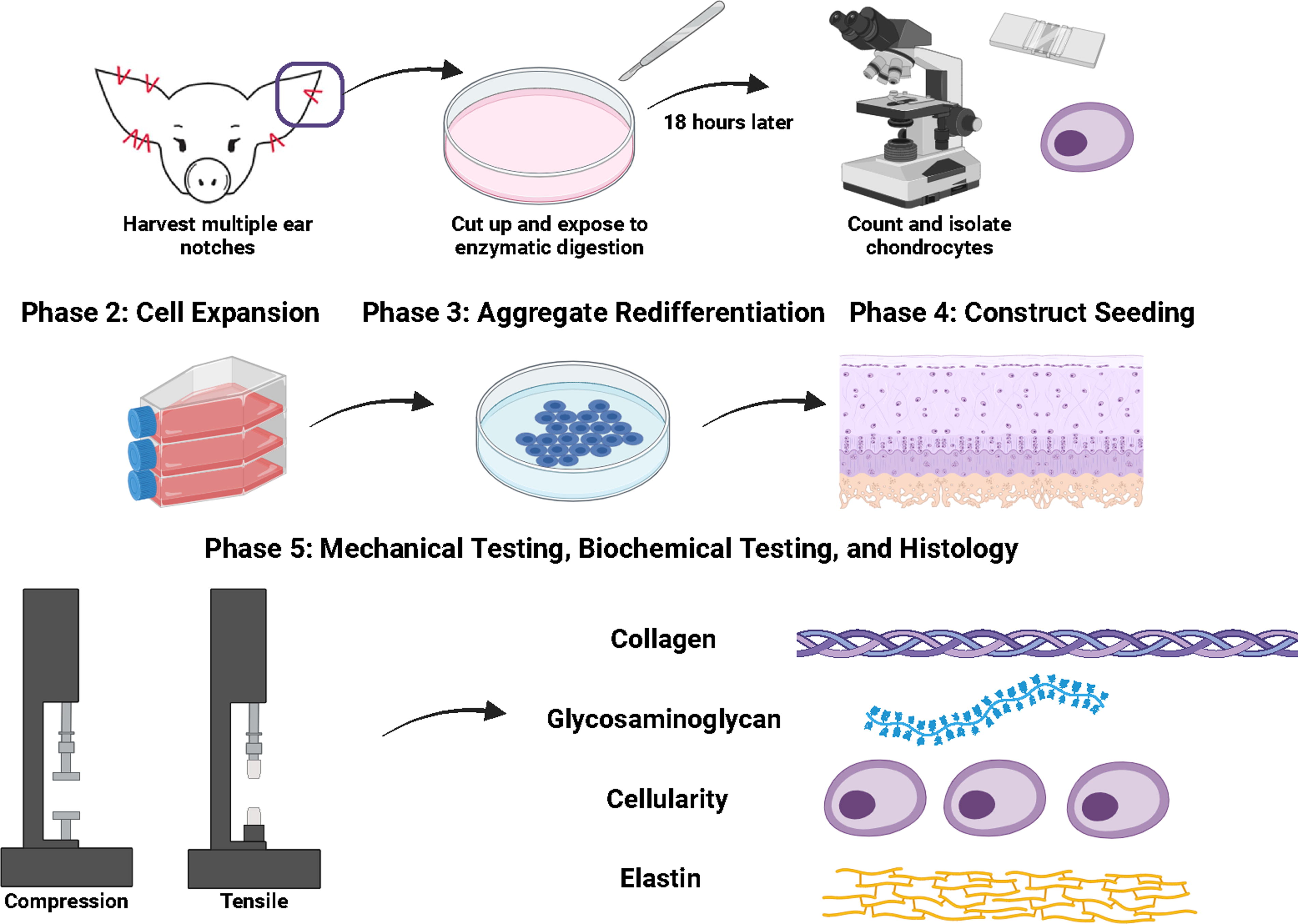
Study design.
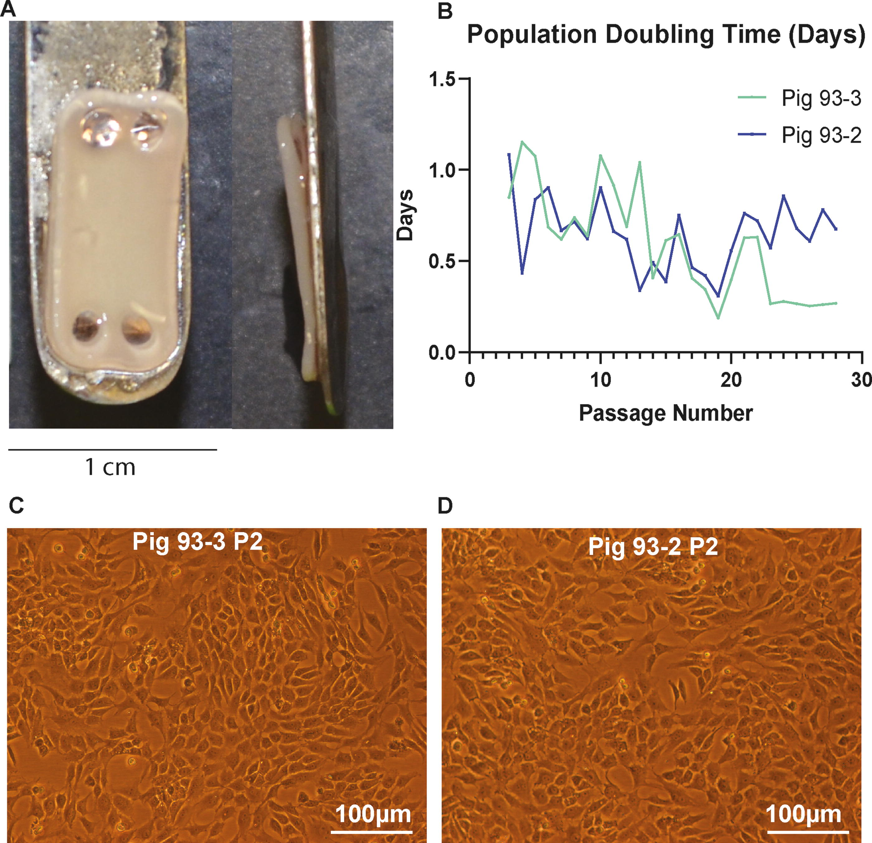
Population doubling
Two pig donors were used to calculate population doubling times. AuCs grew well in monolayer and had comparable population doubling times between donors. AuCs maintained short population doubling times consistently over 25 passages (Fig. 2B).
Histology
With hematoxylin and eosin staining, the microscopic morphology of engineered 3D cartilage tissue was densely cellular, and cells were evenly distributed throughout the section (Fig. 3). The histomorphology of the tissue was consistent with cartilage, where individual chondrocytes reside in the lacunae and are surrounded by a homogeneous basophilic ECM. The intensity of basophilic staining was comparable to native auricular tissue (Fig. 3). With the application of special stains, more specific features of ECM were highlighted. Specifically, safranin O/fast green counterstain labeling highlighted abundant glycosaminoglycans in the ECM (intense orange staining), whereas Picrosirius Red underscored the collagen content (pink color) (Fig. 3).
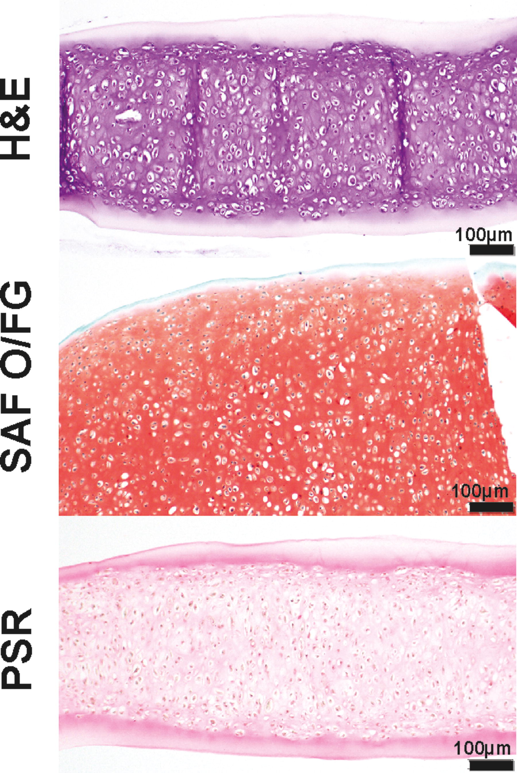
Histology of auricular chondrocyte derived TE constructs. Auricular TE constructs were stained with various stains (from top to bottom): H&E, Safranin-O/Fast Green, and Picrosirius red. H&E staining shows overall cellularity, Safranin-O/Fast shows overall glycosaminoglycan content, and Picrosirius red shows overall collagen content. H&E, hematoxylin and eosin; TE, tissue engineering.
The presence of elastic fibers was interrogated with Virchow Van Gieson stain in various native and engineered tissues for comparison. Elastin fibers were not apparent in the TE construct sections sourced from costal or AuC, but were abundant in the control section of adult porcine pina (Fig. 4). The neonatal ear had 141.5 µg/mL of elastin. Interestingly, very anisotropic elastic fiber distribution was observed in minipig TMJD tissue, where elastin fibers were only microscopically apparent in the posterior and lateral aspects of the disc (Fig. 4).
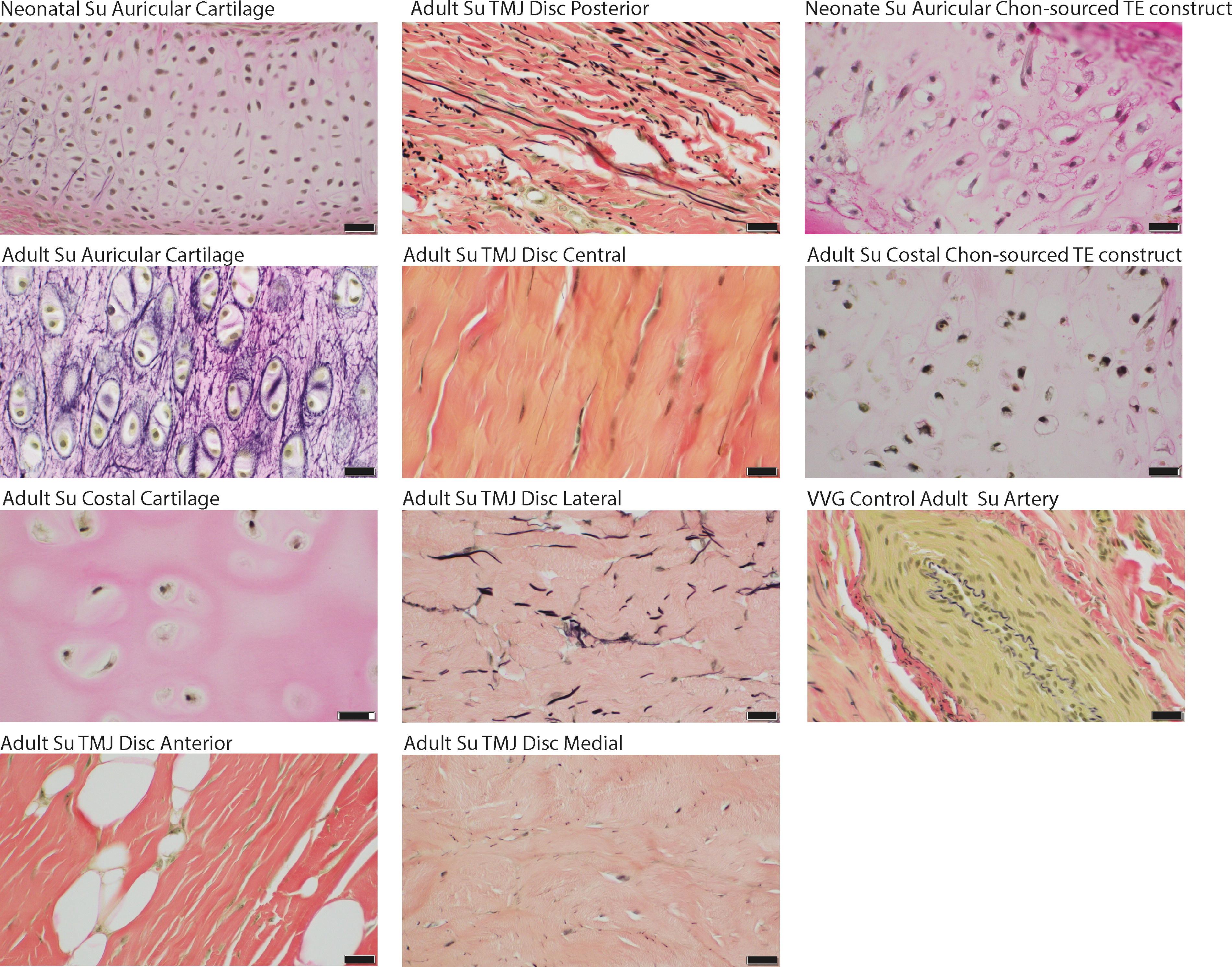
Histology panel of swine (Su) cartilages stained with Verhoeff Van Gieson. The elastic fibers stain black. Panel of images show Su cartilage from various anatomical locations/sources. Cartilage derived from several locations of the TMJ, auricular cartilage, costal cartilage, and auricular chondrocyte-derived TE cartilage constructs. VVG is a histological technique used to visualize elastic fibers in tissues. The size bar for all images is 20 µm. TE, tissue engineering; TMJ, temporomandibular joint; VVG, Verhoeff Van Gieson staining.
Biochemistry
Biochemical analyses supported histological observations. AuC-derived TE constructs contained 0.144 µg ± 0.011 collagen per µg dry weight, 0.185 ± 0.002 µg GAG per µg dry weight, DNA 0.384 ± 0.432 ng per µg dry weight, and 1.7e-3 µg elastin per µg dry weight. The elastin content in the TE constructs was not significantly different from native neonatal auricular tissue p = 0.3523 (Fig. 5). The average water content of all AuC-derived TE constructs was 81% ± 1.18%.

Colorimetric elastin (Fastin) assay comparing the elastin content in the TE cartilage vs. native neonatal auricular cartilage n = 3, p = 0.3523. TE, tissue engineering.
Mechanical properties
We found that constructs derived from AuCs had an Ei at 10% strain of 200.5 ± 43.34 kPa, 471.9 ± 92.8 kPa at 20% strain as well as 36.99 ± 6.47 kPa Er at 10% strain and 110.3 ± 16.99 kPa Er at 20% strain. The Young’s modulus was 2.0 ± 0.63 MPa, and the UTS was 0.619 ± 0.177 MPa.
Discussion
This study is the first to explore the utility of auricular cartilage-sourced chondrocytes for scaffold-free engineering of neocartilage constructs. A small tissue sample was sufficient for isolating primary AuC, which were expanded to hundreds of millions using expansion and redifferentiation methods previously effectively applied to costal and articular chondrocytes.5,32 Histology (Fig. 3) confirmed the cartilage morphology and highlighted ECM features that agreed with all quantitative biochemical tests. Mechanically, AuC-derived constructs were robust and had comparable properties to native porcine TMJ.11,32,34 Cumulatively, these results demonstrate the functional properties of engineered cartilage akin to native tissue and the feasibility of AuCs for scaffold-free cartilage TE. This methodology can be further explored for clinical applications in TMJD and auricular reconstruction and regeneration.
Costal chondrocytes were previously described as an attractive cell source for TMJD and other cartilaginous tissue reconstruction and regeneration.9,35,36 Indeed, constructs generated from this cell source were evaluated in a minipig TMJD thinning model where a successful defect closure was achieved. 11 Interestingly, despite some biochemical and biomechanical discrepancies with the native tissue, the costal cartilage-sourced constructs were able to remodel upon implantation and assume properties more similar to native. 11 Although this study did not compare costal constructs to auricular, their mechanical properties are similar. For instance, in the study conducted by Huwe et al., the instantaneous modulus of the engineered cartilage sourced from costal chondrocytes was ∼300 kPa, 37 as compared with 200 and 400 kPa at 10% and 20% strain, respectively, in the present study. Similarly, the Young’s modulus and UTS of the constructs from Huwe’s measured 1.4 MPa and 0.32 MPa, respectively, as compared with Young’s modulus of 2 MPa and UTS of 0.6 MPa in the current study. 37 There were slight but not dramatic differences in the biochemical properties. In the study conducted by Huwe et al., 37 the collagen content of the engineered cartilage sourced from costal chondrocytes was ∼1.8 collagen/wet weight (%), as compared with 2.6 collagen/wet weight (%) in the present study. Similarly, the GAG content of Huwe’s constructs was ∼7.5 GAG/wet weight (%), as compared with 3.34 GAG/wet weight (%) in the present study. 37 This discrepancy could be attributed to a difference in the proportion between number of cells ECM, but a specific comparison study would be needed to make final conclusions.
Auricular cartilage is an elastic type of cartilage, which means that AuC are able to synthesize elastin. 38 TMJD is a unique type of fibrocartilage with anisotropic fiber direction and site-specific distribution of elastic fibers. 23 Specifically, it was determined that the bilaminar zone (i.e., posterior disc attachment zone) of the human TMJD has the highest elastin fiber concentration. 23 Similar distribution difference in elastic fibers is apparent in the TMJD of the minipigs 24 (Fig. 4). No intense elastin staining was observed on TE AuC-sourced histological sections or costal chondrocyte-sourced TE constructs. However, elastin was detectable in the AuC-sourced constructs with Fastin biochemical assay. Precisely, a concentration of 1.7e-3 µg per µg of dry tissue was detected. In comparison with native neonatal auricular cartilage, which histologically did not have apparent elastin staining with Virchow van Gieson, this concertation was not significantly different statistically (Fig. 5). Provided that the elastic fibers mature through the formation of the lysyl-oxidase-mediated cross-linking process,39–41 it is possible that once formed, elastin fibers continue to mature via the cross-linking mechanism. The reports on elastin maturation are outnumbered by reports on elastin aging and degradation with age, inflammation, and environmental damage. 40 Studies on elastin maturation state that elastin formed during neonatal development remains with the individual throughout its life 42 and that genes for elastin and fibrillin pick up during neonatal development and then silenced for good.43,44 It remains to be determined how structural maturation progresses with age. In the case of scaffold-free cartilage TE, it was postulated that the phase of self-assembling recapitulates the stage of mesenchymal condensation in embryonic development and that neocartilages engineered with this method continue to mature via collagen fiber cross-linking beyond 4 weeks of the manufacturing process. 45 Collectively, it is possible that AuC-sourced TE constructs need more time to mature and cross-link elastin fibers to be detectable with standard histological methods.
It is crucial to note that dozens of auricular neocartilage constructs were manufactured from a very small stock of primary cells. This manufacturing success is because of the one critical step in the scaffold-free TE process, where the cells are cultured for an additional 10 days in suspension following monolayer expansion. It is postulated that during the monolayer expansion step, chondrocytes undergo de-differentiation, i.e., assume fibroblastic phenotype instead of chondrocytic.46–48 However, during the suspension culture, where chondrocytes are unable to adhere to the substrate and proliferate, redifferentiation takes place, and chondrocytes resume their chondrocytic properties. This aggregate redifferentiation step allows to conduct the extensive in vitro expansion of a very small initial stock of primary cells, overcoming a huge hurdle of autologous tissue availability. 38 The advantages are clear. Using the smallest tissue biopsy of autologous auricular cartilage, followed by a sufficient expansion to hundreds of millions of cells, makes autologous cell-sourced TE an achievable reality. The population doubling study reported here indicates that the cell replication speed does not change for at least 20 passages (Fig. 2); thus, cellular senescence is not a concern. An additional advantage of AuC for elastic cartilage TE is that the morbidity of the collection site in the ear is much less than in the autologous rib collection procedure.49,50 Lastly, unlike costal cartilage, auricular cartilage does not have a tendency to ossify with age, presenting a wider timeframe for tissue collection.
In summary, this study is the first to demonstrate the proof of concept and the feasibility of AuCs’ suitability for scaffold-free cartilage TE. Extending this study to a side-by-side comparison to neocartilages engineered from costal chondrocytes would be essential in further determining this cell source’s utility and possible advantage for the regeneration of cartilaginous tissues such as TMJD and pinna. A scale-up to true-size pinna or TMJD tissue can follow. Lastly, a translation of these methods to human cells and the conduction of engraftment transplantation experiments would be necessary for this method to be applied in clinics.
Footnotes
Authors’ Contributions
N.G. contributed to the writing of the article, data acquisition, and analysis. C.G. contributed to the writing of the article, data acquisition, and analysis. I.R. contributed to data collection, analysis, and figure design. H.M. executed the experiments. N.V. conceptualized the idea, acquired funding, analyzed data, and prepared the article.
Disclosure Statement
The authors declare no conflict of interest and confirm the originality of this work.
Funding Information
The study was funded by NIH/NIDCR grant R03 DE030900-01A1.