
Abstract

Anterior cervical nodules that move with swallowing and are confirmed as thyroidal by ultrasonography of the neck are not necessarily of thyroidal origin.
We report a patient who was found to have primary hypothyroidism in 1990, at age 64, and was placed on levothyroxine. A thyroid sonogram dated February 2001 showed “little change in the overall size of the gland from prior study of 10/06/1999. There is, however, one nodule in the posterior medial aspect of the left lobe which has increased in size and now measures 1.1 × 1.6 × 1.4 cm.” The patient was referred to us for fine-needle aspiration (FNA) of this thyroid nodule. We saw him in April 2001. On physical examination we palpated a 1.0-cm nodule in the left lobe of the thyroid gland. We performed a palpation-guided FNA and obtained three samples in usual manner (1) using 22- and 23-gauge, 1.5-inch needles. Some smears were fixed in ethanol and stained with Papanicolaou stain; others were air-dried and stained with Diff-Quik stain (and three were evaluated for adequacy while the patient was still in the room). Smears contained birefringent material (consistent with cellulose/vegetable fibers; Fig. 1), skeletal muscle fibers (meat; Fig. 2), benign squamous epithelial cells, and bacteria (cocci and bacilli; Fig. 3). We made a diagnosis of esophageal diverticulum and suggested a barium swallow. This confirmed the presence of a “diverticulum projecting off the left aspect of the proximal most cervical esophagus slightly anteriorly.” The patient has remained asymptomatic and continues on thyroid replacement therapy (last office visit in September 2008).

Vegetable fiber (coiled structure on the left) and amorphous debris (on the right side). Papanicolaou stain, high magnification.

Skeletal muscle fiber (notice cross-striations). Papanicolaou stain, high magnification.
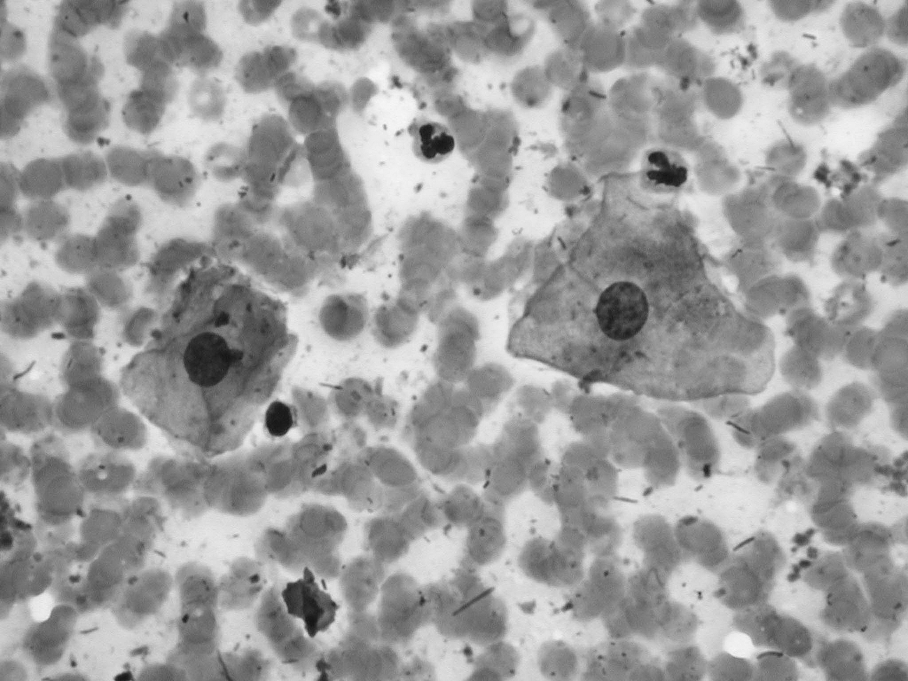
Benign squamous epithelial cells, bacteria (bacilli and cocci), and erythrocytes in the background. Diff-Quik stain, high magnification.
Appropriate management of a patient depends on correct diagnosis. Anterior neck nodules or masses that are clinically considered of thyroidal origin (because they move with swallowing) and are ostensibly confirmed by ultrasonography, in fact, may not be thyroidal (2). The differential diagnosis includes thyroglossal duct cysts which may be located laterally (3) and the correct preoperative cytologic diagnosis will allow the surgeon to perform, as appropriate, the Sistrunk procedure rather than an inappropriate local resection. Two types of oropharyngeal diverticuli have been described: Zenker's diverticulum and Killian–Jamieson diverticulum. Both are rare entities and there are few reports on their ultrasound findings (4). There is one case report in which “on the basis of the cytologic findings, origin from the oropharynx or a paraesophageal diverticulum was suggested” (5). Rekhtman et al. (6) reported on an ultrasound-guided FNA of a nonpalpable thyroid nodule. The authors did not state how the aspirate was diagnosed but mentioned that “Subsequent clinical and further radiologic workup revealed a Killian–Jamieson diverticulum.”
There might be some concern as to how safe it is to aspirate an esophageal diverticulum and cause infection in the surrounding area. For over 30 years we have performed FNAs of palpable lesions from all body sites and in the last 11 years we have aspirated over 13,000 thyroidal lesions (7). We have interpreted aspirates from deep organs performed by radiologists. We have not seen infections from FNAs of pancreatic lesions, even though the needle goes through intestinal lumen and bowel contents may be aspirated. Hence, we attest to the safety of the procedure.
FNA is the recommended diagnostic test in the initial evaluation of thyroid nodules (8,9). As we report in the present case, FNA can also be useful to diagnose mimics of thyroidal lesions.
Footnotes
Acknowledgments
The authors are grateful to Ms. Ivonne Rivadeneira for photographic assistance and to Drs. Kenneth D. Burman and Leonard Wartofsky for reviewing the manuscript and offering valuable suggestions.
Disclosure Statement
The authors declare that no competing financial interests exist.