
Abstract
Background
Methods:
We report the case of a 20-year-old woman with TGDC. We implemented a modified approach to TGDC removal through a frenotomy incision of the mouth using an endoscope system.
Results:
The patient received a modified approach to TGDC removal. The total operative time was 60 minutes. She remains free of disease 12 months after her surgery.
Conclusion:
We describe, in a single patient, a procedure in detail for endoscope-assisted transoral TGDC excision using an intraoral frenotomy incision. The advantage of this approach is the avoidance of a neck scar. Our experience with this patient indicates that resection of a TGDC appears to be feasible through a transoral endoscope-assisted approach using a frenotomy incision in the mouth. Further experience with this procedure is required.
Introduction
The Sistrunk operation is the treatment of choice for symptomatic or clinically apparent TGDC, which includes the removal of the midportion of the hyoid bone in continuity with the TGDC along with excision of a block of tissue between the hyoid bone and the foramen cecum (3 –9). Although major complications are rare in the Sistrunk operation (recurrence develops in <10% of patients) (2,3,5,7 –12), the transcutaneous resection that occurs during the procedure may inevitably result in a scar of ∼5 to 10 cm.
Considering that most patients who undergo surgical resection are young (<30 years), it is desirable to develop surgery that avoids the external neck scar for TGDC. Thanks to advancements in medical technology, we were able to develop a modified endoscope-assisted approach through a frenotomy incision of the mouth.
This approach passes through the midline incision in the floor of the mouth and uses the natural midline dehiscence present between the genioglossus muscles. This area has been shown to be a relatively avascular dissection plane. So, this method has advantages of minimizing the access trauma and allowing for improved preservation of adjacent structures.
Additionally, this method is considered to be endoscopic surgery through a natural opening of the human body (natural orifice transluminal endoscopic surgery). A consideration in developing this technique was that it might also be applied to transoral thyroid and parathyroid surgery. In this report, we describe this new technique and the surgical outcome.
Case Report
A 20-year-old woman was admitted for the chief complaint of a mass at the anterior neck, detected 6 months previously. Since detection its size had increased gradually and she was uncomfortable during swallowing due to the sensation of a lump in throat. On palpation, a painless, movable, soft mass (diameter=3.0 cm) was noted just inferior to the hyoid bone. The mass moved on tongue protrusion and during swallowing. Laryngoscope examination did not show a protruding mass at the base of the tongue. The patient's medical history was unremarkable. She had no history of thyroid disease.
The neck computed tomography imaging of the thyroid verified that the thyroid was in the normal location. With contrast enhancement, there was a cystic mass ∼2.0×1.0 cm below the hyoid bone (Fig. 1). Aspiration cytology and computed tomographic scan suggested an infrahyoid TGDC. The patient was a young woman and she did not want a surgical scar remaining in the neck area. Therefore, the surgery was performed with her consent by a transoral approach using an endoscope.

Computed tomography scan confirmed the 2-cm mass found just inferior to the level of the hyoid bone.
Surgical Technique
With the patient under general anesthesia, the mouth was opened and the tongue was retracted to an upper position. An ∼2 cm incision was made in the oral mucosa including the frenulum (Fig. 2A, B). After careful dissection of soft tissue in the floor of the mouth, we found the genioglossus muscles, separated them in the midline, and retracted them bilaterally (Fig. 2C). With the aid of an endoscope (rigid, 10 mm, 0°; Olympus), after retraction of the genioglossus muscles, we found some midline longitudinal tissue (Fig. 2D). This tissue extended from the midline of the hyoid bone toward the tongue base. This tissue appeared to be the TGDC tract. After dissection of the TGDC tract we cut it between the tongue base and the hyoid bone (Fig. 2E, F). We then identified the hyoid bone and strap muscles (Fig. 2G). We transected the strap muscles with ultrasonic scissors (Harmonic scalpel 300; Ethicon Johnson & Johnson Company) ∼0.5 cm apart from the hyoid bone. We then cut the body of the hyoid bone out with an Osteo Punch Rongeur (1 mm, 40°; KOROS; Fig. 2H). We also transected the infrahyoid muscles attached to the body of the hyoid bone. While pulling the hyoid bone upward, we were able to immediately identify the cystic mass inferiorly; it was attached to the hyoid bone by a stalk (Fig. 2I). We removed it along with the hyoid bone en bloc through careful dissection of the cyst. After the removal of the mass, the thyroid cartilage was present below (Fig. 2J). The operative field was irrigated, a drain was inserted through the floor of the mouth, and it was sutured to the edge of the opened mucosa. Afterward, the genioglossus muscles were sutured with 4-0 Vicryl, and the opened oral mucosa was sutured. The total operation time was ∼60 minutes. The drain was removed on postoperative day 2. We encouraged frequent oral gargling with 0.02% chlorhexidine and allowed a normal diet on postoperative day 3. The patient was discharged home on postoperative day 5. Twelve months have passed since the operation and there have been no problems developing in the operative field. She is currently being followed up and observed in our outpatient clinic. The patient was satisfied with the surgical outcome.
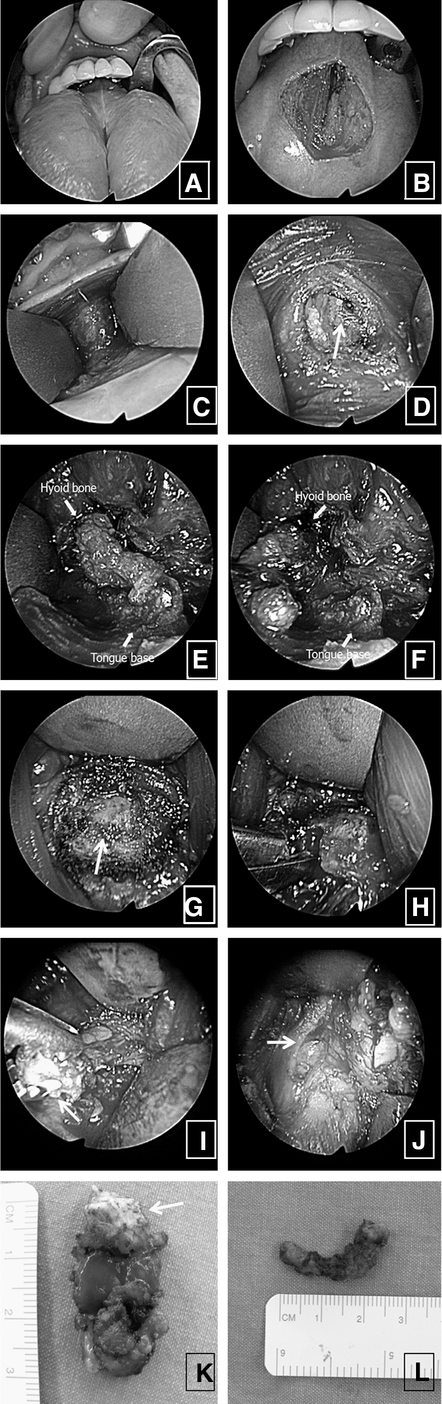
Transoral approach through a frenotomy incision of the mouth.
Discussion
The most effective treatment for a TGDC is the Sistrunk operation. However, this operation requires a transcervical approach, which results in an external neck scar (13). Depending on the location of the TGDC, various surgical resection methods have been reported. There have been several reports of a TGDC located in the base of the tongue or larynx, which have been treated through transoral endoscopic surgery (13). However, these methods have a possibility of damage of the mouth floor structure. On the other hand, in our case, the risk of damage was relatively low because we used anatomical dehiscence through frenotomy incision. Previously, Kim et al. published a paper about the safe excision of a dermoid cyst in the mouth floor through the frenotomy incision and discussed the potential application of this method to TGDC (13).
In contrast to the surgical removal of other benign neck masses, TGDC surgery should include removal of TGDC and its possible tract (2,3,6,9 –12,14). This means the middle part of hyoid bone and a block of tissue extending to the foramen cecum must be removed. This technique became technically feasible with the advancements in endoscope system and ultrasonic scissors (harmonic scalpel) (9). This approach passes through the midline incision in the floor of the mouth and uses the natural midline dehiscence present between the genioglossus muscles (13). This area has been shown to be a relatively avascular dissection plane. If the space between the genioglossus muscle is spread out using a tractor, then the mylohyoid muscle can be reached. When we traced the posterior line of the mylohyoid muscle, the hyoid bone could be reached. When we reached the hyoid bone, we cut the suprahyoid muscles attached to the middle portion of the hyoid bone (1.0 cm in width). To remove the possible TGDC tracts, we found some midline tissues extending from the midline of the hyoid bone toward the foramen cecum, and then removed them to the base of the tongue.
We used the ultrasonic scissors to remove a cuff of tissue (10 mm in width) in the middle portion of the hyoid bone. This method was concordant with the modified Sistrunk operation (3,5,6,8,9). We than cut the hyoid bone and resected the TGDC placed beneath the hyoid bone. The patient was quite satisfied with the surgical outcome.
The transoral approach is relatively easy and it also has the advantage of minimizing trauma associated with access and seems to allow improved preservation of adjacent structures (13). The surgery is made feasible by the development of the endoscope and other medical instrument technologies.
The satisfaction level of our patient after operation was good, and significant swelling or bleeding was not seen. Because the neck exploration runs the risk of turning an aseptic operation to a potentially infectious surgical procedure by spreading of oral flora, steps should be taken to minimize this complication. Therefore, we used antibiotics and an oral gargle and the drain was removed 2 days after operation. Follow-up observation was performed for 12 months after surgery without recurrence or complications. However, experience with this surgery in a series of patients is needed to determine its efficacy. In addition to postoperative infection, there is the theoretical possibility that the risk of TGDC rupture may be greater with this surgical approach that for the Sistrunk operation.
Footnotes
Acknowledgments
Disclosure Statement
No financial or material support has been received for this work; moreover, the authors declare no financial interests in companies or other entities that could have an interest in the information within this contribution. None of the authors has any conflict of interest, financial or otherwise.