
Abstract
Background:
Proteomics and mass spectrometry are useful tools for peptide screening in body fluids. In thyroid-associated orbitopathy (TAO), evidence for lacrimal gland involvement with altered composition of tears has been reported. Our objective was to detect and evaluate potential changes in the proteomic patterns of tear fluid in TAO.
Methods:
Tear fluid was collected from 45 patients with TAO and 15 healthy controls. Tear proteins were analyzed using surface-enhanced laser desorption/ionization time-of-flight mass spectrometry, and peptides were identified using matrix-assisted laser desorption/ionization time-of-flight technology.
Results:
Peptides with molecular weights 3808 Dalton (Da, p=0.004), 3734 Da (p=0.034), and 3837 Da (p=0.042), respectively, were downregulated in patients with TAO versus controls. They were identified as proline-rich protein 4 (PRP4) or as its variant nasopharyngeal carcinoma-associated PRP4. The peptide 3837 Da correlated positively with the basal secretory test (r=0.506, p<0.001) and negatively with the clinical activity score (r=−0.334, p<0.05) and age (r=−0.431, p<0.001). Also, a 12,003-Da peptide was downregulated (p=0.019) in patients and identified as ß2-microglobulin. This peptide decreased in tear fluid with increased clinical severity of TAO (p=0.027). In comparison, a 5815-Da peptide was upregulated (p=0.045) and identified as lysozyme C. When differentiating between treated and untreated patients with TAO, an 11,770-Da peptide (p=0.0072) that was also upregulated was identified as cystatin S.
Conclusions:
Altered regulation of proinflammatory and protective proteins in tears of patients with TAO was demonstrated, reflecting an autoimmune- and/or inflammatory-induced dysfunction of the lacrimal gland.
Introduction
In thyroid-associated orbitopathy (TAO), an organ-specific autoimmune disorder, there is evidence for lacrimal gland involvement (4 –7). Orbital computed tomography revealed lacrimal gland enlargement in patients with TAO (8), and imaging using the radionuclide octreotide showed relevant accumulation in the lacrimal gland (9). These structural changes lead to a reduced tear production (10). Also, the composition of tear fluid is reported to be altered in TAO (11,12). Further, impaired cytokine balance has been observed in tears of patients with TAO (13). However, all these studies indicate changes in tear proteins of patients with TAO, without knowing the concrete proteins and their relevance for therapy and diagnosis of TAO.
Since numerous studies have shown evidence of lacrimal gland impairment in TAO, it is of main interest to obtain precise information on the pathological changes in protein expression within the orbit. Therefore, we aimed to analyze proteomic patterns in tears of patients with TAO having the advantages of noninvasive and well-available sample acquisition. In this present study, tear protein profiles in patients with TAO and in healthy controls were investigated using mass spectrometric techniques. In a pilot project, we were able to detect a set of potential protein biomarkers using the surface-enhanced laser desorption/ionization time-of-flight mass spectrometry (SELDI-TOF-MS) technology (14). Using these candidate biomarkers, it was possible to select peptides in tears of TAO patients with 100% specificity. In this article, we aimed to identify these peptides and evaluate their relevance in the pathogenesis and outcome of TAO.
Methods
Patients
A total of 45 patients with autoimmune thyroid disease (Graves' disease or Hashimoto's thyroiditis) and TAO of various clinical activity and severity were included in this study. Fifteen age- and sex-matched euthyroid healthy persons (median age 45 years, range 33–74 years; 13 women and 5 smokers) served as controls. All patients and controls gave their written informed consent. All protocols were approved by the Institutional Ethics Committee in accordance with the ethical standards laid down in the Declaration of Helsinki.
In the joint thyroid eye clinic of the Gutenberg University Medical Center, complete endocrine and ophthalmic investigations were performed before tear sampling. As previously described and published (15), the clinical activity score (CAS) of TAO consists of seven items: spontaneous pain behind the globe, pain on attempted up gaze, redness of the conjunctiva, redness of the eyelid, chemosis, swelling of the lacrimal caruncle, and eyelid swelling. One point is added for each item present. The CAS score ranges from 0 to 7. Further information was obtained using a questionnaire retrieving patients' clinical history and previous treatment. Patients suffering from additional diseases or receiving medication that could affect the tear film constitution (eye drops, antihistamines, β-blockers, antispasmodics, diuretics, and some psychotropic drugs) were excluded from the study. Methimazole, levothyroxine, and/or tear substitutes without preservatives were accepted, only.
Samples
To obtain samples of tear fluid from all 60 subjects, the basic secretory test (BST, also called Schirmer II) was performed under local anesthesia, and the Schirmer strips were stored at −80°C until use. The Schirmer strips were eluted, and the samples were treated as previously described (14).
Data acquisition
The arrays were analyzed the SELDI-TOF-MS technology using a PBS-IIc Protein Chip Reader equipped with an Autoloader using Protein Chip Software version 3.2. The instrument settings were used as previously described (14). The mass-to-charge ratio (m/z) of the proteins was evaluated according to external calibrators. Normalization was performed by total ion current to an external normalization factor of 0.2. Ciphergen Express Data 2.1 Database Software was used to perform automatic peak detection. Using these detected peaks, clusters were generated across multiple spectra. A peak cluster was created if the peak was found in 10% of all spectra for a given condition. The mass window for peak clustering was set at 0.3% of the peak mass for low laser intensity readings and at 2% of the peak mass for high intensity readings.
Data analysis
The cluster lists containing the normalized peak intensity values for each sample were exported to Statistica (version 6.2; StatSoft), and an analysis of variance based on combinations of multiple peaks was performed. This analysis calculates the most significant peptides that discriminate best between patients and controls. Such analysis was performed for each condition (surface type, laser energy, and elution protocol), generating a list of peaks that differed significantly between the two groups. To evaluate the diagnostic power of this peptide panel, an automated neural network was trained using these markers as input values. The output values classified the patient as TAO or not. The complete dataset (differing peptide intensities) was randomly split into two subsets: one was used for training; the other was used as the test data set since it was previously not known to the neural network, to assure the validity of the method-performance. With this test data set, a receiver-operating characteristic (ROC) curve was generated by plotting sensitivity against 1-specificity.
Peptide identification
To identify the peptides, the samples were separated on a sodium dodecyl sulfate (SDS)–polyacrylamide gel electrophoresis. The gel slice where the peptide was assumed was cut out and split in two pieces. From one-third, the intact protein was eluted for 2 hours using an elution buffer containing 50% formic acid, 25% acetonitrile, 15% isopropanol, 10% water, and 0.1% SDS. The eluted sample was re-profiled with the SELDI-TOF-MS to prove the presence of the peptide. With the other two-thirds of the gel slice, an in-gel digestion with trypsin was performed. The samples were applied onto an anchor chip target (Bruker) using a cinnamic acid matrix (0.02 g cinnamic acid/10 mL in 50% acetonitrile). Data acquisition was accomplished using a matrix-assisted laser desorption/ionization time-of-flight (MALDI TOF) mass spectrometer (Ultraflex TOF/TOF; BrukerDaltonics). After acquiring the digest spectra with 100 laser shots averaged from five sample positions in the linear mode, peptides below m/z 4000 Da with good peak intensity were selected for a fragmentation analysis using a reflector mode. Peptide fragmentation was performed using collision-induced dissociation (CID), and 50 laser shots from five sample positions were summed up for each parent ion. All spectra were externally calibrated by using the peptide calibration standard (Angiotensin II 1047, 19 Angiotensin I 1297.49, Substance P 1348.64, Bombesin 1620.86, ACTH clip 1–17 2093.08, ACTH clip 18–39 2465.19, and Somatostatin 28 3147.47; BrukerDaltonics). Data processing of raw spectra, peak detection, and protein identification was performed using Bruker software (Flex Analysis 2.4 and BioTools 3.1) and MASCOT. The MALDI spectra obtained were used for database searches with MASCOT using NCBI (National Institutes of Health) and SwissProt (Swiss Institute of Bioinformatics) databases. MASCOT compares the peptide and lift spectra against peptide patterns in the databases and searches for homologies. In this case, BioTools Software, which is linked with the MASCOT server, was used. If the data of the spectra matched the data in the database, the probability was measured, if this was a contingency. The smaller the probability was, the more valid the identification.
Results
Demographic and serological data
A total of 45 patients (37 women, median age 50 years, range 22–77 years, 23 smokers, 15 untreated patients, and 30 previously treated with steroids) with thyroid eye disease of various clinical activity (17 active and 28 inactive according to CAS scoring) and clinical severity (19 mild and 26 severe) were included in the study. Forty-three patients with TAO had Graves' disease, of whom 37 were on either methimazole (2.5–10 mg/day) or on levothyroxine therapy (50–125 μg/day) subsequent to thyroid surgery or radioactive iodine therapy. Six Graves' patients were euthyroid and off treatment after a successful course of methimazole therapy. The two patients with Hashimoto's thyroiditis were on levothyroxine. All in all, 39 of 45 patients were euthyroid, whereas 4 were subclinical hypothyroid (normal free thyroxine [T4] and free triiodothyronine [T3] with thyrotropin [TSH] between 4 and 8) and two subclinical hyperthyroid (normal free T4 and free T3 with TSH below 0.3 mU/L). The median length of the BST (Schirmer test) was 16 mm, range 0–30 mm.
Protein patterns
Complex protein patterns were detected in all samples analyzed with SELDI-TOF-MS. Up to 1000 peaks per sample were comprised over all conditions. Over 150 peaks could be consistently clustered in patients and controls. The peptides whose intensities significantly differed between the patient and control groups are summarized in Table 1. All peptides, except those at 3759, 5087, 5815, and 11,770 Da, were downregulated in the patient group. A ROC curve with an area under the curve of 0.99, a sensitivity of 93%, and a specificity of 100% underscored the applicability of this method (Fig. 1). Using the MALDI-TOF-MS/MS technology (Fig. 2), numerous peptides were identified with the help of the MASCOT data base.
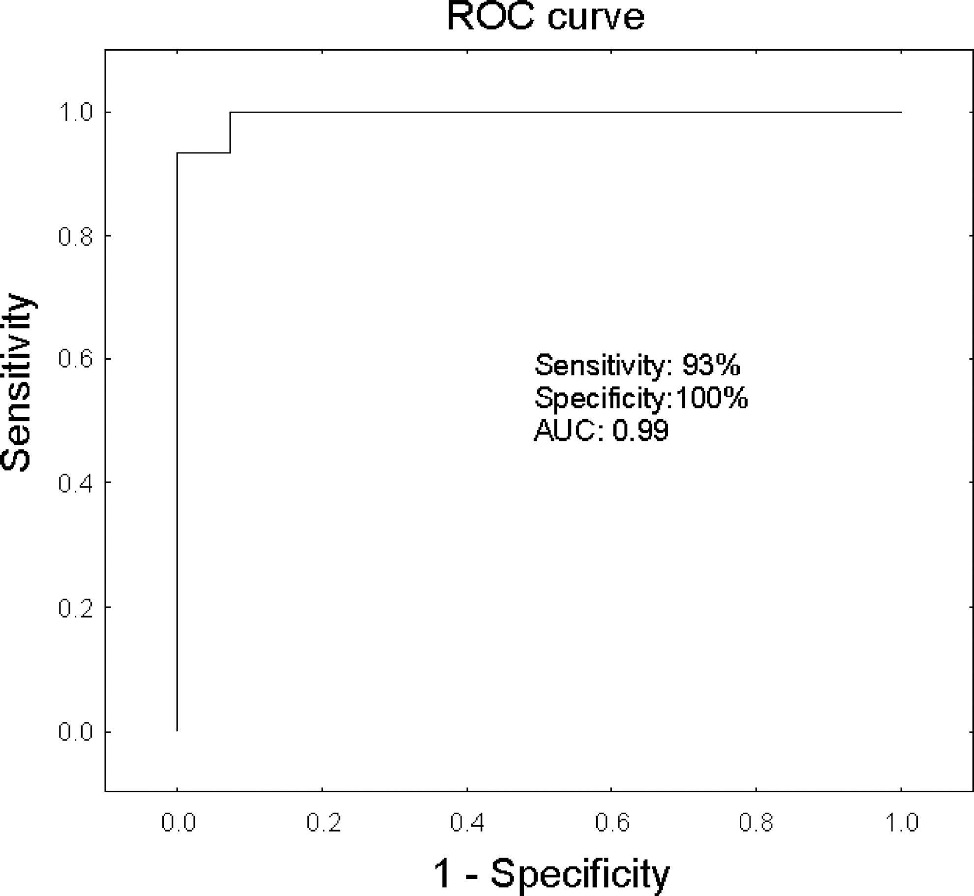
Receiver-operating characteristic (ROC) curve of tear samples. Displayed is the sensitivity versus 1−specificity.
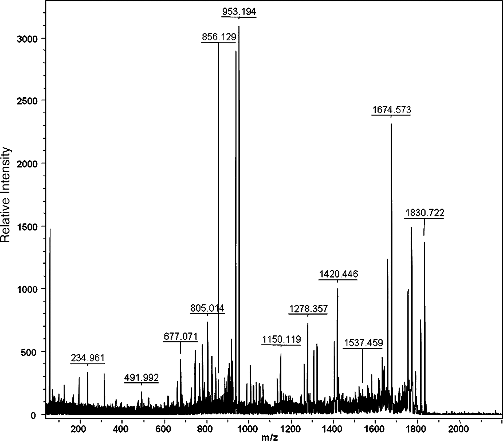
Matrix-assisted laser desorption/ionization time-of-flight mass spectrometry of the tear fluid samples.
TAO, thyroid-associated orbitopathy; PRP4, proline-rich protein 4; NCAPRP, nasopharyngeal carcinoma-associated PRP4.
Proline-rich protein 4
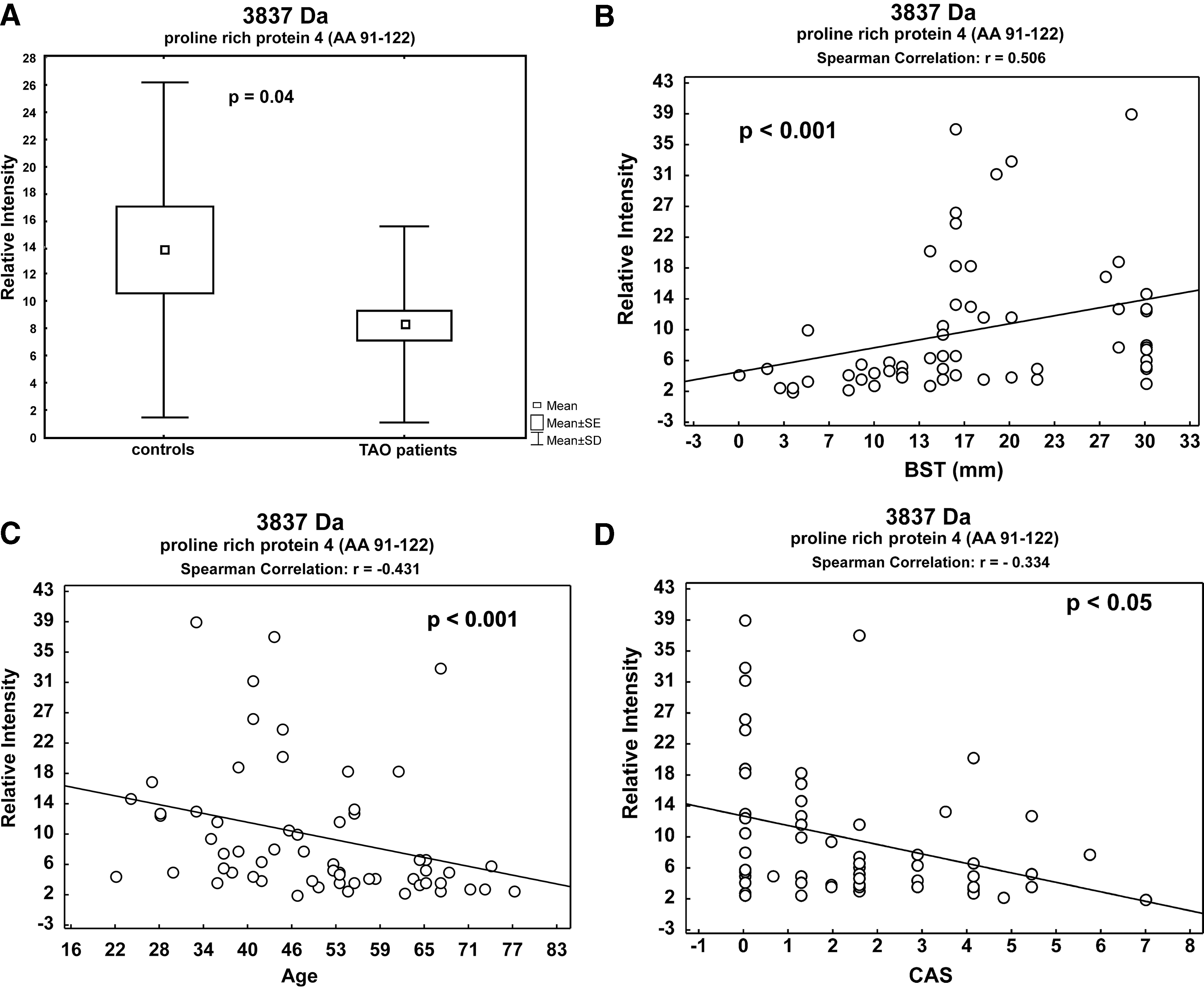
Compared to controls, three significantly downregulated peptide peaks at 3734, 3808, and 3837 Da (Table 2) were identified in the patient group as either proline-rich protein 4 (PRP4, peptide 3837 Da, Fig. 3A) or as nasopharyngeal carcinoma-associated PRP4 (peptides 3734 and 3808 Da; NCAPRP). NCAPRP is a variant of PRP4. These two proteins are almost identical, except for an amino acid exchange (Arginine to Serine at position 102). The peptide 3837 Da negatively correlated with age (r=−0.431, p<0.001) and with the CAS of TAO (r=−0.334, p<0.05). A positive correlation was noted with the BST value (r=0.506, p<0.001, Fig. 3B–D).
β2-microglobulin
The peptide with the molecular weight of 12,003 Da (p=0.019, Table 1) was downregulated in the patient group and was identified as β2-microglobulin.The peptide levels decreased with enhanced clinical severity of TAO (p=0.027, Fig. 4).
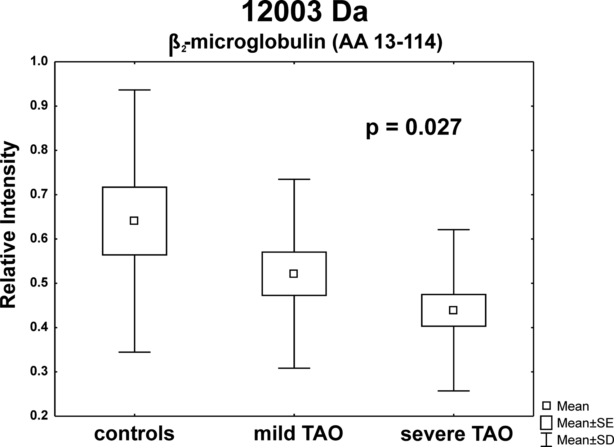
Box-and-whisker plots of the different degrees of severity.
Lysozyme C
The peptide 5815 Da significantly discriminated between patients and controls (p=0.045). It was upregulated in the patients' group and was identified as lysozyme C (Tables 1 and 2).
Impact of therapy and smoking habits on the peptide pattern
When dividing the patient group in treated and untreated subjects, an additional peptide with a molecular weight of 11,770 Da significantly differentiated between controls and patients with TAO (p=0.0072). This peptide was identified as cystatin S and was significantly upregulated in the treated patient group (Table 2). Smoking had a significant impact neither on the distribution nor on the content of protein markers in the tear fluid of patients with TAO.
Discussion
This original article reports on the proteomic profiling of tear fluid in a relatively large collective of patients with autoimmune thyroid eye disease of various degrees of clinical severity and clinical activity and in an age- and sex-matched control group. Proteomics revealed impaired tear protein profiles in patients in contrast to healthy controls. Numerous peptides were identified, and some of these correlated with the clinical activity and severity scores of TAO.
Protein profiles of the tears reflect the processes in the eye and/or orbit due to the close spatial relations of the lacrimal gland. The tear film regulates the homeostasis of the ocular surface, and pathological processes lead to disequilibrium. A variety of systemic diseases, that is, blepharitis (16), dry eye syndrome (3), diabetes (17 –19), and cancer (20), may impair the tear film. In the past, most studies performed to analyze tear protein profiles used one-dimensional (1D) gel electrophoresis combined with high-performance liquid chromatography (HPLC) (21), two-dimensional (2D) gel electrophoresis (22), or chromatographic techniques (23). The SELDI-TOF-MS technology used in this study offers many advantages, for example, the high sensitivity for proteins and peptides <25 kDa (16,24) and the possibility of a high throughput. When using different chromatographic surfaces, peptides can be enriched or washed away from the spot depending on their physical and chemical characteristics. Furthermore, this method allows the analysis of very small sample volumes with a high accuracy.
Numerous reports indicate changes in tear protein profiles using standard proteomic techniques, for example, HPLC and 1D gel electrophoresis. With the help of the SELDI-TOF-MS technology, we were able to generate a complex and reproducible tear protein profile of TAO patients. Statistical analysis between the spectra of TAO patients and healthy controls revealed eight potential protein biomarkers that significantly differed between the two groups. All peptides found in this study have a molecular weight below 15 kDa, demonstrating a very high sensitivity of the method in this mass range. The peptidome is composed of small proteins, for example, proteases and chemokines; however, fragments of proteins with higher molecular weight can also be found (25). These fragments are produced through secreted or membrane-standing proteases or by intracellular proteolysis. In addition, pathological processes can impact enzyme and protease activity. The fragments resulting out of these processes allow for the drawing of conclusions about the cell function in affected tissues (26).
Three of the peptides could be identified as the C-terminal fragment of PRP4 and its variant NCAPRP, respectively (16,27). The present investigation revealed significantly downregulated peptide peaks in the patient group compared to controls. PRPs are mainly expressed in the parotid and submandibular glands (28). Also, PRP4 is highly expressed in acinar cells and can be regarded as marker for acinar cell function. Lacrimal PRPs exert a protective function through modulating the microflora of the eye (29). Other substitutes of the PRP family have neuroprotective properties (30). In patients with dry eye syndrome, two fragments of PRP4 are also downregulated (31), implying that downregulation of protective proteins might be influenced by inflammatory processes in the orbit. In line with this is the negative correlation of the peptide 3837 with the CAS. The higher the CAS values and the degree of inflammation, the lower were the PRP4 levels, probably indicating a progressive inflammatory-induced dysfunction of the lacrimal gland in TAO.
Furthermore, the peptide 12,003 Da identified as β2-microglobulin was decreased in patients. This protein is part of the major histocompatibility complex (MHC) class I expressed in almost all nucleated cells (32). β2-microglobulin plays a critical role in immune processes. The MHC I/β2-microglobulin complex seems to be a requirement for the antigen detection via cytotoxic T-cells (33). Variations of β2-microglobulin levels seem to indicate an altered function of the immune system. The decreased values of β2-microglobulin indicate an increased expression of β2-microglobulin-free MHC class I molecules in TAO. The role of this β2-microglobulin-free MHC class I molecules is not fully understood, but it is granted that they play a role in T-cell activation and may serve as antigens (34).
In contrast, lysozyme C was upregulated in the patient group compared to controls. It is a proteolytic protein that is ubiquitous in many tissues and endocrine secretions (35). With lipocalin and lactoferrin, it represents 85% of the tear protein content. Lysozyme C hydrolyzes peptidoglycans; hence, it destroys the cell wall of microorganisms. It is also associated with the monocyte and macrophage system and plays a role in the immune response (36). The increased values of lysozyme C may indicate inflammatory processes of the orbit and the lacrimal gland, respectively. Increased lysozyme values in serum and saliva of patients with various autoimmune diseases have also been reported (37).
When separating the patients with TAO into treated and untreated patients, we identified the 11,770-Da peptide as cystatin S (36). Cystatine S, cystatine SN, and cystatine C belong to a family of cysteine proteinase inhibitors usually found in the tear film (38). The balance of proteases and protease inhibitors is important to preserve the homeostasis of the tear film (39). Cystatines fulfill protective functions by controlling cysteine proteases, preventing uncontrolled proteolysis and tissue destruction (40). Elevated serum cystatin S levels have been reported in patients with autoimmune diseases after systemic glucocorticoid treatment (41), probably reflecting the beneficial effect of steroids.
In conclusion, the altered regulation of proinflammatory and protective proteins was demonstrated in patients with TAO, reflecting an autoimmune and/or inflammatory-induced dysfunction of the lacrimal gland. Administered steroid treatment seems to have a beneficial impact on the tear film composition.
Footnotes
Acknowledgments
This work was financially supported by a grant of the Johannes Gutenberg University School of Medicine (MAIFOR). We thank the lab technicians Michael Kanitz (Thyroid Research Laboratory) and Nelli Wehrwein (Experimental Ophthalmology) for their appreciated logistic help.
Disclosure Statement
None of the authors has any potential financial conflict of interest related to this article.