
Abstract
Background:
Although anticancer treatment with the tyrosine kinase inhibitor (TKI) axitinib frequently causes thyroid dysfunction, the associated mechanism and clinical features have not been elucidated.
Methods:
Six patients were treated with axitinib for metastatic renal cell carcinoma at the Hamamatsu University School of Medicine between 2008 and 2010. We reviewed their thyroid function results and compared them to those of patients treated with two other TKIs, sunitinib or sorafenib, and to those of subjects with normal hypothalamic–pituitary–thyroid (HPT) function.
Results:
Axitinib-induced thyroid dysfunction was observed in all patients, and two patterns were observed: increased serum thyrotropin (TSH) levels within one month after administration occurred in five patients and transient thyrotoxicosis due to destructive thyroiditis occurred in five patients within 7 months of treatment. Four patients exhibited both. When the relationship between the serum TSH and thyroid hormones was evaluated using plots of TSH versus both free thyroxine and free triiodothyronine, four patients showed an inappropriate elevation of serum TSH during administration of axitinib. Their values apparently shifted against the regression line compared to data from patients with a normal HPT function. A similar tendency, though weaker, was observed in some patients treated with sunitinib or sorafenib.
Conclusion:
This is the first study to report an inappropriate elevation of serum TSH levels in patients treated with axitinib.
Introduction
Patients and Methods
Six patients were included in two clinical trials (7,9) and treated with axitinib (6–14 mg/day) for metastatic RCC between 2008 and 2010. Written informed consent was obtained from all patients to have their data analyzed retrospectively. They were all referred to the Division of Endocrinology and Metabolism at Hamamatsu University School of Medicine for monitoring of thyroid function. The results of their thyroid function tests (TFTs) were reviewed and compared to those of subjects with normal hypothalamic–pituitary–thyroid (HPT) function or those of patients treated with two other TKIs, sunitinib (25–50 mg/day) or sorafenib (800 mg/day). To evaluate the set point of the HPT axis before and after the administration of TKIs, common logarithm values of thyrotropin (TSH) were plotted against free thyroxine (FT4) and free triiodothyronine (FT3) using GraphPad Prism 5 (GraphPad Software, Inc., La Jolla, CA). The regression line and the 95% confidence interval lines were determined from plotted data from 105 patients who were euthyroid and 45 patients with primary hypothyroidism who were found to have a stable thyroid status, normal HPT function, and no exposure to drugs that could cause interference (e.g., dopamine, steroids, or TKIs), at this hospital. The institutional review board approved this study.
The serum FT4, FT3, and TSH were measured using the Elecsys platform (Roche Diagnostics K.K., Tokyo, Japan) for routine follow-up studies during regular outpatient examinations. The reference ranges used were as follows: FT4, 0.9–1.6 ng/dL; FT3, 2.3–4.0 pg/mL; TSH, 0.5–5.0 μIU/mL. Thyroid volumes were calculated based on data from routine computed tomography (CT) scans.
Patient #1
Axitinib treatment (10 mg/day) was initiated as a second-line treatment for cytokine-refractory RCC in a 55-year-old man with lymph node and brain metastases. One month later, the TSH level increased from 5.2 to 23.0 μIU/mL, which was not accompanied by a decrease in the thyroid hormone levels (Table 1 and Fig. 1). Subsequently, the patient developed a destructive thyroiditis, which was followed by profound thyroid atrophy. Five months after the first administration of axitinib, an unusually high elevation of the TSH levels, reaching more than 50 μIU/mL, was observed; the FT4 and FT3 remained within normal limits or decreased only slightly below the lower limit of normal. Although the patient had no complaints suggestive for thyroid dysfunction, levothyroxine (LT4) supplementation (50 μg/day) was initiated after 8 months of axitinib treatment. As the TSH levels remained high during the following 3 months, the LT4 was increased to 100 μg, which resulted in a decrease in the TSH level (from 52.8 to 10.3 μIU/mL). Of note, the TSH levels transiently decreased within 2–4 weeks after interruption of the therapy with axitinib due to other adverse effects (e.g., hand-foot syndrome or diarrhea).

Clinical course of six patients treated with axitinib. The shaded region is the reference range for serum thyrotropin (TSH), free thyroxine (FT4), and free triiodothyronine (FT3) levels. Each value is plotted relative to the reference range. Tg, thyroglobulin; TGO, Tegafur/gimeracil/oteracil potassium; ACTH, adrenocorticotropic hormone.
Parentheses show months after axitinib treatment.
TFT, thyroid function test; TSH, thyrotropin; FT4, free thyroxine; FT3, free triiodothyronine; TPO-Ab, anti–thyroid peroxidase antibody; Tg-Ab, anti-thyroglobulin antibody; PFS, progression-free survival.
Patient #2
A 65-year-old man with pulmonary metastases from RCC was started on axitinib as a second-line treatment. One month later, TSH levels reached a maximum of 30.0 μIU/mL in spite of normal thyroid hormone levels and a slightly decreased thyroid volume. After 2 months of axitinib treatment, the patient developed destructive thyroiditis. As the patient complained of diarrhea at that time, axitinib was temporarily discontinued for one month. After treatment with axitinib was started again at a 40% reduced dose, a steep increase in TSH levels was observed. Subsequent LT4 treatment improved the TFTs.
Patient #3
Axitinib was started as a second-line treatment for cytokine-refractory RCC in an 80-year-old man with pulmonary metastases. One month later, his TSH level had increased from 3.6 to 51.2 μIU/mL in spite of slightly decreased FT4 and FT3 levels. Despite the administration of LT4 with an initial dose of 50 μg, the TSH levels remained elevated for one month. Four months after the first administration, axitinib was discontinued because of disease progression documented by imaging. A cervical CT showed thyroid nodules in both lobes of the thyroid, which were diagnosed as thyroid metastases from RCC by ultrasound-guided fine-needle aspiration biopsy. After another TKI, sorafenib, was started as a third-line treatment, the TSH levels increased and remained slightly above the upper limit of normal.
Patient #4
A 55-year-old man with pulmonary metastases from RCC was started on therapy with axitinib. One month later, his TSH level had increased from 2.1 to 35.9 μIU/mL, accompanied by a slight decrease in thyroid volume and FT3 level. Adrenocorticotropic hormone and cortisol levels also increased at that time, but showed fluctuating changes after axitinib interruption. The patient developed transient thyrotoxicosis after 5 months of treatment, followed by persistent hypothyroidism.
Patient #5
A 77-year-old woman with lymph node, pulmonary, mammary, and brain metastases from RCC started to receive axitinib as a second-line treatment. The TFTs showed negligible changes; there was a transient thyrotoxic phase at 5 months of axitinib treatment. One month after axitinib was stopped because of a convulsive seizure, possibly associated with suspected encephalitis, the patient died due to disease progression.
Patient #6
Axitinib was initiated as a second-line treatment for sunitinib-refractory RCC in a 77-year-old man with lymph node metastases. A moderate increase in his TSH levels was observed after one month of axitinib treatment. A transient thyrotoxic phase was then observed 8 months after the first administration. After 10 months of treatment, axitinib was withdrawn because of disease progression. Although everolimus was started as a third-line treatment, the patient died 3 months later.
Results
Evaluation of thyroid function at baseline revealed a euthyroid status in five patients and subclinical hypothyroidism in one patient (#1). During axitinib treatment, thyroid dysfunction was observed in all patients; five patients (#1–#4 and #6) exhibited increased serum TSH levels within one month after the first administration of axitinib, and five patients (#1, #2, and #4–#6) had a transient thyrotoxic phase within 7 months after treatment. No patients developed complaints compatible with thyroid dysfunction.
When the relationship between serum TSH and thyroid hormones was evaluated, four patients (#1–#4) treated with axitinib showed an inappropriate elevation of serum TSH during axitinib administration, with their values apparently shifted to the right against the regression line, compared to the data from patients with normal HPT function (Fig. 2). Some patients treated with sunitinib or sorafenib had a similar, but weaker, tendency.
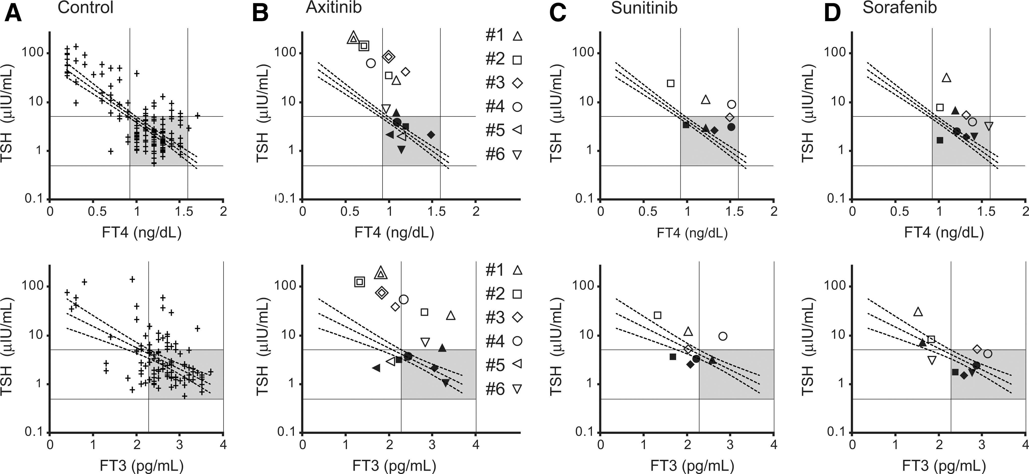
Correlation between thyrotropin (TSH) and thyroid hormone levels in control patients with normal hypothalamic-pituitary-thyroid function
Discussion
Axitinib-related thyroid dysfunction was first reported by Mukohara and colleagues in 2010 (6). Most of their patients exhibited increased TSH levels within one month of axitinib treatment, which rapidly normalized or returned to almost the upper limit of the reference range after interruption of the therapy with axitinib. A transient decrease in TSH, suggesting a thyroiditis-induced thyrotoxic phase, was also identified in some of their patients. Several subsequent studies have confirmed a high frequency of axitinib-related thyroid dysfunction, ranging from 88% to 100% (2,7,8). Consistent with earlier reports, the six patients reported here developed thyroid dysfunction. The progression of their TFTs can be classified into two patterns: elevated TSH levels early during the course of treatment (patients #1–#4 and #6), and transient thyrotoxicosis with subsequent hypothyroidism (patients #1, #2, and #4–#6). De Groot et al. and Brassard et al. reported that an increased dose of LT4 was required to maintain a normal TSH during treatment with TKIs in a thyroidectomized patient (10,11). In agreement with this finding, elevated TSH levels in four of the patients (#1–#4) reported here were resistant to LT4 treatment. Abdulrahman et al. proposed that TKIs might cause consumptive hypothyroidism via increased deiodination by type-3 iodothyronine deiodinase (DIO3) (4). Kappers et al. confirmed increased hepatic DIO3 activity in sunitinib-treated rats and hypothesized that the induction of DIO3 may result from an increased activity of hypoxia-induced factor 1, which may be upregulated because of the antiangiogenic effects of TKIs (12).
Regarding TSH changes during the early course of treatment, this is the first study to evaluate the correlation between TSH and thyroid hormone levels. The results showed that four out of six patients had inappropriate TSH elevations relative to thyroid hormone levels. Since their TFTs were shifted on the plots of TSH versus thyroid hormone, compared to data from patients with normal HPT function, an extrathyroid effect may have contributed to the inappropriately high TSH elevations, in addition to the well-known direct effects of TKIs on the thyroid gland, such as thyroid capillary regression, reduction of thyroid volume, and interference with T4/T3 metabolism (4,12,13). In mice treated with axitinib, Kamba et al. reported significant capillary regression not only in the thyroid gland but also in several other organs, including the anterior pituitary, brain cortex, and pancreatic islet cells (14).
Since feedback was impaired at the pituitary level, and because there was a concomitant resistance to TSH at the thyroid level, the axitinib-induced inappropriate TSH elevation seems to be caused by a decreased biological activity of TSH. One possible mechanism to be considered may consist in reduced thyrotropin-releasing hormone (TRH) secretion from the paraventricular hypothalamic nucleus (PVN) via decreased production of nitric oxide (NO). It is well recognized that VEGF exerts a stimulatory effect on NO production by upregulating NO synthase expression (15). On the other hand, Bains and Ferguson reported that NO specifically depolarizes parvocellular neurons within the PVN (16). As the parvocellular region of the PVN is the source of TRH, the decreased production of NO, caused by a VEGF inhibitor (e.g., axitinib), may lead to reduced secretion of TRH from the PVN, resulting in an attenuated biological activity of TSH. Uribe et al. found an inhibited TRH release in the PVN of rats treated with N(G)-nitro-
Thyroid dysfunction has also been described with other antiangiogenic agents, such as sorafenib and sunitinib, although at lower rates (1,3,8). In this study, the relationship between TSH and thyroid hormone levels was also evaluated in patients treated with sunitinib and sorafenib; the results showed a weaker tendency compared to the axitinib-treated patients. There appears to be an association between the occurrence of thyroid dysfunction and the efficacy of TKIs (20). In a recent phase III trial of patients with treatment-refractory metastatic RCC, axitinib treatment resulted in a significantly longer progression-free survival (PFS) than did sorafenib (9). Marked elevations of TSH may reflect the high efficacy rather than the unwanted toxicity of axitinib. This possibility is supported by this study, where three out of four patients (#1, #2, and #4) with inappropriate TSH elevation had better PFS than the other patients (Table 1).
It remains unclear whether all patients with TKI-related thyroid dysfunction should be treated with LT4. The serum TSH measurements cannot be used to determine the adequacy of LT4 therapy in central hypothyroidism as it can in primary hypothyroidism, because of the attenuated TSH biological activity (21). Given that TKIs can cause inappropriate TSH elevations via an impaired HPT axis, the correct dosage should be determined by clinical response and by measurements of the serum thyroid hormone with some biochemical indices of hormone action. Further studies are needed to evaluate the appropriate indices for the treatment of TKI-related thyroid dysfunction.
Footnotes
Acknowledgment
The study of axitinib was sponsored by Pfizer, Inc.
Disclosure Statement
T.T. and S.O. received research funding from Pfizer. K.O., H.M., A.M., S.S., Y.O., and H.N. declare that they have no conflicts of interest.