
Abstract
Background:
Goiter, or benign enlargement of the thyroid gland, can be asymptomatic or can cause compression of surrounding structures such as the esophagus and/or trachea. The options for medical treatment of euthyroid goiter are short-lived and are limited to thyroxine hormone suppression and radioactive iodine ablation. The objective of this statement article is to discuss optimal surgical management of goiter.
Methods:
A task force was convened by the Surgical Affairs Committee of the American Thyroid Association and was tasked with writing of this article.
Results/Conclusions:
Surgical management is recommended for goiters with compressive symptoms. Symptoms of dyspnea, orthopnea, and dysphagia are more commonly associated with thyromegaly, in particular, substernal goiters. Several studies have demonstrated improved breathing and swallowing outcomes after thyroidectomy. With careful preoperative testing and thoughtful consideration of the type of anesthesia, including the type of intubation, preparation for surgery can be optimized. In addition, planning the extent of surgery and postoperative care are necessary to achieve optimal results. Close collaboration of an experienced surgical and anesthesia team is essential for induction and reversal of anesthesia. In addition, this team must be cognizant of complications from massive goiter surgery such as bleeding, airway distress, recurrent laryngeal nerve injury, and transient hypoparathyroidism. With careful preparation and teamwork, successful thyroid surgery can be achieved.
Introduction
Statement of Clinical Problem
T
The clinical problem that is addressed in this article is the optimal surgical management of goiter. In addition, a thorough understanding of the medical management is equally important. The medical options for treatment of goiter consist primarily of iodine replacement, thyroid hormone replacement, thyroid hormone suppressive therapy, and radioactive iodine (5,6). Preoperative assessment, laboratory testing, imaging, and intraoperative and postoperative management will be discussed.
Iodine deficiency
In areas of severe iodine deficiency, iodine replacement therapy can be associated with reduction in goiter size, but this is not effective in areas of iodine sufficiency. The use of thyroid hormone (levothyroxine) may result in goiter shrinkage between 15% and 40% within about a 3-month period in patients who are iodine deficient. However, the available literature indicates that after discontinuation of thyroid hormone supplementation, the goiter can be expected to rebound to pretherapy size (7).
Thyroid hormone
Levothyroxine replacement can be used to normalize elevated TSH levels related to hypothyroidism, which can be associated with reduction in goiter size and is routinely recommended. In contrast, levothyroxine can also be used to suppress a serum TSH level below the lower limit of the normal range, which in turn causes involution of normal thyroid tissue to a much greater extent than pathological thyroid tissue; thus, the effectiveness in large goiters is limited and suppressive therapy is not routinely recommended. Previous studies on diffuse goiter, multinodular goiter, and toxic nodular goiter have reported different degrees of effectiveness. A nonrandomized study evaluating the effect of levothyroxine therapy on nontoxic multinodular goiter reported that 30% of patients achieved >50% reduction in goiter volume and another 23% achieved a 20–50% reduction in volume (5). Another study comparing the effect of levothyroxine versus radioactive iodine found only a 7% one-year and 1% two-year decrease in goiter size in 43% of subjects placed on thyroid hormone therapy in comparison to a 38% one-year and 44% two-year reduction in patients receiving radioactive iodine treatment (8). Patients receiving levothyroxine treatment had a higher rate of thyrotoxic symptoms (>30%) and a 3.6% reduction in bone mineral density at the lumbar spine compared to the group receiving radioactive iodine (6). Therefore, levothyroxine therapy is generally not recommended for long-term treatment of nontoxic multinodular goiter (7,9). Lifelong suppression is required and such long-term TSH suppression therapy is associated with increased risk for adverse side effects such as atrial fibrillation and osteoporosis—especially in postmenopausal women (7,9 –11).
Radioactive iodine
Radioactive iodine therapy has been widely utilized to reduce goiter size in patients with nontoxic multinodular goiter. Patients who are considered to be poor surgical candidates and who have goiters ranging in size from modest to large with compressive symptoms may be appropriate candidates for radioactive iodine (6,9,12). Radioactive iodine ablation has been associated with a 40–60% reduction in volume within two years of therapy, but the majority of shrinkage occurs within the first few months of treatment (9). Doses greater than 30 mCu have been used in some series. Recombinant human TSH (rhTSH) has also been successfully utilized to enhance radioactive iodine uptake in euthyroid goiters (13 –15). In addition, prestimulation with rhTSH has been shown to improve the long-term effect of radioactive iodine therapy for multinodular goiter (13) and reduce the needed dose of radioactive iodine therapy (14,16). Younger age, smaller goiter size, shorter duration of goiter, cervical goiter (rather than substernal), and higher radioactive iodine treatment dose and uptake appear to be associated with improved goiter shrinkage (17,18). Complications associated with therapy include (i) initial goiter swelling after therapy (potential 15–25% increase in size), an important issue to consider in cases where significant tracheal narrowing is present and where any exacerbation in the tracheal lumen size could be dangerous; (ii) transient thyrotoxicosis from radiation thyroiditis; (iii) subsequent development of subclinical or overt hypothyroidism (8,9,19); and (iv) the occurrence of secondary hyperthyroidism/Graves' disease and radiation-induced malignancies, which have been reported in patients receiving radioactive iodine for ablation of euthyroid goiter (8). For example, patients who have received radioiodine treatment have been reported to have an increased risk of breast cancer (20).
In summary, medical management of enlarging or symptomatic goiter may include iodine or thyroid hormone replacement for selected patients with iodine deficiency or hypothyroidism. Thyroid hormone suppressive therapy is less effective than radioiodine and associated with side effects associated with iatrogenic thyrotoxicosis. Radioactive iodine therapy may result in about a 50% volume reduction, and pretreatment with rhTSH may reduce the required dose, potentially reducing the risk of complications to other organs from radiation exposure. This use of rhTSH is an off-label use.
Indications for surgery
In contrast to medical therapy, which at best results in partial reduction in goiter volume, surgery offers the possibility of definitive treatment with removal of the goiter, although at risk of laryngeal nerve injury, hypoparathyroidism, and bleeding or hematoma. Indications for surgery include symptoms related to compression of the trachea or esophagus (dyspnea and/or dysphagia), suspicion for malignancy, prevention of complication from progressive enlargement or mediastinal extension (tracheal narrowing, superior vena cava syndrome), and occasionally for cosmesis.
Discussion
A. Preoperative Assessment
The symptoms and signs associated with a goiter depend on the size and location of the goiter. Direct compression of the trachea or esophagus by the goiter (most commonly by substernal goiter) can cause worsening dysphagia to solids, positional dyspnea, and dysphonia. Shin et al. reported positive correlations between thyroid size and presence of shortness of breath and globus sensation (21). Other conditions in the differential diagnosis for goiter with similar symptoms include upper aerodigestive tract disorders such as neoplasms, acid reflux, dysmotility, diverticulum, stricture, and allergy/asthma, among others.
Dyspnea
Shen et al. found in a retrospective review of 60 substernal goiter patients that dyspnea (59%) was the most common symptom, occurring as orthopnea in 22% of patients and as shortness of breath in 37% (22). Similarly, Shin et al. reported 50% of patients having shortness of breath (21). Using a unique prospective management algorithm, Stang et al. found that positional dyspnea, defined as difficulty breathing at rest improved by position change, was reported by 10.8% of patients presenting for thyroid surgical evaluation and 18% of patients who then underwent thyroidectomy for any reason (23). Furthermore, Stang et al. reported that positional dyspnea is more commonly associated with substernal goiters (75.5%), and is highly associated with tracheal compression on cross-sectional imaging (23). There is also a high prevalence of orthopnea with substernal goiters as measured by a reduction of end expiratory lung volume and flow reserve. In addition, obesity compounds this effect (24).
Thyroidectomy is effective in relieving positional dyspnea (23,25,26). Stang et al. systematically examined response to thyroidectomy for goiter and found that preoperative positional dyspnea was improved or resolved in 82.4% of cases overall, in 65% of patients with cervical goiter, and in 88% of patients with substernal extension (23). A resected gland weight >100 g was associated with improvement or resolution in 97.9% of total thyroidectomy patients. For unilateral goiter, a resected lobe weight >75 g was associated with response in 100% of patients. One study reported the successful management of pulmonary hypertension and heart failure caused by long-standing substernal goiter by performing a thyroidectomy (27).
Dysphagia
Dysphagia, that is, difficulty swallowing solids, is commonly associated with goitrous compression, but is also common in the elderly population for a variety of reasons and may ultimately require endoscopy or a swallow study to evaluate. Completing a thorough history of the patient's dysphagia symptoms may help discriminate extrinsic compression of the goiter from other causes of dysphagia (Fig. 1), such as laryngopharyngeal reflux. Similar to dyspnea, dysphagia is more commonly seen in substernal than in cervical goiters (21). Hedayati and McHenry reported that among 116 patients undergoing surgery for substernal goiter, 32% presented with dysphagia (1). In a retrospective review of 60 patients operated on for substernal goiter, dysphagia was the second most common symptom (43%) after dyspnea (22). Thyroidectomy appears to improve dysphagia, but the data are mixed and are measured with qualitative parameters. For example, Lombardi et al. reported that among 257 individuals, 47% had dysphagia, which increased to 74% at 1 week postoperatively and then decreased to 20% at 1 year postoperatively for a p-value of <0.001 (28). Greenblatt et al. reported a qualitative improvement of swallowing function after thyroidectomy using a validated swallowing quality-of-life questionnaire, the SWAL-QOL (29). Of 146 eligible patients, 116 (79%) completed the study. Mean preoperative scores were below 90 for 9/11 domains. Postoperatively, there was significant improvement in eight of the domains. However, if recurrent laryngeal nerve injury was evident, there was a dramatic decrease in the score. Thus, among patients with uncomplicated thyroidectomy, self-reported swallowing scores were improved (29).
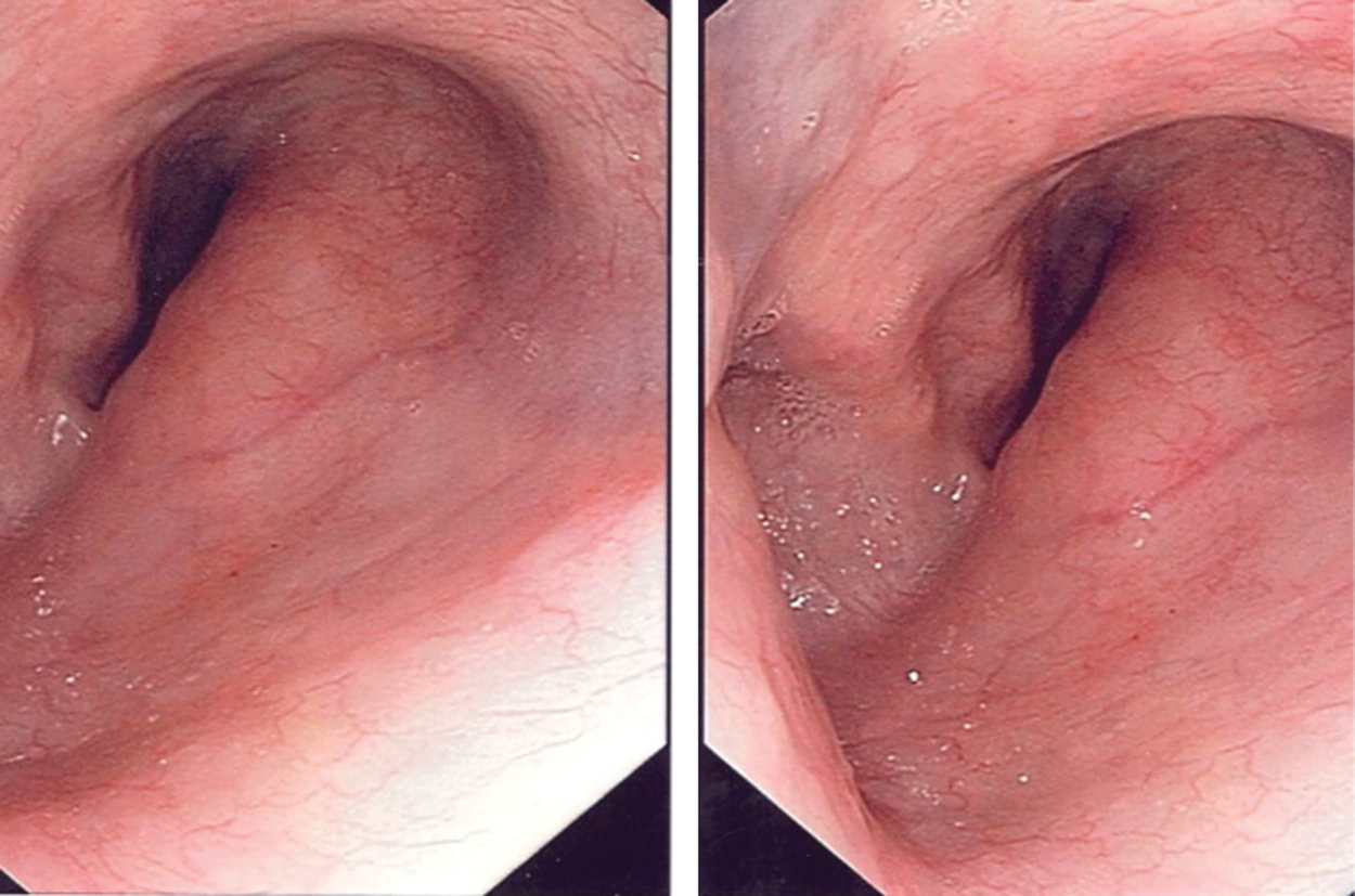
Esophageal compression.
Dysphonia
Hoarseness or dysphonia is a change in quality or volume of voice. Preoperative laryngoscopy to assess laryngeal nerve function should be considered for patients undergoing thyroidectomy for goiter, especially in the setting of prior thyroid or other anterior neck surgery (30). Indirect or fiberoptic laryngoscopy should be performed for patients with dysphonia to evaluate laryngeal nerve function and/or causes such as laryngeal neoplasm or otherwise.
B. Signs on Physical Examination
A goiter may be simple with diffuse enlargement or with a uninodular or multinodular pattern. Goiter has been graded by the World Health Organization in the following fashion: Grade 0: Impalpable/invisible Grade 1a: Palpable but invisible even in full extension Grade 1b: Palpable in neutral position/visible in extension Grade 2: Visible but no palpation required to make diagnosis Grade 3: Visible at a distance
Substernal goiter may be diagnosed on physical examination by detection of caudal extension of the thyroid gland below the clavicles or sternal notch. Both sitting and supine, the inferior thyroid pole normally lies cranial to the thoracic inlet. With the patient lying supine in mild neck extension, substernal extension is suggested by an impalpable lower thyroid lobe(s) edge or by bulky thyroid tissue that extends caudally under the sternal notch; that is, even with swallowing, the lower lobe edge cannot be detected in the neck (23). However, White et al. reported that 20–30% of substernal goiters were not palpable in the neck or were barely palpable, especially if the extension is posterior (31). In patients with obesity or other causes of thick neck tissue and in patients with kyphoscoliosis or other causes of poor neck extension, it can be difficult to palpate the lower extent of the thyroid gland; in this situation, cross-sectional imaging may be necessary to determine if inferior extension is present.
Substernal goiter can compress the intrathoracic great vessels. Superior vena cava syndrome occurs as facial or neck venous engorgement at rest with a prevalence in substernal goiter of 5–9% (22,32). The presence of a goiter compressing the great vessels can be identified by having the patient perform Pemberton's maneuver, where the patient raises his or her arms above the head for several minutes (33 –35). This position results in narrowing of the thoracic outlet and a large goiter will inhibit venous return, causing the patient's face to turn reddish blue, which resolves when the arms are lowered. In addition, patients may develop inspiratory stridor. Furthermore, some patients with superior vena cava syndrome will have massive venous collaterals in their upper chest and neck. Cross-sectional imaging of the chest should be done in these individuals.
C. Thyroid Function and Laboratory Testing
TSH determination is an obligatory initial step in goiter evaluation and management to evaluate for thyroid dysfunction. Free thyroxine and triiodothyronine levels can be obtained in patients with a suppressed TSH level. Antibodies to the TSH receptor, thyroid peroxidase, and thyroglobulin may be measured to help elucidate the etiology of thyroid dysfunction if autoimmune disease is suspected. Further, chronic thyroiditis may be a factor complicating thyroid surgery, especially in cervical delivery of a substernal goiter. Measuring serum thyroglobulin does not discriminate benign from malignant disease and is not recommended. Calcitonin measurement can be considered if there is any concern for potential risk of medullary thyroid cancer because of a positive family history, genetic testing, or a suspicious cytology. False-positive calcitonin elevations can be seen in Hashimoto's disease, chronic renal failure, and hypercalcemia, among others (36 –38).
D. Imaging Tests
To evaluate any kind of thyromegaly, thyroid ultrasonography is the standard practice in North America (39). Ultrasonography using high-resolution transducers is the most sensitive method available of assessing gland architecture and detecting nodules (40). Ultrasonography should be used in any patient with a nodular thyroid in order to determine the size of the nodules, the number of nodules, and the need for fine-needle aspiration (FNA) biopsy (39). Certain ultrasonographic features are associated with increased likelihood of malignancy but are less specific on an individual basis (41). These features include microcalcifications, irregular margins, hypoechogenicity, extrathyroidal extension, tall shape, absence of halo, hypervascularity, and abnormal lymph nodes (32,42).
In addition, ultrasonography is useful to follow the size of thyroid nodules over time and to assess for any suspicious lymph nodes. A significant increase in thyroid nodule size, defined as greater than 50% increase in volume, is an indication for FNA sampling/re-sampling of a thyroid nodule (39). Ultrasonography is also utilized for FNA, ensuring optimal placement of the needle tip for nodule sampling.
A chest radiograph is not routinely used to image the thyroid gland, but it can be the first modality indicating a substernal goiter. On chest radiography, substernal goiter is usually seen as a mass associated with tracheal narrowing, tracheal deviation, or superior mediastinal widening (Fig. 2).
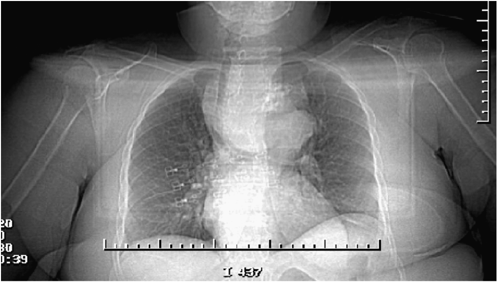
Chest radiograph demonstrating deviation of trachea to the right.
Because ultrasonography is limited in cases with substernal extension, cross-sectional imaging should be considered and is frequently helpful, especially in assessing for mass effect on the trachea and determining the diameter of the trachea. Computed tomography (CT) scan imaging may also help exclude nodal disease and other signs of malignancy such as irregular borders or microcalcifications (21). CT scan imaging for suspected substernal goiter can quantify the caudal extent, show the 3D shape, define any tracheal compression, and pinpoint the mediastinal compartment(s) that are occupied by goiter (anterior/posterior; Fig. 3a, b) (43,44). Noncontrast neck CT scans are usually sufficient unless a goiter is found to extend caudally past the lower edge of the aortic arch. It is helpful to request evaluation of the substernal extent of goiter and degree of tracheal narrowing if present. However, if contrast is given, along with its high iodine content, surgery should be completed within three to four weeks to avoid precipitating a hyperthyroid state (45,46). Intravenous contrast should be avoided in patients with thyroid nodules suspicious for thyroid cancer of follicular cell origin as it will delay radioactive iodine therapy because of the high iodine content.
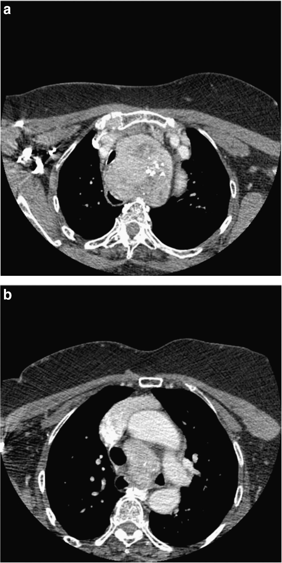
Tracheal compression is described as the greatest percent reduction in tracheal diameter and is more common in substernal than in cervical goiter. Stang et al. found that tracheal narrowing was present in 8.0% of patients presenting for surgical thyroid evaluation and in 13% of patients undergoing thyroidectomy for any reason (23), with between 35% and 97% of substernal goiter demonstrating evidence of tracheal compression on cross-sectional imaging (22,23). Whereas an earlier report recommended thyroidectomy for any degree of luminal narrowing on CT imaging (47), a more recent study found that positional dyspnea improvement or resolution after surgery for substernal goiter varied stepwise with the degree of preoperative tracheal compression (23). With a clear inflection point at 35%, the authors reported that <35% narrowing was associated with 65–70% likelihood of resolution of positional dyspnea, whereas ≥35% narrowing was associated with 95–98% chance of resolution after resection (23). On the basis of the analysis of a prospective management strategy, the authors proposed using ≥35% tracheal compression as a routine indication for substernal goiter resection (23). Surgery for patients with smaller degrees of compression may be reasonable for symptomatic patients, or for younger patients to prevent progression. Asymptomatic, elderly, and/or medically infirm patients may be followed for evidence of progression. Evidence of tracheal compression does not necessitate median sternotomy, most commonly as a partial procedure; in fact, most studies report only a 1% rate of sternotomy (47 –52).
E. Other Testing
FNA biopsy is the standard of care for nodules meeting size and ultrasonography characteristics (39,53). Large-gauge (Tru-cut) needle sampling produces tissue rather than just cells and may occasionally be indicated where fine needle fails to provide sufficient sampling in lesions with rapid growth, invasion, or pain that can suggest anaplastic thyroid cancer or lymphoma, and is useful in such cases as the optimal initial treatment is often not surgical.
F. Intraoperative Management
The surgical management of large goiters and substernal goiters is complex. Surgical outcomes can be optimized when thyroidectomy is performed by an experienced surgeon who performs thyroidectomy in patients with large goiters on a regular basis. At least two studies have reported that improved outcomes are associated with high-volume teams and centers (54,55). Success revolves around issues related to smooth process of the surgical procedure, appropriate extent of thyroidectomy, good communication with the anesthesia team, and avoiding injury to the recurrent laryngeal nerve and parathyroid glands. Both intraoperative and postoperative complications can be serious. Appropriate preparation and evaluation of the extent of the disease, airway status, and medical condition is crucial.
Intubation
The majority of patients with large goiters, including those with tracheal deviation, can be easily intubated since the larynx itself is merely displaced. It is the trachea that may get compressed. Intubation of these patients should be undertaken by an experienced anesthesiologist who is familiar with complex airway management. A partnership between the surgeon and anesthesiologist is essential for successful and safe intubation. Suggested available equipment for management of a difficult airway include a rigid bronchoscope, fiberoptic intubation equipment, video laryngoscopes, and a laryngeal mask airway (through which a fiberoptic intubation can be performed). There are situations where it may be beneficial for the surgeon to be in the room during induction and to assist as necessary. Emergent tracheotomy may be difficult depending upon the location of the goiter. However, most patients with substernal goiters can be intubated without difficulty.
Fiberoptic intubation is primarily helpful when laryngeal exposure is expected to be difficult, due to parapharyngeal involvement with goiter and/or obesity. Severe tracheal compression causing stridor is concerning for an inability to pass the endotracheal tube into the trachea, which is not facilitated by fiberoptic intubation. Having the patient be awake during fiberoptic intubation may be helpful in this situation. The use of a smaller-diameter endotracheal tube may also be helpful. Fiberoptic intubation should be performed by an experienced person. The patient is most commonly awake in an upright seated position so as to maintain his or her own airway. Awake fiberoptic intubation can be carried out via the nasal route or the oral route, with the upper airway adequately anesthetized with topical lidocaine. Again, the surgeon should be immediately available to assist, and the surgical team should be prepared and ready to perform an emergent tracheotomy or rigid bronchoscopy if necessary. Use of a nerve-monitoring endotracheal tube can be considered, although there is no consensus that nerve monitoring decreases complications in the setting of thyroidectomy for goiters (56).
Further, current commercially available endotracheal nerve-monitoring endotracheal tubes are less rigid and have a greater outer diameter than traditional tubes. There are certain situations where nerve monitoring may be useful. For example, after removal of one lobe of a large goiter, one can verify the function of the ipsilateral recurrent laryngeal nerve before moving to do the contralateral thyroid lobe resection. In the event that a nerve signal is lost, consideration can be given to abort and/or stage the contralateral lobectomy.
Preoperative thoracic consultation
The majority of substernal goiters can be easily retrieved through the neck because their blood supply can be controlled in the neck. However, in a small number of cases, it will be important to have a thoracic surgeon on surgical standby. These situations include (i) potential fibrosis or scarring from prior radiation or surgery involving the neck or chest; (ii) suspected malignancy with extrathyroidal extension or invasion; (iii) inferior extension below the arch of the aorta abutting the carina, or involving the posterior mediastinum; and (iv) ectopic mediastinal goiter not connected to the cervical component.
It is important to brief the thoracic surgeon before surgical intervention and that the chest is prepped and draped in case of difficult retrieval of the substernal component or unexpected hemorrhage in the chest. The patient should be well informed of this situation. If the possibility of sternal split is high, a formal consultation should be arranged preoperatively so that the patient is well informed about the sternotomy and its potential complications. If the substernal extension of the goiter is in the posterior mediastinum and cannot be removed through a cervical approach, such patients will require a posterolateral thoracotomy.
Pearls of surgical technique
Even with a large substernal goiter it is important to recognize that the recurrent laryngeal nerve is generally in its normal position, which can be identified carefully in the tracheoesophageal groove (Fig. 4). However, in a posterior mediastinal goiter the recurrent laryngeal nerve may be displaced anterior to the posterior extension of the thyroid. The resulting location can result in a higher risk of injury, especially if not recognized. In general, in large bilateral goiters, a total thyroidectomy is considered rather than subtotal resection. However, a thyroid lobe may be preserved if there is concern about the recurrent laryngeal nerve on the ipsilateral side or for parathyroid preservation. Total thyroidectomy is usually carried out in patients with bilateral goiters, but if the substernal goiter involves only one lobe of the thyroid gland and the contralateral lobe is essentially normal, a lobectomy may be considered to relieve tracheal compression with lower risk of complications (47).
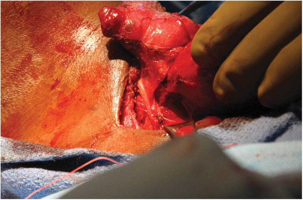
Recurrent laryngeal nerve seen in the right tracheoesophageal groove.
In large goiters, the anterior cervical veins may be congested. In addition, mediastinal vessels such as the internal mammary artery, the thyroidea ima artery, and the innominate vein may be enlarged. To avoid blood loss, a basic principle of goiter resection is prompt and direct control of the superior and inferior vascular pedicles, which are typically surprisingly straightforward to locate in their usual positions. In addition to engorgement of conventional vessels, most large goiters also have additional enlarged arteries and veins, which also require control.
Massive hemorrhage can occur in the delivery of a large substernal goiter, and a plan should be in place to address such a scenario, which may include type and crossmatch, packing the surgical site with lap sponges, using a Foley catheter with a large balloon, clamping and ligating the inferior thyroid pole first before delivering the substernal goiter, and consulting another surgeon to help with exposure and control of hemorrhage.
Delivery of a substernal goiter through a cervical incision can be accomplished with blunt dissection, sharp dissection, and/or mobilization with an instrument such as a spoon or Deaver retractor. Placing sutures, Babcock forceps, or Allis clamps sequentially into the already-delivered thyroid tissue can help hoist the remainder of the substernal component out of the chest. This delivery must be accomplished first before the recurrent laryngeal nerve can be identified. Commercial hemostatic devices may help to cauterize and cut vessels from the lateral side. A drain in the resection bed can be considered at the conclusion of the case, especially if obesity or kyphoscoliosis would make a cervical hematoma difficult to detect on physical examination, or in cases involving a significant dead space or oozing after resection. However, the placement of a drain has never been shown to reduce the risk of life-threatening neck hematoma.
The operative report should reflect the type of intubation as well as the extent of surgery, and any unexpected complications (57).
Extubation
During emergence and subsequent extubation, excellent communication between the surgeon and the anesthesiologist is essential. Ideally, the patient should be extubated in the operating room and observed to ensure that there are no airway issues or stridor and then proceed to the recovery room. The surgeon should be immediately available and prepared to manage any postoperative airway complications such as stridor or obstruction before leaving the operating room. Bilateral recurrent laryngeal nerve injury may lead to vocal cord paresis on both sides leading to acute airway issues. These patients need close observation, and if there is any airway problem such as stridor, the best option is to evaluate the vocal cords with fiberoptic endoscopy and reintubation. Tracheostomy can usually be avoided until re-evaluation of the patient 24–48 hours later. Strong consideration should be given to observing patients for a period of time after resection of a large goiter.
Complications
Complications related to surgery for large substernal goiter are essentially similar to those of standard thyroid surgery. These include bleeding, recurrent laryngeal nerve injury, hoarseness of voice, inability to raise the voice, and temporary or permanent hypoparathyroidism. The incidence of these complications is slightly higher in goiter patients who require extensive dissection and where the status of the parathyroid glands may be difficult to appreciate or recognize because of the large size of the thyroid gland (32,51,58). Wound infection is uncommon. Intraoperative bleeding may be directly related to venous congestion. Detection of hematoma is accomplished by physical examination of the neck (palpation and visualization) performed at regular intervals postoperatively, for example, the night of surgery and the next morning. Floor nurses should be educated on the signs and symptoms of a cervical hematoma. Rarely, pneumothorax may occur and new onset dyspnea or oxygen requirements postoperatively may require a chest film for evaluation.
Tracheomalacia appears to be a well-described condition in patients with large goiter and tracheal compression. However, in current practice, severe tracheomalacia requiring tracheal resection is a rare condition and is usually symptomatic with stridor preoperatively and found in the very largest of goiters (49,59). At the time of surgery, the trachea may be deviated considerably and compressed. However, true weakening of the tracheal wall is very rare and disintegration of the cartilage rings of the trachea is extremely rare. If new onset stridor is evident postoperatively, excluding recurrent laryngeal nerve injury (either unilateral or bilateral) is of utmost priority. Special management of tracheomalacia such as Silastic rings or tracheopexy is rarely necessary in patients with large goiters.
If the parathyroid glands are devascularized during the surgical procedure, frozen-section examination of a portion can be utilized to confirm the presence of parathyroid tissue and then local parathyroid autoimplantation into neck muscle can be performed. There is no need to implant the parathyroid in the forearm. Careful attention must be paid to the calcium levels postoperatively. Obtaining parathyroid hormone in the postoperative period can help with determining the need for postoperative calcium supplementation with or without postoperative calcitriol and the safety of discharge (60,61).
It is also important to start the patient on levothyroxine postoperatively. The usual dose is 1.4–1.6 μg/kg per day (of actual body weight), with plans for TSH testing 4–6 weeks postoperatively and titration as needed. Patient education at the time of discharge should include signs/symptoms of hypocalcemia, hematoma, infection, or airway distress. A follow-up appointment with the surgeon and/or endocrinologist should be included as well.
Conclusions
William Halstead has stated that “the extirpation of the thyroid gland for goiter typifies better than any operation the supreme triumph of the surgeon's art” (62). Goiter, or benign enlargement of the thyroid gland, can be asymptomatic or can cause symptoms related to compression of surrounding structures such as the esophagus and/or trachea. Iodine or thyroid hormone replacement may be helpful in iodine deficiency or hypothyroidism. The options for medical treatment of euthyroid goiter are short-lived and are limited to thyroxine hormone suppression and radioactive iodine ablation. Newer methods of radioactive iodine ablation include prestimulation with rhTSH (currently off-label use) and may result in reduced dosage and less side effects, such as hypothyroidism.
However, for goiters with compressive symptoms, surgical management is recommended. Symptoms of dyspnea, orthopnea, and dysphagia are more commonly associated with thyromegaly, in particular, substernal goiters. Several studies have demonstrated improved breathing and swallowing outcomes after thyroidectomy.
Preoperative testing includes thyroid function assays and imaging (ultrasonography and/or cross-sectional imaging). Thoughtful consideration of the type of anesthesia, type of intubation, extent of surgery, and postoperative care is necessary to achieve optimal results. Close collaboration of an experienced surgical and anesthesia team is essential for induction and reversal of anesthesia. In addition, this team must be cognizant of complications from massive goiter surgery such as bleeding, airway distress, recurrent laryngeal nerve injury, and transient hypoparathyroidism. With careful preparation and teamwork, successful thyroid surgery can be achieved.