
Abstract
Background:
There remain a small number of patients with papillary thyroid cancer (PTC) who suffer recurrence, metastases, or death. While mutation of the BRAF gene, corresponding to the constitutively active BRAFV600E protein, has been associated with worse clinical outcomes in thyroid cancer, the reasons underlying this observation are presently unknown. Disruption of endogenous host immune surveillance and promotion of tumor immune escape is one mechanism by which BRAFV600E tumors may achieve more aggressive behavior. This study evaluated the relationship between BRAFV600E status and known strategies of tumor-mediated immune suppression.
Methods:
Tissue sections of PTC tumors from 33 patients were evaluated by immunohistochemistry for tumor-expressed suppressive ligands and enzymes and effector and suppressor populations of tumor-infiltrating immune cells. Presence of BRAFV600E was evaluated by direct DNA sequencing of PTC specimens and the results correlated with tumor-expressed molecules and tumor-infiltrating immune cell populations, as well as patient characteristics and pathologic findings.
Results:
BRAFV600E tumors more often express high levels of immunosuppressive ligands programmed death ligand 1 (53% vs. 12.5%) and human leukocyte antigen G (41% vs. 12.5%) compared to BRAF wild-type tumors. There was no association between indoleamine 2,3-dioxygenase 1 expression and BRAFV600E status. Furthermore, BRAFV600E tumors demonstrate both lower CD8+ effector to FoxP3+ regulatory T cell, and CD68+ pan-macrophage to CD163+ M2 macrophage ratios, indicating relative increases in suppressive T cell and macrophage components, respectively.
Conclusions:
Overall, BRAFV600E PTC tumors display a broadly immunosuppressive profile and evidence of disturbed host tumor immune surveillance that may contribute to the poorer outcomes observed in this subset of patients with thyroid cancer.
Introduction
T
Along with clinical features that portend a worse prognosis such as age, tumor invasiveness, and metastasis (6), certain genetic mutations may influence thyroid cancer progression. One mutation in the v-Raf murine sarcoma viral oncogene homolog B1 (BRAF) gene, corresponding to the constitutively active BRAFV600E protein, is present in approximately 45% of PTC (9 –11) and has been associated with greater tumor size and invasion, lymph node metastasis, recurrence, and mortality (9 –13), although not in all studies (14,15).
The mechanisms underlying aggressive PTC continue to be elucidated and may include tumor-mediated immune suppression. It is now established that host immune cells can recognize and eliminate malignant cells, but that cancers evolve mechanisms to escape immune destruction, including downregulation of antigen recognition, expression of immune-inhibitory ligands, and recruitment of suppressor immune cell populations. Both the expression of immunosuppressive molecules and specific patterns of tumor infiltrating leukocytes (TIL) have been shown to predict cancer recurrence and survival in solid malignancies (16 –22).
Recently, studies have shown that the BRAFV600E protein mutation is associated with immunosuppressive mechanisms in PTC, such as indoleamine 2,3-dioxygenase 1 (IDO) (23), human leukocyte antigen G (HLA-G) (24), and programmed death ligand 1 (PD-L1) expression (25). Studies of TIL in other cancer types have shown that specific patterns predict cancer behavior, such as the correlation between a low CD8+ effector T cell to FoxP3+ suppressor T cell ratio with greater tumor aggressiveness and worse patient outcomes (17,19 –21). Whether or not these mechanisms of immune suppression occur in isolation or as part of a broader pattern of immune suppression in BRAFV600E PTC has not been well studied. We hypothesized that BRAFV600E is associated with a general suppressive immune profile and disruption of host tumor immune surveillance that could help explain the poorer outcomes observed in this subset of patients with thyroid cancer.
Materials and Methods
Tissue samples
PTC specimens from patients with thyroid cancer with anonymized clinical data were obtained from the University of Southern California (USC) Keck Medical Center Tissue Bank (Institutional Review Board [IRB] protocol HS-11-00215). A pathologist specializing in thyroid cancer reviewed each case to confirm the diagnosis of PTC. Contralateral lobe normal tissue was collected and evaluated in parallel as control tissue when available.
Immunohistochemistry
Formalin-fixed paraffin-embedded (FFPE) tissue sections were deparaffinized, rehydrated, and subjected to heat-induced antigen retrieval (0.01 M citrate, pH 6.0) followed by treatment with 3% H2O2 for 10 minutes to block endogenous peroxidase activity. Sections were incubated overnight at 4°C with primary antibodies against CD3 (PC3/188; Santa Cruz Biotechnology, Santa Cruz, CA), CD8 (C8/144B, Dako, Carpinteria, CA), CD68 (PG-M1, Dako), CD163 (10D6, Abcam, Cambridge, MA), Arginase-1 (H-52, Santa Cruz), FoxP3 (236A/E7, Novus, Littleton, CO), HLA-G (4H84, Santa Cruz), PD-L1 (4059, ProSci, Inc., Poway, CA), or IDO (ab55305, Abcam). Secondary antibody staining and antigen detection with 3,3'-diaminobenzidine was performed using a Vectastain ABC kit (Vector Laboratories, Burlingame, CA). Sections were counterstained with hematoxylin, dehydrated, and mounted. Antibodies for detection of immune cell populations and markers of tumor immune suppression used in this study were previously validated by Russell et al. (19). Positive controls for immunohistochemistry analyses included lymph node or spleen tissues from healthy patients and/or patients with cancer with active disease for immune cell populations (CD3, CD8, CD68, CD163), liver parenchyma (Arg-1), human placental tissues (HLA-G, PD-L1, IDO), and head and neck carcinomas with known overexpression or loss of classic and nonclassic major histocompatibility complex (MHC) class I molecules (19). Negative controls included immunohistochemistry (IHC) using isotype control primary antibodies, secondary antibody in the absence of primary antibody, and known negative tissues for target antigens, including nonlymphoid tissues and lymphoid tissues from healthy donors obtained under IRB-approved protocol HS-11-00215. Correct cellular localization of positive staining was confirmed by pathologists A.L.E. and A.J.C. Hematoxylin and eosin (H&E)-stained sections were provided by the Keck Medical Center Translational Pathology Core.
Scoring of immune markers
An immune infiltrate scoring system to evaluate cancer specimens previously developed in our laboratory was adapted for PTC (19). Areas of tumor and associated TIL intratumorally or at the invading margin were identified on H&E-stained sections, with areas of obvious lymphoid follicle arrangement, necrosis, or hemorrhage excluded. Positively stained leukocytes for CD3, CD8, CD68, FoxP3, CD163, or Arginase-1 were counted in five representative high-powered fields (hpf) for each tumor section (hpf; 400× magnification). Tumor expression of HLA-G, IDO, or PD-L1 was assessed qualitatively as low, moderate, or high. Two independent observers scored each section and the results were pooled, with rare disagreements resolved by a third evaluator.
BRAF mutational analysis
Tumor DNA was isolated from FFPE sections by excision of tumor tissue and DNA purification performed using a Qiagen QIAmp FFPE kit (Qiagen, Valencia, CA). Human BRAF exon 15 was amplified by PCR (forward: TCATAATGCTTGCTCTGATAGGA, reverse: GGCCAAAAATTTA ATCAGTGGA) as described previously (26). PCR DNA amplicons electrophoresed on 1.5% agarose were extracted and purified using a Qiagen MiniElude Gel Extraction kit and sequenced at the USC Genomics Core Facility. Tumor DNA sequences were evaluated for the c.1799T>A mutation in the BRAF gene (Fig. 1) encoding the BRAFV600E mutant protein. Given the rarity and uncertain significance of other BRAF mutation, cases without a BRAF 1799T>A transversion were considered wild-type (BRAFWT).
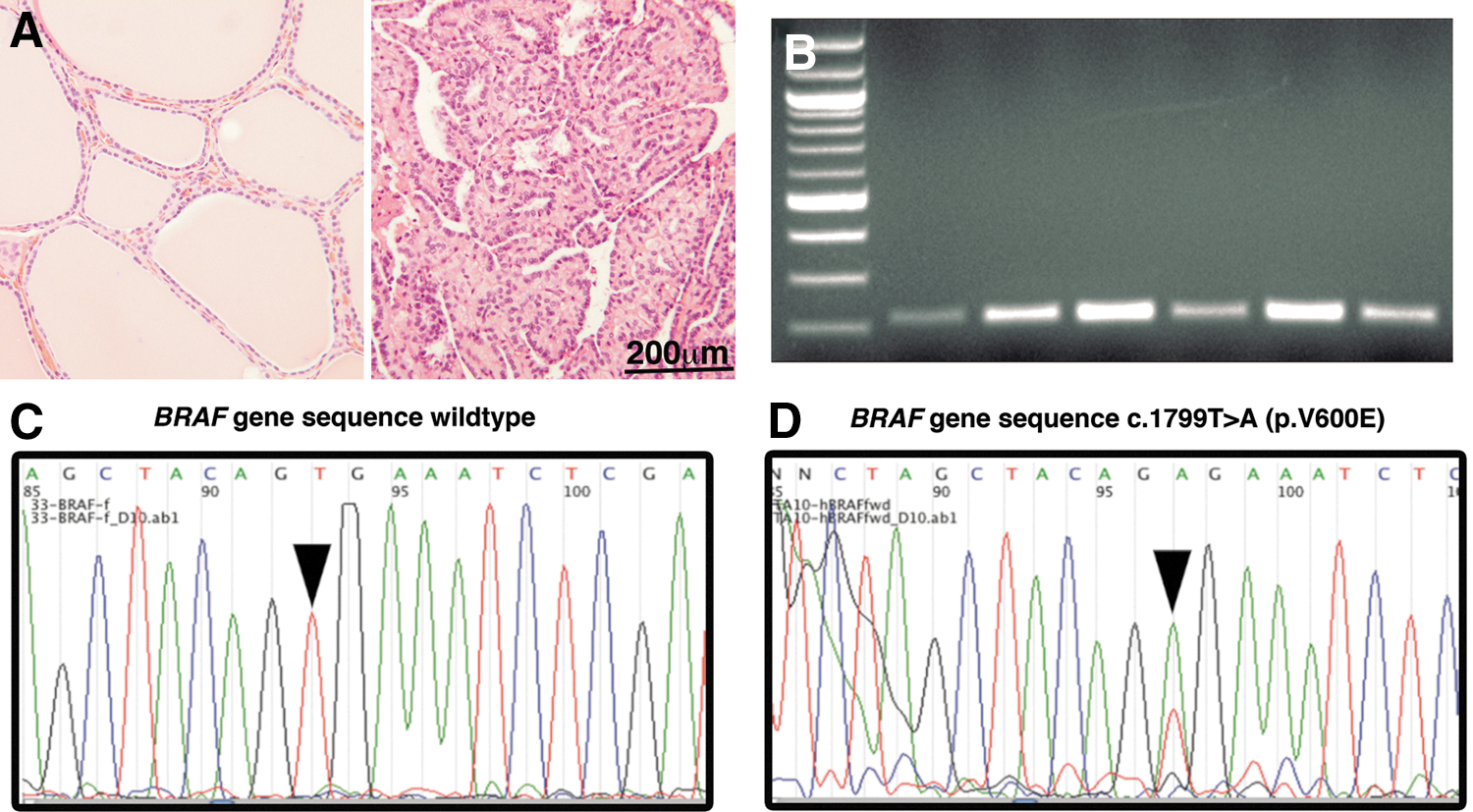
Assessment of BRAFV600E in papillary thyroid carcinoma specimens by polymerase chain reaction (PCR) and nucleotide sequencing. Hematoxylin and eosin staining of thyroid tissue is shown for
Statistical methods
Data are shown as mean±standard error of the mean (SEM) unless otherwise stated. High expression of immunosuppressive markers was defined as the highest quartile of qualitative scores and the correlation with BRAF status analyzed using the Fisher's exact test. The two-sided unpaired Student's t test was used to compare the mean positively stained immune cells per hpf, or the mean value for the CD8+/FoxP3+ or CD68+/CD163+ ratios, respectively, in BRAFV600E and BRAFWT samples. Regression analyses were performed to evaluate markers of immune suppression as predictors of BRAF status, and to evaluate these immune suppression markers in combination with BRAF status as predictors of lymph node metastasis, TNM stage, or tumor invasion, using SPSS Statistics 21.0 (IBM SPSS, Armonk, NY). All other statistical tests were performed using GraphPad Prism 6.0 software (La Jolla, CA) and figures were produced using GraphPad and Abobe Photoshop (San Jose, CA).
Results
Patient characteristics
Tumor specimens from 33 patients with PTC were retrospectively obtained and analyzed. Clinical and pathologic data are summarized in Table 1. Female patients constituted 87.8% (29/33) of the samples. The median age of the patients was 49 years (range, 22–75), with 30.3% (10/33) being younger than 45 years of age. Primary tumors were noninvasive (TNM 1 or 2) in 26 (78.8%) and invasive (TNM 3 or 4) in 7 (21.2%) cases. In these studies, 17 of 33 (51.5%) tumor samples were positive for the BRAF mutation. Lymph node metastases at initial surgery were present in 12 of 33 (35.2%) patients, 9 of whom were in the BRAFV600E group compared to 3 in BRAFWT group (p=0.07). Other than this trend, presence of BRAFV600E was not associated with tumor size, invasion, or any demographic parameter. No patient had known distant metastases at the time of initial surgery.
Fisher's exact test except as indicated.
Unpaired Student's t-test.
BRAFV600E mutation is associated with the suppressive immune molecules PD-L1 and HLA-G
The BRAFV600E mutation was significantly associated with increased expression of immunosuppressive molecules by PTC cells (Fig. 2). PD-L1 staining showed high expression in 9 of 17 (53%) BRAFV600E specimens, compared with only 1 of 16 (12.5%) BRAFWT tumors (p<0.01). Similarly, 41% of BRAFV600E tumors were positive for HLA-G, whereas only 12.5% were positive in BRAFWT specimens (p<0.05). High IDO expression was more common in BRAFV600E specimens but the difference was not significant.
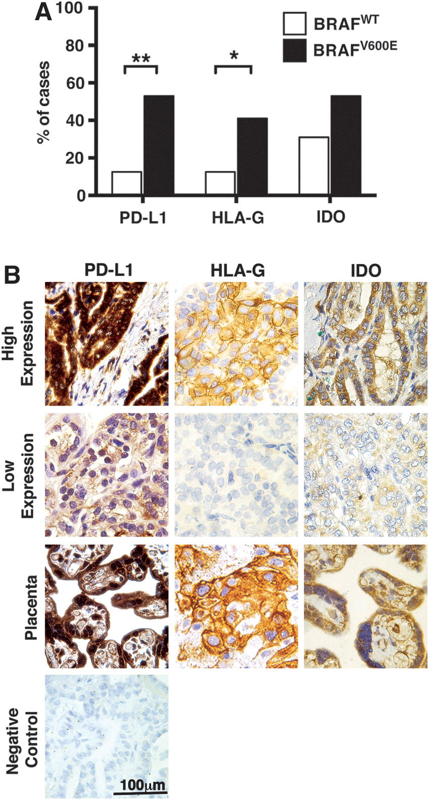
Expression of immunosuppressive mechanisms in BRAFV600E papillary thyroid cancer specimens.
Immune infiltrates in papillary thyroid carcinoma
The frequency and quality of tumor infiltrating effector and suppressor immune cell populations was assessed in tumor sections by IHC, as shown in Figure 3. There was a trend toward greater overall T cell infiltration, measured by intratumoral CD3+ cells, in BRAFV600E tumors compared to BRAFWT (p=0.12). While there was a trend toward increased FoxP3+ Treg cells/hpf in BRAFV600E cases, when FoxP3+ cells were measured in relation to intratumoral effector CD8+ T cells, by calculating a CD8+/FoxP3+ cell ratio, there was a signficantly lower CD8+/FoxP3+ cell ratio in BRAFV600E compared to BRAFWT tumors (8.67±2.23 vs. 30.32±8.84, respectively [p<0.05]). Similarly, while neither the mean number of CD68+ (pan-macrophage) nor CD163+ (type M2 macrophage or tumor-associated macrophages [TAM]) immune cell populations varied significantly between groups, a trend toward a lower mean CD68+/CD163+ cell ratio was seen in BRAFV600E versus BRAFWT tumors, 1.49±0.28 versus 3.41±1.41, respectively (p=0.1). Measurement of arginase-1+ myeloid populations, which includes TAM and MDSC, revealed significantly greater intratumoral accumulation of these cells in BRAFV600E versus BRAFWT tumors (3.46±0.67 vs. 1.53±0.35 cells/hpf, respectively [p<0.05]).
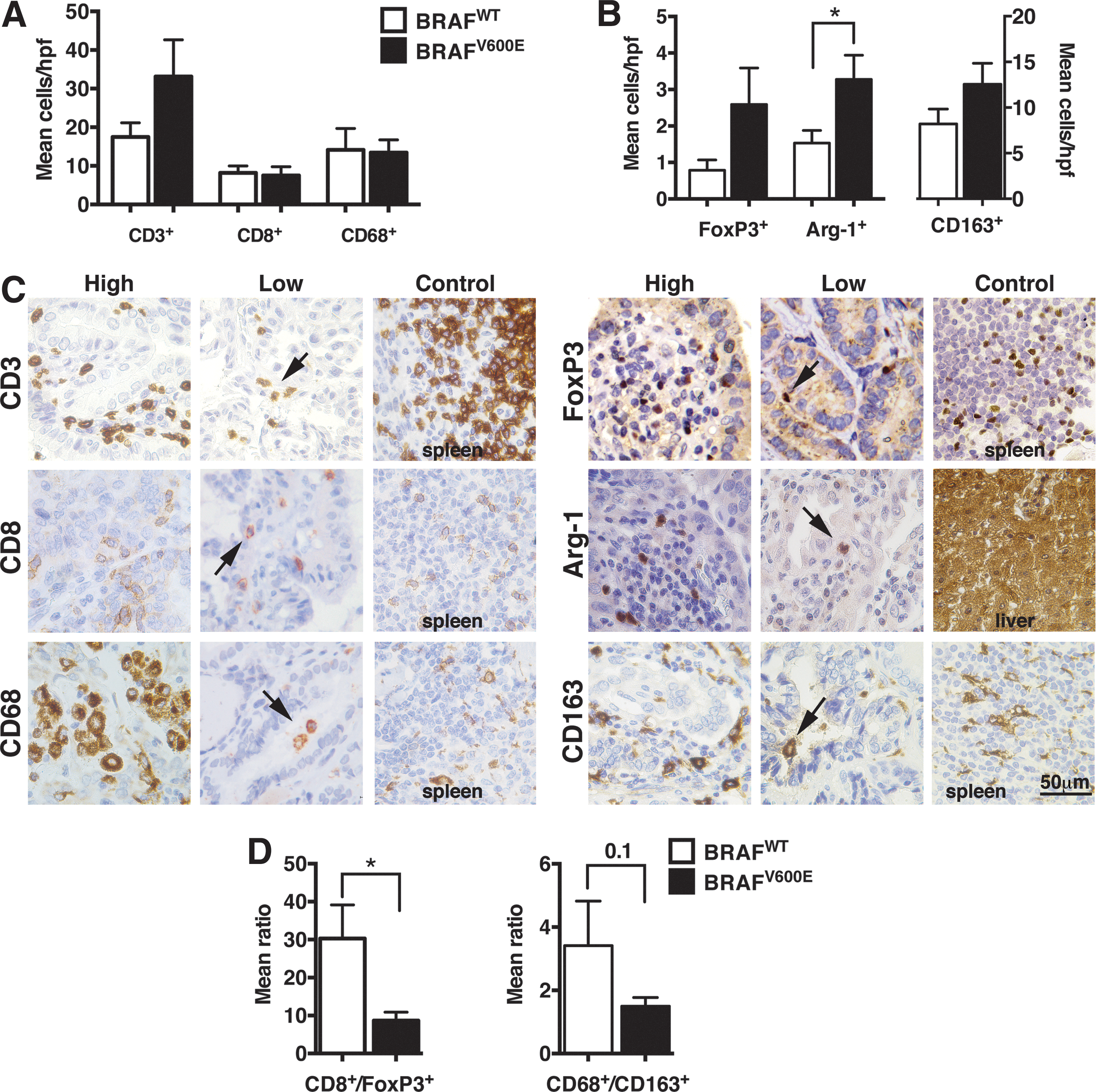
Correlation of BRAF status with tumor-infiltrating leukocytes (TILs) in papillary thyroid cancer (PTC). Mean (±standard error of the mean [SEM]) cells per high-powered field (hpf) for
Regression analysis revealed that markers of tumor immune suppression, namely tumor PD-L1, HLA-G, and IDO expression, decreased intratumoral CD8+/FoxP3+ cell ratio, and increased Arg-1+ tumor infiltrating leukocytes, were together, significant predictors of tumor BRAF status χ2[5]=32.88, p<0.001). The model explained 84.1% (Nagelkerke R 2) of the variance and correctly classified 90.9% of cases. When analyzed independent of BRAF status, the frequency of the studied immune populations in PTC specimens did not vary significantly between specimens stratified by patient age, TNM stage, tumor invasion, or lymph node metastasis. Logistic regression analyses were performed to evaluate markers of tumor immune suppression and BRAF status as predictors of pathologically aggressive disease. The combined model approached statistically significant prediction (p=0.061) only for lymph node metastasis, and no single factor was independently predictive.
Discussion
The BRAFV600E mutation has been shown to predict PTC aggressiveness and patient prognosis (10,13,27,28). Increased BRAF activity appears to augment numerous cancer pathways, including apoptosis resistance, cell proliferation, and angiogenesis (28), but its immunologic effects in PTC are not well studied. Building upon prior reports of single immune markers in PTC, this investigation evaluated multiple aspects of tumor-induced host immune tolerance, including tumor-expressed factors and infiltrating leukocyte populations, concurrently in PTC specimens to identify overarching patterns of tumor–host immune interactions, specifically in relation to tumor BRAF mutation status. BRAFV600E positivity was associated with an immunosuppressive profile, encompassing direct tumor expression of immunosuppressive ligands and recruitment of suppressor cells.
Examination of PTC expression of immune inhibitory ligands and enzymes found that PD-L1 overexpression was frequently found (53% of cases), confirming the findings of Cunha et al. (25) demonstrating increased PD-L1 staining in thyroid cancer specimens compared with benign thyroid tissue. In addition, the rate of high PD-L1 expression was significantly higher in BRAFV600E tumors. PD-L1 functions to inhibit activated immune cells, such as those directed against tumor expressed antigens, and prevents cytotoxic cell destruction in a variety of ways, including binding with the co-inhibitory receptor, PD-1 (29). Since PD-L1 expression in thyroid cancer has been associated with a number of poor prognostic indicators (30), promising PD-1/PD-L1 pathway antagonistic antibodies currently in clinical trials for other solid maignancies (29,31,32) shoud be considered for the immunotherapy of PTC once clinically available.
HLA-G is a nonclassic and inhibitory MHC class I molecule expressed at immuno-privileged sites and by the placenta to induce maternal/fetal tolerance that is known to inhibit antigen-presenting, natural killer, and T cells (33,34). Aberrant expression of HLA-G is a known mechanism of tumor-mediated immune tolerance and data suggest that HLA-G expression correlates with tumor growth and poorer prognosis (35,36). High HLA-G expression was present in a quarter of PTC cases and was more than three times more frequent in BRAFV600E compared to BRAFWT tumors. In two studies of thyroid cancer that did not analyze mutational status, HLA-G expression was assoicated with tumor size larger than 1 cm (37) and lymph node metastasis (38), respectively. Recently, Smallridge et al. (24) demonstrated an association between increased mRNA expression of HLA-G and BRAFV600E in PTC. Here we confirm the association between HLA-G and BRAFV600E at the protein level and place HLA-G in the context of broader immune suppression within BRAFV600E containing PTC.
The immunosuppressive enzyme IDO was found to be upregulated in nearly a third of PTC specimens, confirming recent results by Moretti et al. (23). Although not statistically significant, high IDO expression was more frequently observed in BRAFV600E tumors, suggesting that IDO inhibitors such as 1-methyltryptophan (39) may be a potential target for future therapeutic interventions for aggressive PTC.
Populations of tumor-infiltrating immune cells were also evaluated in relation to tumor BRAF status. It is now recognized that rather than simply being present, the type, location, functional status, and ratios of immune cells are important to predicting antitumor immunity (17,27). Conflicting reports on the prognostic importance of autoimmune thyroid disease (40 –42) may stem in part from not fully elucidating these key factors. Previous evaluations of immune infiltrates in PTC (43 –48) have focused on single suppressive immune populations, such as TAM or Treg, finding significant correlations with cancer aggressiveness. However, to our knowledge there has been little data describing multiple immune cell subtypes in PTC tumors, their relative frequencies, and their association with BRAFV600E positivity. A lower CD8+/FoxP3+ cell ratio, a marker of poorer prognosis in other solid malignancies (17), was found in BRAFV600E compared to BRAFWT thyroid cancers (p<0.05). This finding, as well as the observed greater CD3+ cell infiltration seen in BRAFV600E PTC, are consistent with gene expression data of colon cancers suggesting that activating BRAF mutations have greater immune cell infiltration and a reduced intratumoral CD8+ cell to Treg ratio (17,27,49). Similarly, studies in melanoma and murine models demonstrated TIL with a decreased CD8+ to Treg ratio when a BRAF mutation was present, and further showed that inhibition of the enhanced BRAF signaling modulated the cytokine mileu of the tumor microenvironment to promote anti-tumor immunity (50,51). Congruent with these reports, our findings suggest that aberrant constitutive BRAF activity in PTC promotes immune dysfunction by lowering the effector to suppressor cell ratio.
TAM may also be present within tumors and have been noted in thyroid cancers (30,43,46), but study of this population has been limited due to few distinguishing markers for proinflammatory M1 and suppressive M2 phenotypes. While there is no unique marker for M1 macrophages (52), CD163, a hemoglobin scavenger receptor (53), is now recognized as a marker for M2-polarized macrophages (54). While greater intratumoral TAM accumulation was demonstrated with BRAFV600E-induction in a mouse model (55), no association between TAM infiltration and BRAF status was found in a study of human thyroid cancer specimens (46). In this study, quantification of the relative M2 macrophage component by measurement of a CD68+/CD163+ ratio (52) showed that BRAFV600E tumors had a trend toward increased immunosuppressive M2 phenotype or TAMs. This suggests that, like the CD8+/FoxP3+ ratio, comparisons of macrophage subtypes may be more important than either population measured alone. This shift toward suppressive myeloid cell recruitement was further supported by the finding of an increased number of infiltrating arginase-1+ cells in BRAFV600E compared to BRAFWT tumors. Arginase-1 is another immunosuppressive enzyme expressed by TAM and myeloid suppressor cells (56) that can help to identify functional subsets of tumor-infiltrating myeloid cells. Future investigations with possible markers that are specific for M1 macrophages may further elucidate the contributions of these macrophage subsets to the tumor microenvironment.
In this study, while no single immune feature was independently predictive, the composite of tumor expressed PD-L1, HLA-G, and IDO, decreased intratumoral CD8+/FoxP3+ cell ratio, and increased Arg-1+ tumor infiltrating leukocytes, significantly predicted tumor BRAFV600E status, reinforcing the close relationship between BRAFV600E and a general profile of immune suppression. Irrespective of BRAF status, the frequency of TIL populations within PTC specimens stratified by clinical or pathologic characteristics did not vary significantly, though the analyses are preliminary and likely limited by the small sample size. However, logistic regression analysis indicated that presence of the BRAFV600E mutation combined with markers of tumor immune suppression may be predictive of lymph node metastasis (p=0.061). Larger investigations that concurrently assess multiple features of immune suppression may be necessary to demonstrate that the collective presence of BRAFV600E and immunosuppressive features predicts aggressive pathologic features.
These data show for the first time a significant positive association between BRAFV600E and PD-L1 expression, as well as arginase-1+ immune cell infiltrates. Along with increased expression of HLA-G and IDO, and a shift towards immunosupressive Treg and type M2 macrophages in TIL, our analysis of tumor-host immune interactions demonstrates a strongly immunosuppressive profile within the tumor microenvironment of BRAFV600E-positive PTC. While these associations provide preliminary data for the relationship between presence of BRAFV600E and strong immune suppression in PTC, the current study has a small sample size and by its retrospective nature is limited to correlative analyses.
Based on these data, we hypothesize that mutated BRAF protein antigens presented by BRAFV600E PTC cells may increase tumor antigenicity, prompting a greater immune response, as evidenced by the greater T cell infiltration observed in this study, and necessitating that BRAFV600E tumors evolve immunosuppressive mechanisms including PD-L1 and HLA-G expression, and induction or recruitment of suppressive immune cell populations in order to disrupt host immune surveillance and allow escape from immune-mediated tumor destruction. These findings suggest that further investigations using experimental in vitro and in vivo models are warranted to elucidate causative mechanisms by which BRAFV600E action promotes immune suppression and define its contribution to thyroid cancer immune escape.
Footnotes
Acknowledgments
The authors thank Dr. Peter Singer of the Keck Medical Center of USC, Los Angeles, California, for his assistance in the preparation of this manuscript, and Dr. Rikki B. Sevell, Mr. Connor H. Church, and Ms. Lillian Young, all of the Keck Medical Center of USC, for contributions to immunohistochemistry studies. This work was supported in part by the USC Steven's Institute for Innovation Ideas Empowered Program, National Institutes of Health grants 3T32GM067587-07S1 (M.G.L.) and P30CA014089, Department of Defense grant W81XWH-11-1-0466 (A.L.E), Cancer Therapeutics Laboratories, Inc. (Los Angeles, CA), of which A.L.E is a cofounder, and by the National Center for Research Resources and the National Center for Advancing Translational Sciences, National Institutes of Health (NIH), through Grant Award Number TL1TR000132. Julie K. Jang is a TL1 Trainee awarded under the TL1 (Pre-doctoral) Training Award through Southern California Clinical and Translational Science Institute at University of Southern California, Keck School of Medicine.
Author Disclosure Statement
All authors have no conflicts of interest to declare.