
Abstract
Background:
A recent clinical trial has shown a beneficial effect of the antioxidant agent selenium in Graves' orbitopathy (GO). In order to shed light on the cellular mechanisms on which selenium may act, this study investigated its effects in cultured orbital fibroblasts.
Methods:
Primary cultures of orbital fibroblasts from six GO patients and six control subjects were established. Cells were treated with H2O2 to induce oxidative stress, after pre-incubation with selenium-(methyl)selenocysteine (SeMCys). The following assays were performed: glutathione disulfide (GSSG), as a measure of oxidative stress, glutathione peroxidase (GPX) activity, cell proliferation, hyaluronic acid (HA), and pro-inflammatory cytokines.
Results:
H2O2 induced an increase in cell GSSG and fibroblast proliferation, which were reduced by SeMCys. Incubation of H2O2-treated cells with SeMCys was followed by an increase in glutathione peroxidase activity, one of the antioxidant enzymes into which selenium is incorporated. At the concentrations used (5 μM), H2O2 did not significantly affect HA release, but it was reduced by SeMCys. H2O2 determined an increase in endogenous cytokines involved in the response to oxidative stress and GO pathogenesis, namely tumor necrosis factor alpha, interleukin 1 beta, and interferon gamma. The increases in tumor necrosis factor alpha and interferon gamma were blocked by SeMCys. While the effects of SeMCys on oxidative stress and cytokines were similar in GO and control fibroblasts, they were exclusive to GO fibroblasts in terms of inhibiting proliferation and HA secretion.
Conclusions:
Selenium, in the form of SeMCys, abolishes some of the effects of oxidative stress in orbital fibroblasts, namely increased proliferation and secretion of pro-inflammatory cytokines. SeMCys reduces HA release in GO fibroblasts in a manner that seems at least in part independent from H2O2-induced oxidative stress. Some effects of SeMCys are specific for GO fibroblasts. These findings reveal some cellular mechanisms by which selenium may act in patients with GO.
Introduction
G
Selenium is a trace mineral and an essential nutrient for selenocysteine synthesis (16). The latter is incorporated into several selenoproteins, most of which are enzymes. Within selenoproteins, selenium acts as a reduction-oxidation center and exerts antioxidant actions. In this regard, several in vitro studies have shown that increased generation of oxygen free radicals plays a role in the pathogenesis of GO (17 –19). To investigate whether selenium has a beneficial effect in GO, Marcocci et al. carried out a multicenter, randomized, double-blind, placebo-controlled trial of selenium versus placebo, and found that treatment with selenium is associated with an improvement in eye signs and symptoms and in the quality of life of patients with mild GO (15).
The beneficial effects of selenium in GO may reflect either a direct effect in orbital tissues, where selenium may counteract oxidative stress, or an effect on the immune system, which has been shown to take place both in GD and autoimmune thyroiditis (16,20 –25). This study investigated some of the possible mechanisms responsible for the action of selenium in GO using primary cultures of orbital fibroblasts subjected to oxidative stress.
Materials and Methods
Primary cultures of fibroblasts
Orbital adipose tissue samples were collected from six GO patients who underwent orbital decompression. Normal orbital tissue samples were collected from six patients who underwent eye surgery for conditions other than GO not involving fibroadipose tissue. Signed, fully informed consent was obtained from all patients. Shortly prior to surgery, GO patients underwent, among others, the following assessments and measurements: (i) ophthalmological evaluation, including evaluation of the Clinical Activity Score (CAS) according to Mourits et al. (26); and (ii) measurement of serum autoantibodies against the thyrotropin receptor (TRAb; RSR Ltd., Cardiff, United Kingdom).
Primary cultures of orbital fibroblasts were prepared as described (27,28). Briefly, tissue samples were minced, dispersed in Medium 199 (Sigma, St. Louis, MO) containing 20% fetal bovine serum (Invitrogen Corp., Carlsbad, CA), penicillin (Sigma), and gentamycin (Sigma). Tissue samples were then kept in 10 cm Petri dishes in cell incubators at 37°C for two weeks. Cells attached to dishes were detached by trypsinization and seeded onto 10 cm Petri dishes (2 × 10⁶ cells per dish) in Medium 199 containing 10% fetal bovine serum and antibiotics. Medium was replaced by fresh medium on alternate days. Every passage was performed at confluence, which occurred every 5–10 days, depending on the cells.
For all the experiments, cells were seeded onto 96-well plates (5000 cells/well) in complete medium. Experiments were started after 24 hours, when ∼50% confluence had been reached.
Induction of oxidative stress and treatment with selenium
To induce oxidative stress, cells were incubated for 24 hours at 37°C with fresh, complete medium containing H2O2 at various concentrations, after which the medium was replaced with fresh complete medium and cells were kept in cell incubators at 37°C for 48 hours.
In experiments aimed at assessing the effects of selenium, cells were pre-incubated for two days in cell incubators at 37°C with complete medium without compounds, or with medium containing selenium in the form of Se-(methyl)selenocysteine hydrochloride (SeMCys; Sigma) or, as a control, methylcysteine (MCys; S-[methyl]-L-cysteine; Sigma) at various concentrations. Then, cells were treated with 5 μM of H2O2, as described above.
Preparation of cell extracts
Cells were washed with phosphate-buffered saline and incubated on ice for one hour in lysis buffer (1% Triton X-100, 1% deoxycholate in H2O). Samples were spun for 10 minutes at 10,000 g, pellets were discarded, and supernatants were collected. Protein concentrations were measured with the Bradford method.
Cell assays
After incubation with SeMCys or MCys, and/or H2O2, performed as detailed above, media were collected and cell vitality was assessed using a colorimetric reagent (Invitrogen Corp.), according to the manufacturer's instructions. Glutathione disulfide (GSSG) was measured in cell extracts using a commercial assay (Trevigen, Gaithersburg, MD), according to the manufacturer's instructions. Glutathione peroxidase (GPX) activity was measured using a commercial assay (Enzo Life Scinces, New York, NY), according to the manufacturer's instructions. Cell proliferation was measured using a commercial assay (Roche Applied Science, Mannheim, Germany), according to the manufacturer's instructions. Hyaluronic acid (HA) was measured in cell media using a commercial enzyme-linked immunosorbent assay (ELISA; Echelon Sciences, Salt Lake City, UT), according to the manufacturer's instructions. Tumor necrosis factor alpha (TNF-α), interleukin 1 beta (IL-1β), and interferon gamma (IFN-γ) were measured in cell media using commercial ELISAs (Invitrogen Corp.), according to the manufacturer's instructions.
Data presentation and statistics
Results were normalized for the amounts of proteins in cell extracts, unless otherwise specified. Data are presented as median and interquartile range. The following tests were performed as appropriate: (i) chi-square test; (ii) t-test; (iii) Wilcoxon's test; (iv) Friedman's test; and (v) analysis of variance for repeated measures.
Results
Preliminary tests
Primary cultures of orbital fibroblasts were established from six patients with GO and six patients without GO who underwent eye surgery. The demographical and clinical features of the patients are reported in Table 1. GO and control patients were similar in terms of age and sex distribution. All GO patients had long-standing, inactive eye disease, as shown by clinical activity score values ≤2/7 and by undetectable, or in two cases low, serum TRAb concentrations. All GO patients underwent orbital decompression for disfiguring proptosis. Control patients underwent eye surgery for eyelid ectropion or entropion. There were no patient-related differences in the results reported below (not shown).
Age is reported as mean ± SD. p-Values were obtained by chi-square (sex) and t-test (age).
GO, Graves' orbitopathy; TRAb, anti-TSH-receptor autoantibodies.
In order to induce oxidative stress, fibroblasts were incubated with H2O2. To determine the optimal concentrations of H2O2 to be used, cell vitality assays were performed after treatment of fibroblasts with increasing concentrations of H2O2 for 24 hours. As shown in Figure 1A, the majority of cells survived up to a H2O2 concentration of 5 μM. At this concentration, which was the lowest at which significant oxidative stress was observed (see below), the median vitality was 86.2% in GO and 79.4% in control fibroblasts. Overall, H2O2 had a significant, dose-dependent effect on cell vitality regardless of the cell origin, reflecting cytotoxicity at concentrations ≥10 μM. The effect of H2O2 on cell vitality was similar in GO and control fibroblasts.

(
GSSG was used as a measure of oxidative stress following incubation of fibroblasts with H2O2. As shown in Figure 1B, after incubation with increasing concentrations of H2O2, GSSG was detectable in the cell extract. Although there was some GSSG increase already at 2.5 μM, the effect became quite evident at a 5 μM concentration of H2O2, which was slightly lower than the 6.25 μM concentration used in a previous study (29). The effect somehow reached a plateau between 5 and 10 μM. Overall, the effect of H2O2 on GSSG was dose dependent and statistically significant. The apparent difference between GO and control fibroblasts was not statistically significant. In view of the findings obtained in cell vitality and GSSG release experiments, a 5 μM concentration of H2O2 was used in the experiments reported below.
In order to determine the optimal concentration of SeMCys to be used, the effects of SeMCys on cell vitality were assessed by incubating fibroblasts with increasing concentrations of the compound. As shown in Figure 1C, SeMCys was overall very poorly cytotoxic. Thus, a very slightly reduced cell vitality could be observed only at concentrations ≥500 μM. Overall, no statistically significant effect was observed, and there was no difference between GO and control fibroblasts. The highest vitality was observed at a 10 μM concentration of SeMCys for both GO and control fibroblasts, corresponding to a concentration of atomic selenium of 4.3 μM. Therefore, this concentration was used in the experiments reported below.
Selenium prevents oxidative stress
To determine the effects of SeMCys on the oxidative stress caused by H2O2, GSSG was measured in cell extracts following incubation of fibroblasts with H2O2, in cells pre-incubated or not with SeMCys. As shown in Figure 2A, the increase of GSSG induced by H2O2 was abolished by pre-incubation with SeMCys in a statistically significant manner in both GO and control fibroblasts. MCys, used as a negative control, had no effect. The apparent difference between GO and control fibroblasts was not statistically significant.
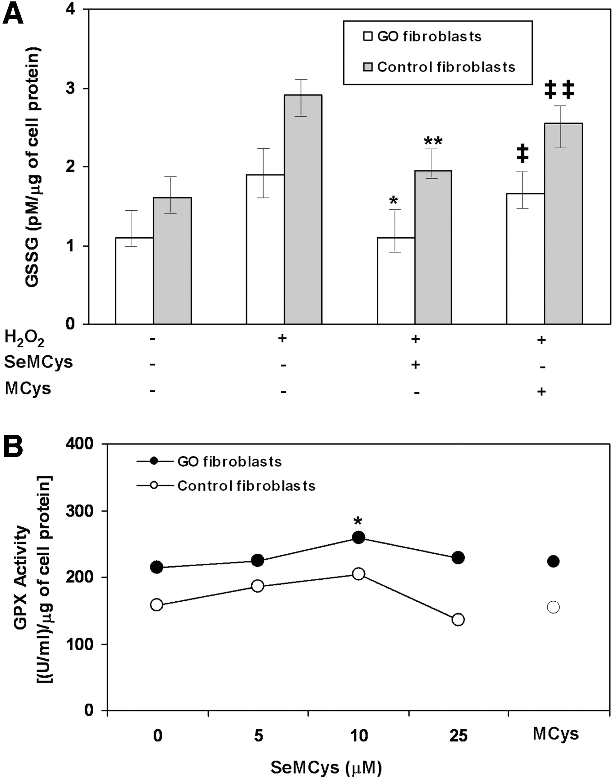
(
Selenium is known to exert its functions after incorporation into several selenoproteins, especially GPX (16,24–25). As shown in Figure 2B, following incubation of fibroblasts with H2O2, treatment with SeMCys, but not with MCys, resulted in a significant, dose-dependent increase in GPX activity, with a peak at a SeMCys concentration of 10 μM (the concentration used in all the experiments reported below), followed by a plateau. The apparent difference between GO and control fibroblasts was not statistically significant.
Selenium prevents cell proliferation
Fibroblast proliferation is probably the major pathogenetic mechanism of GO (1). Therefore, the effects of H2O2 and SeMCys on cell proliferation were investigated. As reported previously (29), in spite of its cytotoxic effects at high concentrations (see Fig. 1A), the relatively low concentration of H2O2 used (5 μM), which as reported above was not cytotoxic, determined a significant increase in cell proliferation in GO but not in control fibroblasts (Fig. 3). SeMCys, but not MCys, abolished the increased proliferation induced by H2O2 in GO fibroblasts. Overall, proliferation was significantly greater in GO than it was in control fibroblasts, regardless of incubation conditions.
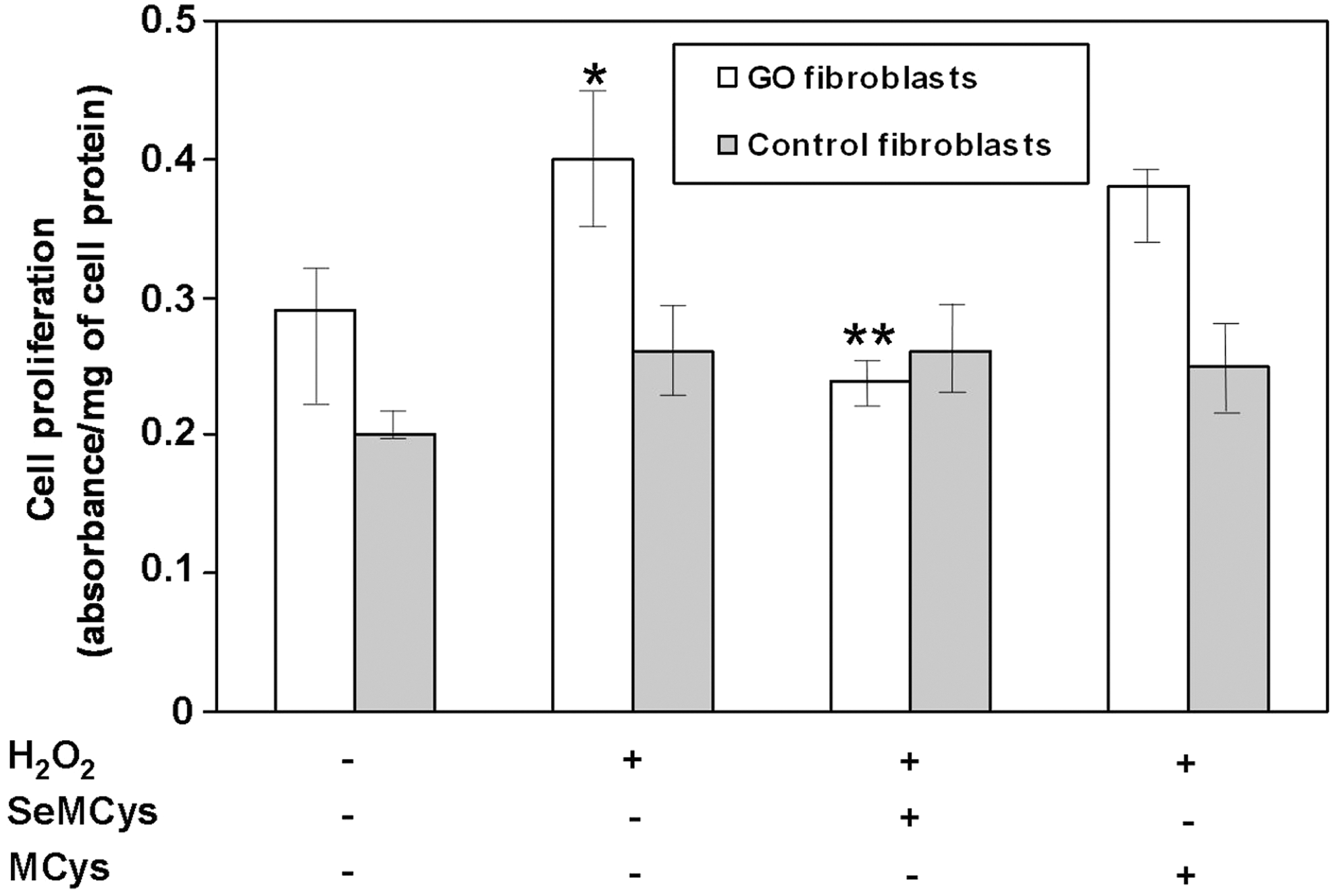
Combined effects of H2O2 (5 μM), SeMCys (10 μM) or its control MCys (10 μM) on cell proliferation in fibroblasts from patients with Graves' orbitopathy (GO fibroblasts) or from control subjects. *p = 0.02 versus untreated cells; **p = 0.02 versus H2O2; p = 0.003 between GO and control fibroblasts by ANOVA for repeated measures.
Selenium prevents HA release
Associated with their increased proliferation, orbital fibroblasts in GO release great amounts of glycosaminoglycans, especially HA, which contributes to the increased orbital content (1). The effect of H2O2 and SeMCys on HA release by orbital fibroblasts was investigated. As shown in Figure 4, HA in cell media was significantly greater in GO than it was in control fibroblasts, regardless of the incubation conditions. H2O2 did not significantly affect HA in GO or in control fibroblasts. However, SeMCys significantly reduced HA compared with cells treated with H2O2 in GO fibroblasts, whereas there was no effect in control fibroblasts. MCys had no effect in either cell type.
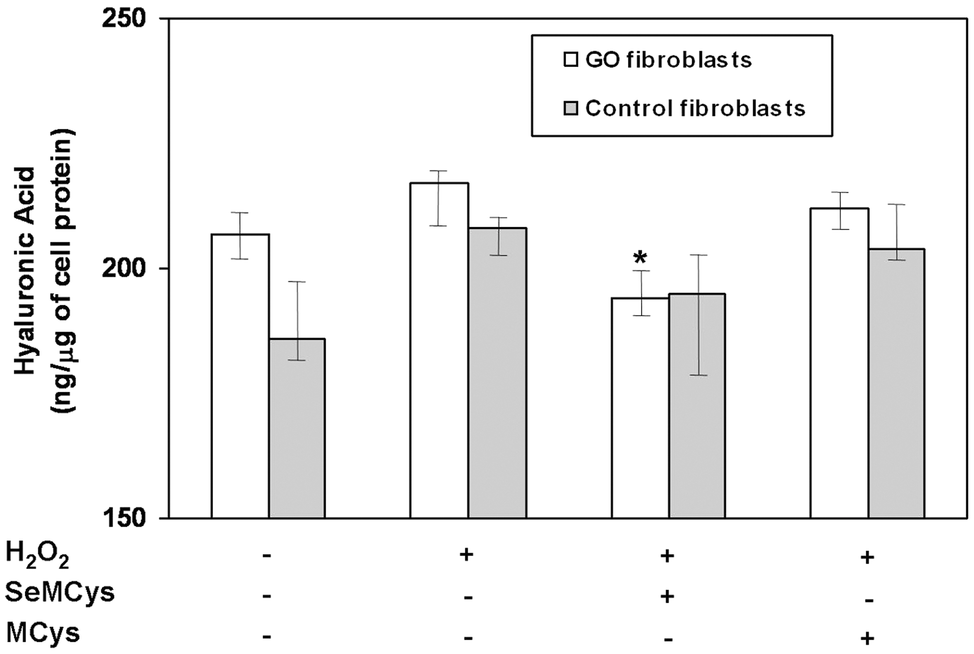
Combined effects of H2O2 (5 μM), SeMCys (10 μM), or its control MCys (10 μM) on hyaluronic acid (HA) release in fibroblasts from patients with Graves' orbitopathy (GO fibroblasts) or from control subjects. *p = 0.02 versus H2O2; p = 0.02 between GO and control fibroblasts by ANOVA for repeated measures.
Effect of selenium on pro-inflammatory cytokines
Oxidative stress is known to be associated with increased release of pro-inflammatory cytokines by fibroblasts, which is likely one of the mechanisms through which oxidative stress contributes to GO pathogenesis (1,30,31). The effects of SeMCys on cytokine release following treatment with H2O2 were investigated. For this purpose, TNF-α, IL-1β, and IFN-γ were measured in the cell medium. As shown in Figure 5A, TNF-α increased significantly upon H2O2 treatment, in both GO and control fibroblasts. The effect was virtually abolished by SeMCys, but not by MCys, in both cell groups to a significant extent, with no difference between GO and control fibroblasts.
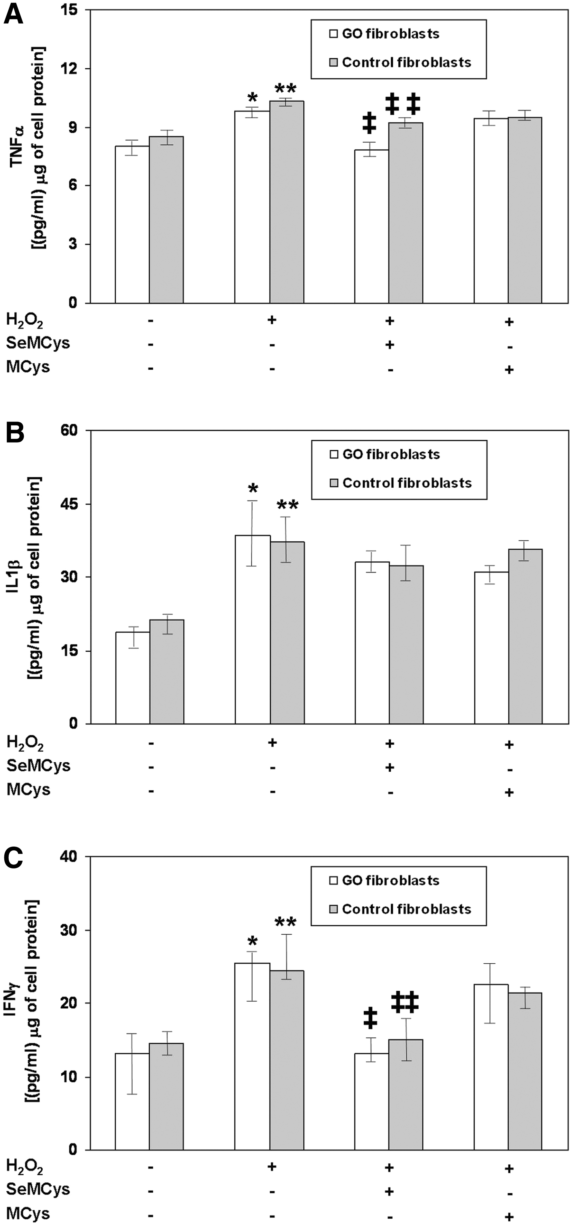
Combined effects of H2O2 (5 μM), SeMCys (10 μM), or its control MCys (10 μM) on pro-inflammatory cytokines in fibroblasts from patients with Graves' orbitopathy (GO fibroblasts) or from control subjects. (
In contrast, SeMCys had no effect on IL-1β. Thus, although treatment with H2O2 was followed by a significant increase of IL-1β in both GO and control fibroblasts, SeMCys, and also MCys, did not affect this cytokine (Fig. 5B). There was no difference between GO and control fibroblasts.
Finally, similar experiments were performed to investigate the effects of H2O2 and SeMCys on IFN-γ. As shown in Figure 5C, IFN-γ was significantly increased by H2O2 in both GO and control fibroblasts. SeMCys, but not MCys, abolished the effect of H2O2 in both cell groups to a significant extent, with no difference between GO and control fibroblasts.
Discussion
The present study shows an antioxidant action of selenium, in the form of SeMCys, in orbital fibroblasts, which provides a cellular basis for the effects of selenium in vivo in patients with GO (15). Selenium is known to be a quite potent inhibitor of oxidative stress (16), one of the mechanisms involved in the pathogenesis of GO (17 –19,30). This study found that administration of SeMCys to primary cultures of orbital fibroblasts from GO patients or control subjects reduces oxidative stress, which is associated with protection from increased cell proliferation, one of the major pathogenetic mechanisms of GO (1,30). In addition, SeMCys reduces HA release, another major mechanism involved in GO pathogenesis (1,30). Finally, SeMCys prevents some of the micro-environmental changes induced by oxidative stress, namely the increased release of some of the cytokines involved in GO pathogenesis (1,29 –31), which is likely one of the mechanisms through which oxidative stress, and therefore selenium, act on GO. The findings supporting the conclusions are summarized below.
H2O2 had an oxidative effect in orbital fibroblasts, reflected by a dose-dependent increase in GSSG, a known measure of cell response to free radicals (32). Pre-incubation of cells with SeMCys abolished the effects of H2O2, therefore providing evidence for an antioxidant action of SeMCys in orbital fibroblasts. The effects of SeMCys were observed in both fibroblasts from GO patients and from subjects without GO, suggesting that fibroblasts are susceptible to oxidative stress and to protection by SeMCys, regardless of the underlying conditions and/or of their genetic background. Treatment of cells with SeMCys was followed by an increase in GPX activity in both GO and control fibroblasts. As mentioned above, selenium acts after incorporation into various selenoproteins, including GPX, where selenium is located on the catalytic site of the enzyme, which is an essential component of its antioxidant action (16,24,25). These findings indicate that GPX is very likely involved in protection from oxidative stress by selenium in orbital fibroblasts. It remains to be investigated whether other selenoproteins are also involved. This was beyond the scope of the present study.
As mentioned above, fibroblast proliferation is one of the two major mechanisms responsible for the increased orbital volume in GO (1,30). In confirmation of a previous study (29), a relatively low concentration of H2O2 (5 μM) resulted in a significant increase in fibroblast proliferation, indicating that oxidative stress induces cell proliferation. The findings indicate that, concerning this particular feature and as reported below also concerning the release of HA, cells from GO patients are somehow different. This may reflect a different response to an initial pathogenetic event, presumably an autoimmune insult, or a different genetic background that renders them more proliferative. Obviously, these explanations are entirely speculative, and further studies are needed to clarify the mechanisms responsible for this different cell behavior. In GO fibroblasts, the effect of H2O2 in terms of proliferation was counteracted by SeMCys, which brought proliferation back to the levels observed in untreated cells. It is worth noting that H2O2 seems to exert a dual effect on fibroblasts. If used at doses >5 μM, it displays cytotoxic actions, as observed in cell vitality experiments, reflecting its well-known direct cytotoxic effect on cells. On the contrary, if used at concentrations ≤5 μM, H2O2 is poorly cytotoxic, and it rather stimulates cell proliferation, presumably indirectly, as a consequence of the induction of oxidative stress. Similar findings were obtained by Tsai et al., who used H2O2 in a concentration of 6.25 μM (29).
H2O2 did not significantly affect HA release in cultured fibroblasts. Nevertheless, in GO fibroblasts, SeMCys significantly reduced HA compared with cells treated with H2O2, bringing HA release to levels that were even lower than those observed in untreated cells. The finding seems to indicate that SeMCys acts on HA release, at least in part, regardless of the oxidative stress induced by H2O2, through mechanisms that remain to be investigated. SeMCys had no effect on orbital fibroblasts from subjects without GO. A possible explanation may be that it is identical to the mechanism suggested above for the effect on proliferation.
To gain some insights into the mechanisms induced by oxidative stress and involved in GO pathogenesis on which selenium may act, the release of pro-inflammatory cytokines upon oxidative challenge (treatment with H2O2) in orbital fibroblasts was investigated. In a previous study, it was shown that in addition to immunocompetent cells, fibroblasts are also capable of releasing pro-inflammatory cytokines, in particular TNF-α, IL-β, and IFN-γ, in response to oxidative stress (29). In turn, these cytokines stimulate HA synthase, leading to an increase in HA release (30). In addition to these actions, IFN-γ is known to regulate fibroblast growth and collagen biosynthesis, and TNF-α is able to stimulate fibroblast proliferation (30). As reported previously (29), treatment with H2O2 resulted in an increased release of TNF-α, IL-1β, and IFN-γ in both GO and control fibroblasts. The effect of H2O2 on TNF-α and IFN-γ, but not on IL-1β, was abolished by SeMCys in both cell types, suggesting that SeMCys acts on the oxidative stress-induced release of some pro-inflammatory cytokines, which may be responsible for some of the beneficial effects of selenium in GO fibroblasts. The different behavior of pro-inflammatory cytokines in response to selenium is quite intriguing and requires further investigations to shed light on the responsible mechanisms. In this regard, a highly speculative hypothesis involves the IL1-receptor antagonist (IL1-RA), a natural competitor for binding to the IL1-receptor (IL1-R), which is synthesized and released by the same cells that produce IL1 (31). Selenium may independently cause an increase in IL1-RA, which, by competing for IL1-R, may result in the persistence of increased IL-1β in the cell medium following exposure of the cells to H2O2. A similar phenomenon cannot occur for TNF-α and IFN-γ, as they do not have natural antagonists. The fact that IL1-RA itself has antioxidant actions is also quite intriguing (32). Clearly, further studies are needed to investigate this hypothesis. Because the respective role and relative importance of the various pro-inflammatory cytokines in the cellular and biochemical milieu of orbital tissue in GO is not established, it is not known whether the different effects of selenium on pro-inflammatory cytokines have any implications on its actions in vivo in patients with GO. IL-1β is more effective in eliciting HA synthase in skin fibroblasts compared with TNF-α and especially with IFN-γ (29). However, to the authors' knowledge, no data are available in orbital fibroblasts, nor is it known whether and to what extent orbital tissue cytokines of GO patients are affected by selenium.
This study carries the obvious limitations of in vitro investigations. In the cell system, only orbital fibroblasts were present, unlike in vivo where immune and inflammatory cells determine a much more complex environment, which probably includes the presence of autoantibodies against the thyrotropin receptor, which is largely considered the most important autoantigen in GO, among other important actors (1). A further development of these investigations may foresee the use of more complex culture systems, for example with the addition of the well-known monoclonal antithyrotropin receptor antibody M22 (33).
As mentioned above, selenium was used in the form of SeMCys. However, this is only one of the various selenium-containing compounds that are known to be metabolized by different biochemical cell pathways and have eventually different extents of action in cells (16,23,24,34). Thus, it is possible that different forms of selenium may be less or more effective than SeMCys in fibroblasts, which will be the subject of further studies. Of note, selenium is known to be incorporated into selenoproteins, which in turn act as reduction-oxidation centers (16,24,34). This study demonstrates an increased GPX activity induced by selenium in orbital fibroblasts, but additional studies are needed to investigate whether other selenoproteins contribute to the actions of selenium.
As mentioned above, these investigations were based on the observation of a beneficial effect of selenium compared with placebo in patients with GO, as shown by a multicenter, double-blind, randomized clinical trial conducted by the European Group on Graves' Orbitopathy (EUGOGO) (15). Treatment with selenium did result in an improvement of the overall GO outcomes, as illustrated by an amelioration of soft-tissue involvement and eyelid aperture. The quality of life of patients increased accordingly. The EUGOGO study included only patients with mild GO (15), but in view of the findings indicating a direct action in orbital fibroblasts, it is postulated that selenium may also exert some beneficial effects in moderate to severe GO. Further studies are needed to investigate this possibility.
In conclusion, this study provides insights into some cellular mechanisms that may explain the beneficial effects of selenium observed in patients with mild GO (15). Selenium has a direct action in orbital fibroblasts, presumably the major target of the immune system in GO. It remains to be investigated whether the effects of selenium in patients with GO also reflect its known actions on the immune system (16,24,25).
Footnotes
Acknowledgment
This study was unconditionally supported by IBSA Farmaceutici Italia, Lodi, Italy.
Author Disclosure Statement
The authors declare that they do not have any commercial association that might create a conflict of interest in connection with this manuscript.